. 2024 Jun:72:103148.
doi: 10.1016/j.redox.2024.103148. Epub 2024 Apr 5.

Selenite selectively kills lung fibroblasts to treat bleomycin-induced pulmonary fibrosis

Affiliations

Selenite selectively kills lung fibroblasts to treat bleomycin-induced pulmonary fibrosis

Jiun-Han Lin et al. Redox Biol. 2024 Jun.

Abstract

Background: Interstitial lung disease (ILD) treatment is a critical unmet need. Selenium is an essential trace element for human life and an antioxidant that activates glutathione, but the gap between its necessity and its toxicity is small and requires special attention. Whether selenium can be used in the treatment of ILD remains unclear.

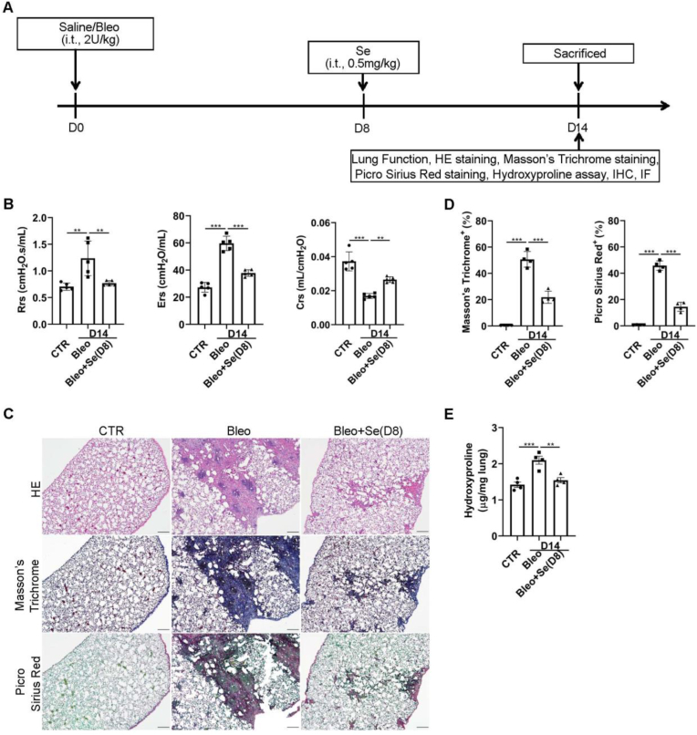
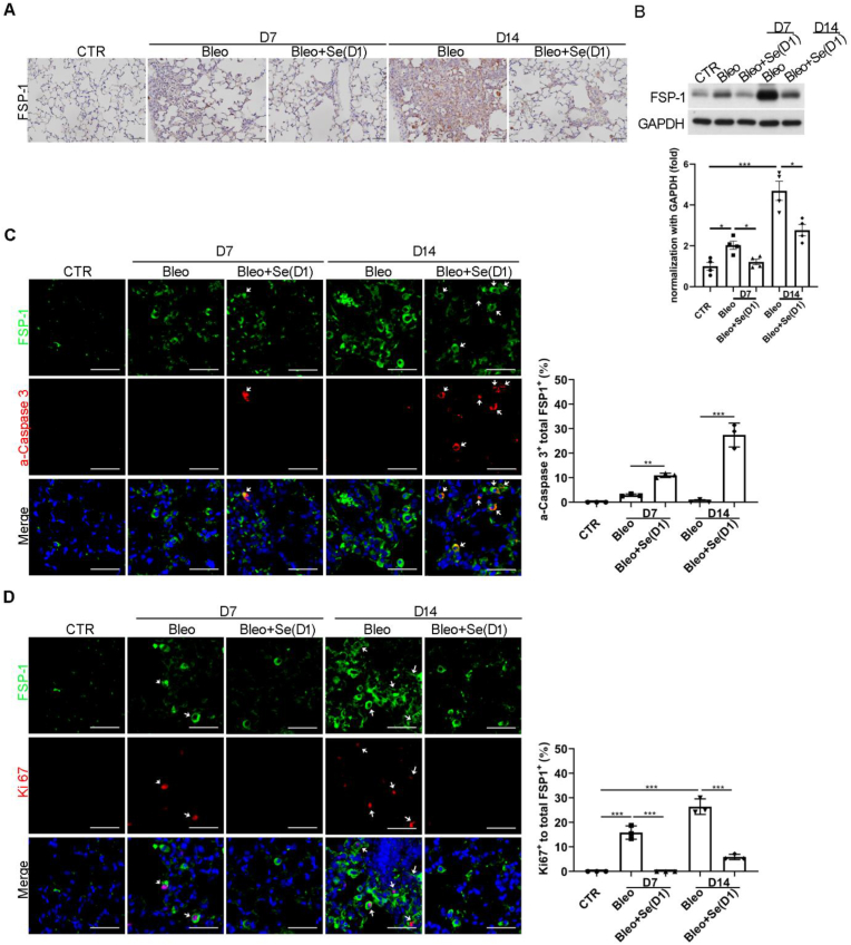
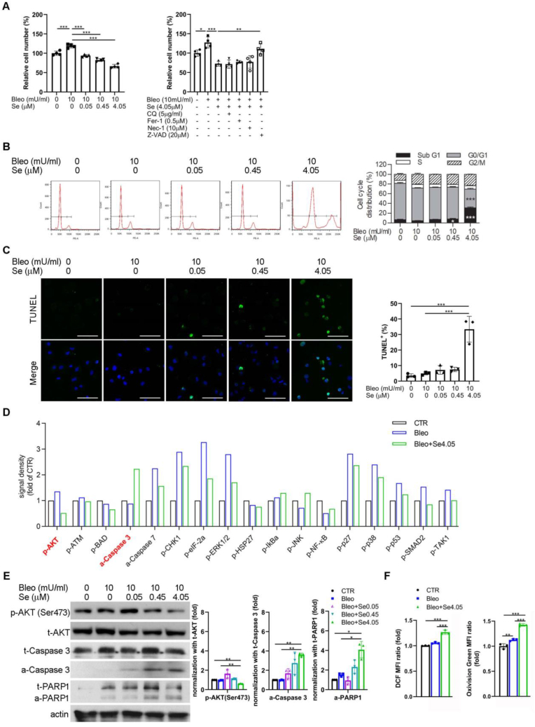
Methods: We investigated the prophylactic and therapeutic effects of selenite, a selenium derivative, in ILD using a murine model of bleomycin-induced idiopathic pulmonary fibrosis (IPF). We further elucidated the underlying mechanism using in vitro cell models and examined their relevance in human tissue specimens. The therapeutic effect of selenite in bleomycin-administered mice was assessed by respiratory function and histochemical changes. Selenite-induced apoptosis and reactive oxygen species (ROS) production in murine lung fibroblasts were measured.

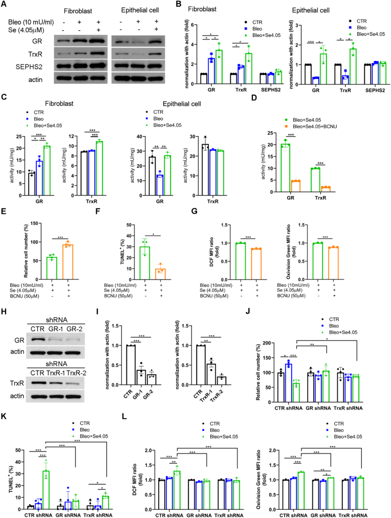
Results: Selenite, administered 1 day (inflammation phase) or 8 days (fibrotic phase) after bleomycin, prevented and treated deterioration of lung function and pulmonary fibrosis in mice. Mechanistically, selenite inhibited the proliferation and induced apoptosis of murine lung fibroblasts after bleomycin treatment both in vitro and in vivo. In addition, selenite upregulated glutathione reductase (GR) and thioredoxin reductase (TrxR) in murine lung fibroblasts, but not in lung epithelial cells, upon bleomycin treatment. GR and TrxR inhibition eliminates the therapeutic effects of selenite. Furthermore, we found that GR and TrxR were upregulated in the human lung fibroblasts of IPF patient samples.

Conclusions: Selenite induces ROS production and apoptosis in murine lung fibroblasts through GR and TrxR upregulation, thereby providing a therapeutic effect in bleomycin-induced IPF.

Keywords: Apoptosis; Glutathione reductase; Pulmonary fibrosis; ROS; Selenium; Thioredoxin reductase.


Conflict of interest statement

Declaration of competing interest The authors declare that they have no known competing financial interests or personal relationships that could have appeared to influence the work reported in this paper.

Figures

Fig. 1
Fig. 1
Selenite prevented pulmonary fibrosis and improved lung function in the bleomycin model of IPF mice. (A) Flow chart of the experimental protocol. Bleomycin (2 U/kg) or saline (vehicle control, CTR) was intratracheally (i.t.) administrated on day 0 (D0). Selenite (0.5 mg/kg, i.t.) was intratracheally administered on day 1 (D1). Mice were sacrificed on day 7 (D7) for the preventive effect or on day 14 (D14) for the antifibrotic effect of selenite. (B) Lung function test performed on D7 or D14 before sacrifice. Rrs, airway resistance; Ers, lung tissue elastance; Crs, lung tissue compliance. Scatter plots representing individual mice. n = 3∼5 per group. (C) Lung tissue histological analysis with HE, Masson’s trichrome staining, picrosirius red staining. (D) The quantification of Masson’s trichrome and picrosirius red staining. Scatter plot showing the percentage of the positive area of staining in lung tissue. n = 4 mice per group. (E) The quantification of hydroxyproline. Scatter plot representing a single mouse. n = 5 per group. Scale bar, 200 μm *p < 0.05, **p < 0.01, ***p < 0.001. (For interpretation of the references to colour in this figure legend, the reader is referred to the Web version of this article.)
Fig. 2
Fig. 2
Selenite improved pulmonary fibrosis and lung function in the bleomycin model of IPF mice. (A) Flow chart of the experimental protocol. Bleomycin (i.t., 2 U/kg) or saline (CTR) was administrated on day 0. Selenite (i.t., 0.5 mg/kg) was administered on day 8 (D8) and mice were sacrificed on day 14. (B) Lung function test performed on day 14 before sacrifice. Scatter plot representing a single mouse. n = 5 per group. (C) Lung tissue histological analysis with HE, Masson’s trichrome staining, picrosirius red staining. (D) The quantification of Masson’s trichrome and picrosirius red staining. Scatter plot showing the percentage of the positive area of staining in lung tissue. (E) The quantification of hydroxyproline. Scatter plots representing individual mice. n = 4 per group in D and E. Scale bar, 200 μm *p < 0.05, **p < 0.01, ***p < 0.001. (For interpretation of the references to colour in this figure legend, the reader is referred to the Web version of this article.)
Fig. 3
Fig. 3
Selenite treatment in the inflammatory phase of the bleomycin model of IPF mice inhibited proliferation and induced apoptosis of lung fibroblasts. Mice administrated with bleomycin (i.t., 2 U/kg) or saline (CTR) on day 0 were treated with or without selenite (0.5 mg/kg, i.t.) on day 1. Mice were sacrificed on day 7 or day 14 and lung specimens were harvested for (A) immunohistochemical (IHC) analysis with FSP-1 staining, (B) Western blot analysis with densitometry (lower panel) of FSP-1, (C) immunofluorescent (IF) staining with FSP-1 and a-caspase-3 followed by quantification, (D) IF staining with FSP-1 and Ki67 followed by quantification. The arrow indicated an example of a cell double staining for these markers. Scatter plot showing the percentage of the positive area of staining in lung tissue. n = 3∼4 mice per group. Scale bar, 50 μm *p < 0.05, **p < 0.01, ***p < 0.001.
Fig. 4
Fig. 4
Selenite treatment in the fibrotic phase of the bleomycin model of IPF mice inhibited proliferation and induced apoptosis of lung fibroblasts. Mice administrated with bleomycin (i.t., 2 U/kg) or saline (CTR) on day 0 were treated with or without selenite (i.t., 0.5 mg/kg) on day 8. Mice were sacrificed on day 14 and lung specimens were harvested for (A) IHC analysis with FSP-1 staining, (B) Western blot analysis with densitometry (lower panel) of FSP-1, (C) IF staining with FSP-1 and a-caspase-3 followed by quantification, (D) IF staining with FSP-1 and Ki67 followed by quantification. The arrow indicated an example of a cell double staining for these markers. Scatter plot showing the percentage of the positive area of staining in lung tissue. n = 3∼4 mice per group. Scale bar, 50 μm *p < 0.05, **p < 0.01, ***p < 0.001.
Fig. 5
Fig. 5
Selenite induced apoptosis and ROS of murine lung fibroblasts under bleomycin treatment. (A) Left panel, cell number of murine lung fibroblasts treated with or without selenite (0.05, 0.45, 4.05 μM) under bleomycin treatment (10 mU/ml) for 24 h; right panel, cell number of fibroblasts pretreated for 1 h with chloroquine (CQ, 0.5 μg/ml), Ferrostatin-1 (Fer-1, 0.5 μM), Necrostatin-1 (Nec-1, 10 μM), Z-VAD-FMK (Z-VAD, 20 μM), and then treated with selenite (4.05 μM) and bleomycin for 24 h. Scatter plot showing the results for independent samples examined. n = 4 per group. (B) Cell cycle analysis of fibroblasts treated with or without selenite under bleomycin treatment for 48 h. Fibroblasts were treated with or without selenite under bleomycin treatment for 24 h, then subjected to (C) TUNEL assay with quantification, (D) protein array of apoptosis-related signaling pathways, (E) Western blot analysis with densitometry (right panel) of p-AKT, t-AKT, t-caspase-3, a-caspase-3, total and active poly (ADP ribose) polymerase (t-PARP and a-PARP). Densitometry for p-AKT and active caspase 3 and PARP1 are normalized to total AKT, caspase 3, and PARP1, respectively. (F) ROS and H2O2 measurement of fibroblasts treated without or with bleomycin alone or with selenite (4.05 μM) for 3 h n = 3 per group in B, C, E, and F. Scale bar 100 μm *p < 0.05, **p < 0.01, ***p < 0.001.
Fig. 6
Fig. 6
Selenite improved the cell survival and reduced apoptosis and ROS of murine lung epithelial cells under bleomycin treatment. Murine lung epithelial cells were treated with bleomycin (10 mU/ml) or selenite (4.05 μM) for 24 h, then subjected to (A) cell number and (B) TUNEL assay with quantification. Scale bar, 100 μm. (C) ROS and H2O2 measurement of epithelial cells treated without or with bleomycin alone or with selenite for 3 h n = 4 per group in A. n = 3 per group in B and C. *p < 0.05, **p < 0.01, ***p < 0.01.
Fig. 7
Fig. 7
Selenite upregulated GR and TrxR to induce apoptosis and ROS accumulation in murine lung fibroblasts treated with bleomycin. Murine lung fibroblasts and epithelial cells were treated with bleomycin (10 mU/ml) or selenite (4.05 μM) for 24 h, then subjected to (A) Western blot analysis with (B) densitometry of GR, TrxR, and SEPHS2. (C) GR and TrxR activity of fibroblasts and epithelial cells treated with bleomycin or selenite for 3 h. Fibroblasts were treated with or without BCNU (50 μM) under bleomycin and selenite treatment for 24 h, then subjected to (D) GR or TrxR activities, (E) cell number, and (F) TUNEL assay with quantification. (G) ROS and H2O2 measurement of fibroblasts treated with or without BCNU under bleomycin and selenite treatment for 3 h. (H) Western blot analysis and (I) densitometry of fibroblasts with GR or TrxR knockdown. CTR, scramble control. Fibroblasts with GR or TrxR knockdown were treated with bleomycin or selenite for 24 h and then subjected to (J) cell number and (K) TUNEL assay with quantification. (L) ROS and H2O2 measurement of fibroblasts with GR or TrxR knockdown treated with bleomycin or selenite for 3 h. Scale bar, 100 μm. n = 3 per group in B, C, D, E, F, G, I, and L. n = 4 per group in J and K. *p < 0.05, **p < 0.01, ***p < 0.001.
Fig. 8
Fig. 8
Inhibition of GR and TrxR eliminated the therapeutic effects of selenite in the bleomycin model of IPF mice. In the bleomycin model of IPF, mice were treated without (CTR) or with bleomycin on day 0, then sacrificed on day 14. The lung tissue section and homogenates were subjected to (A) IHC analysis and (B) Western blot analysis with densitometry (right panel) of GR and TrxR, (C) IF staining of GR, TrxR, FSP1, and CK18. The ratio of GR or TrxR positive cells in FSP1 positive or CK18 positive cells was counted according to IF staining. n = 4 per group in IHC and n = 3 per group in IF. To inhibit the therapeutic effect of selenite in bleomycin model of IPF, bleomycin (i.t., 2 U/kg) and BCNU (i.t., 1 mg/kg) were administrated in mice on day 0. Selenite (i.t., 0.5 mg/kg) was administered on day 1, and mice were sacrificed on day 14. (D) Lung function measurement was performed on day 14 before sacrifice. n = 4∼5 per group. (E) Lung tissue histological analysis with HE, Masson’s trichrome staining, picrosirius red staining, and IHC analysis with FSP-1 staining. (F) The quantification of Masson’s trichrome and picrosirius red staining. (G) The quantification of hydroxyproline. (H) Western blot analysis with densitometry (lower panel) of FSP-1. n = 3∼4 per group in F, G, and H. Scale bar for IHC and IF staining, 50 μm; scale bar for HE, Masson’s trichrome, and picrosirius red staining, 200 μm *p < 0.05, **p < 0.01, ***p < 0.001. (For interpretation of the references to colour in this figure legend, the reader is referred to the Web version of this article.)
Fig. 9
Fig. 9
GR and TrxR expressions were increased in human lung fibroblasts of IPF patients. (A) Masson’s trichrome staining, picrosirius red staining, and IHC analysis of GR and TrxR expression in lung tissue of normal people and IPF patients. (B) Percentages of GR positive or TrxR positive cells in human fibroblasts in scRNA-seq database. (C) IF staining of GR, TrxR, and FSP1 in lung tissue of normal people and IPF patients. The ratio of GR or TrxR positive cells in FSP1 positive. n = 3 in each group. Scale bar, 50 μm. (For interpretation of the references to colour in this figure legend, the reader is referred to the Web version of this article.)

References

    1. Kalchiem-Dekel O., Galvin J.R., Burke A.P., Atamas S.P., Todd N.W. Interstitial lung disease and pulmonary fibrosis: a practical approach for general medicine physicians with focus on the medical history. J. Clin. Med. 2018;7(12) - PMC - PubMed
    1. Ley B., Collard H.R. Epidemiology of idiopathic pulmonary fibrosis. Clin. Epidemiol. 2013;5:483–492. - PMC - PubMed
    1. King T.E., Jr., Bradford W.Z., Castro-Bernardini S., Fagan E.A., Glaspole I., Glassberg M.K., et al. A phase 3 trial of pirfenidone in patients with idiopathic pulmonary fibrosis. N. Engl. J. Med. 2014;370(22):2083–2092. - PubMed
    1. Richeldi L., du Bois R.M., Raghu G., Azuma A., Brown K.K., Costabel U., et al. Efficacy and safety of nintedanib in idiopathic pulmonary fibrosis. N. Engl. J. Med. 2014;370(22):2071–2082. - PubMed
    1. Ambardar S.R., Hightower S.L., Huprikar N.A., Chung K.K., Singhal A., Collen J.F. Post-COVID-19 pulmonary fibrosis: novel sequelae of the current pandemic. J. Clin. Med. 2021;10(11) - PMC - PubMed

Publication types

MeSH terms

LinkOut - more resources