. 2024 May 1:394:110993.
doi: 10.1016/j.cbi.2024.110993. Epub 2024 Apr 9.

Biochemical, structural, and computational analyses of two new clinically identified missense mutations of ALDH7A1

Affiliations

Biochemical, structural, and computational analyses of two new clinically identified missense mutations of ALDH7A1

David A Korasick et al. Chem Biol Interact. .

Abstract

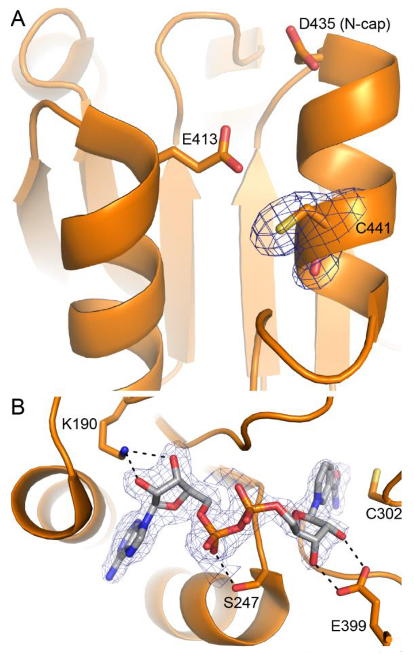
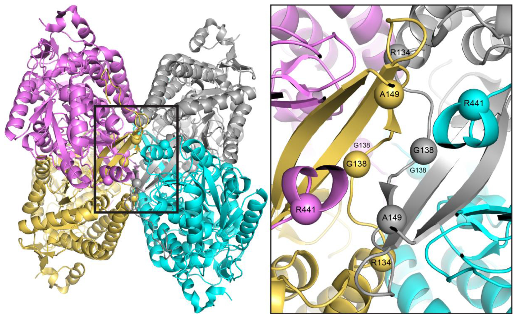
Aldehyde dehydrogenase 7A1 (ALDH7A1) catalyzes a step of lysine catabolism. Certain missense mutations in the ALDH7A1 gene cause pyridoxine dependent epilepsy (PDE), a rare autosomal neurometabolic disorder with recessive inheritance that affects almost 1:65,000 live births and is classically characterized by recurrent seizures from the neonatal period. We report a biochemical, structural, and computational study of two novel ALDH7A1 missense mutations that were identified in a child with rare recurrent seizures from the third month of life. The mutations affect two residues in the oligomer interfaces of ALDH7A1, Arg134 and Arg441 (Arg162 and Arg469 in the HGVS nomenclature). The corresponding enzyme variants R134S and R441C (p.Arg162Ser and p.Arg469Cys in the HGVS nomenclature) were expressed in Escherichia coli and purified. R134S and R441C have 10,000- and 50-fold lower catalytic efficiency than wild-type ALDH7A1, respectively. Sedimentation velocity analytical ultracentrifugation shows that R134S is defective in tetramerization, remaining locked in a dimeric state even in the presence of the tetramer-inducing coenzyme NAD+. Because the tetramer is the active form of ALDH7A1, the defect in oligomerization explains the very low catalytic activity of R134S. In contrast, R441C exhibits wild-type oligomerization behavior, and the 2.0 Å resolution crystal structure of R441C complexed with NAD+ revealed no obvious structural perturbations when compared to the wild-type enzyme structure. Molecular dynamics simulations suggest that the mutation of Arg441 to Cys may increase intersubunit ion pairs and alter the dynamics of the active site gate. Our biochemical, structural, and computational data on two novel clinical variants of ALDH7A1 add to the complexity of the molecular determinants underlying pyridoxine dependent epilepsy.

Keywords: Aldehyde dehydrogenase; Analytical ultracentrifugation; Enzyme kinetics; Missense mutation; Molecular dynamics simulations; X-ray crystallography.


Conflict of interest statement

Declaration of competing interest The authors declare that they have no known competing financial interests or personal relationships that could have appeared to influence the work reported in this paper.

Figures

Fig. 1.
Fig. 1.
Structural context of the mutations R134S and R441C. A, The reaction catalyzed by ALDH7A1 and a cartoon drawing of one protomer of wild-type ALDH7A1 (PDB code 4ZUL). The domains are colored as follows: light blue; NAD+-binding; green, catalytic; and wheat, oligomerization. The active site gate corresponds to the C-terminal 12 residues (500-511) and is colored red. The mutations studied here target Arg134 (purple sticks) and Arg441 (orange sticks). B, The ALDH7A1 dimer-of-dimers tetramer, viewed down one of its 2-fold symmetry axes. Each chain has a different color: gray, cyan, violet, and gold. C, The structural context of Arg134 and Arg441 in the dimer-dimer interface of the wild-type ALDH7A1 tetramer. Each chain of the tetramer has a different color, as in panel B. The active site gate of one of the protomers is colored red. The catalytic cysteine (Cys302) and the product α-aminoadipate (AA) are shown in cyan sticks. The black dashes denote hydrogen bonds and ion pairs.
Fig. 2.
Fig. 2.
Steady-state kinetic analysis. (A) Kinetic data for wild-type ALDH7A1 (black) and the variants R134S (purple) and R441C (orange) as a function of AASAL concentration at fixed NAD+ concentration of 2.5 mM. The circles, squares, and triangles represent three replicate experiments performed at the same AASAL concentration. Wild-type ALDH7A1 was assayed at an enzyme concentration of 0.07 μM. Due to their low catalytic activities, the variants were assayed at an enzyme concentration of 5 μM. The black and orange curves represent global fits to the Michaelis-Menten equation for wild-type ALDH7A1 and R441C, respectively. The purple line is from linear regression of the R134S data. Kinetic parameters from curve fitting are listed in Table 2. (B) Expanded view of the data for the two variants. Note the vertical axis spans only about one-tenth of the scale of panel A.
Fig. 3.
Fig. 3.
Determination of the oligomeric states in solution from sedimentation velocity analytical ultracentrifugation for (A) wild-type ALDH7A1, (B) R134S, and (C) R441C. In each panel, the dashed curve is the distribution of sedimentation coefficients, c(S), for the enzyme in the absence of NAD+, and the solid curve is the c(S) in the presence of 1 mM NAD+.
Fig. 4.
Fig. 4.
Electron density from the structure of R441C complexed with NAD+. (A) Electron density for the mutated residue, verifying cysteine at residue 441. (B) Electron density and interactions for NAD+ bound to R441C. In both panels, the blue mesh represents a polder omit map contoured at 4σ.
Fig. 5.
Fig. 5.
Dynamics of the active site gate. (A) Structures of the active site gates in the open (PDB 4ZUK, salmon) and closed (PDB 4ZUL, cyan) states, highlighting the motion of the gate inferred from crystal structures. (B) Histogram of the active site gate status, defined as the distance between the Cα atoms of Gly461 and Ala505. The values observed in the crystal structures representing the open and closed states are noted. The bin size is 0.3 Å.
Fig. 6.
Fig. 6.
Histogram of Lys500-Asp435 ion pair formation, as defined as the distance between NZ of Lys500 and CG of Asp435. The peak at 3-4 Å indicates formation of the ion pair. The bin size is 0.1 Å.
Fig. 7.
Fig. 7.
Examples of the intersubunit Lys500-Asp435 ion pair formed during MD simulations. (A) Two frames from the wild-type simulation viewed down a two-fold axis. In one of the frames, an ion pair linking Lys500 of chain A and Asp435 of chain C is observed. The curved arrow shows the rotation of Arg441 in chain C that accompanies ion pair formation. (B) Typical frame from the R441C simulation, showing two Lys500-Asp435 ion pairs formed across the 2-fold symmetry axis. The backbones of Arg/Cys441 are highlighted in red in both panels.
Fig. 8.
Fig. 8.
Locations of Arg134, Gly138, Ala149, and Arg441 near a two-fold axis of the tetramer (spheres denote Cα atoms). Left: the tetramer is viewed down the two-fold axis that passes between oligomerization flaps of adjacent protomers (PDB 4ZUK). Inset: close-up view of the region of interest.

References

    1. Coughlin CR 2nd, Swanson MA 2nd, Spector E Meeks NJL Kronquist KE Aslamy M Wempe MF van Karnebeek CDM Gospe SM Jr. Aziz VG Tsai BP Gao H Nagy PL Hyland K van Dooren SJM Salomons GS Van Hove JLK, The genotypic spectrum of ALDH7A1 mutations resulting in pyridoxine dependent epilepsy: A common epileptic encephalopathy, Journal of inherited metabolic disease, 42 (2019) 353–361. - PMC - PubMed
    1. Tamaura M, Shimbo H, Iai M, Yamashita S, Osaka H, Seizure recurrence following pyridoxine withdrawal in a patient with pyridoxine-dependent epilepsy, Brain Dev, 37 (2015) 442–445. - PubMed
    1. Haidar Z, Jalkh N, Corbani S, Fawaz A, Chouery E, Megarbane A, Atypical pyridoxine dependent epilepsy resulting from a new homozygous missense mutation, in ALDH7A1, Seizure, 57 (2018) 32–33. - PubMed
    1. Korasick DA, Tanner JJ, Impact of missense mutations in the ALDH7A1 gene on enzyme structure and catalytic function, Biochimie, 183 (2021) 49–54. - PMC - PubMed
    1. Pena IA, Marques LA, Laranjeira AB, Yunes JA, Eberlin MN, Arruda P, Simultaneous detection of lysine metabolites by a single LC-MS/MS method: monitoring lysine degradation in mouse plasma, Springerplus, 5 (2016) 172. - PMC - PubMed

Substances

Supplementary concepts

LinkOut - more resources