Review
. 2024 May 7;57(9):1238-1253.
doi: 10.1021/acs.accounts.3c00794. Epub 2024 Apr 11.

Chemical Strategies for the Detection and Elimination of Senescent Cells

Affiliations
Review

Chemical Strategies for the Detection and Elimination of Senescent Cells

Jessie García-Fleitas et al. Acc Chem Res. .

Abstract

Cellular senescence can be defined as an irreversible stopping of cell proliferation that arises in response to various stress signals. Cellular senescence is involved in diverse physiological and pathological processes in different tissues, exerting effects on processes as differentiated as embryogenesis, tissue repair and remodeling, cancer, aging, and tissue fibrosis. In addition, the development of some pathologies, aging, cancer, and other age-related diseases has been related to senescent cell accumulation. Due to the complexity of the senescence phenotype, targeting senescent cells is not trivial, is challenging, and is especially relevant for in vivo detection in age-related diseases and tissue samples. Despite the elimination of senescent cells (senolysis) using specific drugs (senolytics) that have been shown to be effective in numerous preclinical disease models, the clinical translation is still limited due to the off-target effects of current senolytics and associated toxicities. Therefore, the development of new chemical strategies aimed at detecting and eliminating senescent cells for the prevention and selective treatment of senescence-associated diseases is of great interest. Such strategies not only will contribute to a deeper understanding of this rapidly evolving field but also will delineate and inspire new possibilities for future research.In this Account, we report our recent research in the development of new chemical approaches for the detection and elimination of senescent cells based on new probes, nanoparticles, and prodrugs. The designed systems take advantage of the over-representation in senescent cells of certain biomarkers such as β-galactosidase and lipofuscin. One- and two-photon probes, for higher tissue penetration, have been developed. Moreover, we also present a renal clearable fluorogenic probe for the in vivo detection of the β-galactosidase activity, allowing for correlation with the senescent burden in living animals. Moreover, as an alternative to molecular-based probes, we also developed nanoparticles for senescence detection. Besides, we describe advances in new therapeutic agents to selectively eradicate senescent cells using β-galactosidase activity-sensitive gated nanoparticles loaded with cytotoxic or senolytic agents or new prodrugs aiming to increase the selectivity and reduction of off-target toxicities of current drugs. Moreover, new advances therapies have been applied in vitro and in vivo. Studies with the probes, nanoparticles, and prodrugs have been applied in several in vitro and in vivo models of cancer, fibrosis, aging, and drug-induced cardiotoxicity in which senescence plays an important role. We discuss the benefits of these chemical strategies toward the development of more specific and sophisticated probes, nanoparticles, and prodrugs targeting senescent cells.


Conflict of interest statement

The authors declare no competing financial interest.

Figures

Figure 1
Figure 1
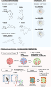
Representation of galacto-activable nanosystems for senescence cell detection and elimination. A) Molecular probes or drugs can be modified with a galactose moiety to obtain an inactive product. B) Mesoporous silica nanoparticles (MSN) are loaded with fluorophores or drugs, and the external surface is coated with the hexagalactosaccharide (galactan). C) These systems are triggered by the activity of senescence-associated β-galactosidase (overexpressed in senescent cells), where the enzyme catalyzes the hydrolysis of galactan into monosaccharides, while in proliferative cells they remain inactive.
Scheme 1
Scheme 1. Summary of Chemical Strategies, Molecular Probes, and Nanoparticles, Developed to Detect Cellular Senescence in Different Scenarios
Figure 2
Figure 2
A) Activation of AHGa in senescent cells (schematic representation). B) Tumors of SK-Mel-103 a) control or b) SK-Mel-103 senescent tumors after SAβGal staining. C) Confocal images of SK-Mel-103 control cells from nonsenescent tumors c) vehicle, d) after injection of the AHGa probe; and SK-Mel-103 from senescent tumors e) vehicle, f) after injection of the AHGa probe. Adapted with permission from ref (1). Copyright 2017 American Chemical Society.
Figure 3
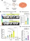
Figure 3
A) Representation of selectivity senescence detection and renal clearance of the Cy7Ga probe. B) At the top, in vivoCy7Ga-associated fluorescence IVIS imaging in bladders of BALB/cByJ female mice with 4T1 breast cancer tumors and treated orally with palbociclib (0, 10, 50, and 100 mg/kg), compared to mice treated with the highest dose of palbociclib but not injected with Cy7Ga. Using the same animal model injected with the WOS-Cy7Ga probe, we compared those receiving 100 mg/kg of palbociclib to mice not receiving any treatment. At the bottom, the IVIS readout of Cy7 average radiant efficiency in urine from mice. C) Cy7 urine fluorescence from 2- vs 14-m BALB/cByJ mice. D) Cy7 urine fluorescence of 7-m SAMP8 and SAMR1 mice. Adapted with permission from ref (35), with the Creative Commons CC BY license http://creativecommons.org/licenses/by/4.0/. Copyright 2024, the authors of the original publication.
Figure 4
Figure 4
Detection of a senescent cell by binding lipofuscin to a Sudan Black B derivative containing an azide moiety (SBB-N3) and consequent reaction with a fluorophore having a cyclooctene ring in its structure (BODIPY). Adapted with permission from ref (38). Copyright 2023, John Wiley and Sons.
Figure 5
Figure 5
A) Representation of Gal-MSN(NB) activation in the 4T1 breast cancer tumoral model. After tumor formation, mice were treated with palbociclib (resulting in senescence tumors). Subsequently, they received Gal-MSN(NB) treatment, allowing in vivo detection of cellular senescence. B) In vivo imaging system (IVIS) captured images at various time points depicting BALB/cByJ female mice with 4T1 breast tumors. From left to right are control mice treated with Gal-MSN(NB) and mice treated with palbociclib and Gal-MSN(NB). Adapted with permission from ref (2). Copyright 2020 Wiley-VCH.
Scheme 2
Scheme 2. Summary of Chemical Strategies, Nanoparticles, and Prodrugs, Developed for Eliminating Senescent Cells in Different Preclinical Models of Disease
Figure 6
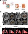
Figure 6
A) Representation of lung fibrosis therapy with Gal-MSN(Dox). B) Representative images of computerized tomography of the indicated treatments at days 11 and 29 postbleomycin injury. Both images, at days 11 and 29, are for the same mouse. C) 3D isocontour-based volume rendering of a representative lung before and after treatment with Gal-MSN(Dox). Fibrotic lesions are shown in gray, and healthy lung tissue is shown in red. Ventral and lateral views are shown. Adapted with permission from reference (3) with the Creative Commons CC BY license http://creativecommons.org/licenses/by/4.0/. Copyright 2018, the authors of the original publication.
Figure 7
Figure 7
A) Representation of cancer combined therapy of palbociclib with Gal-MSN(Nav). In TNBC tumors. B) The relative volume change of Balb/cByJ female mice orthotopically injected with 4T1 breast cancer cells and treated daily in comparison to its baseline before treatment. C) Quantification of lung metastasis and representative H&E lung sections. Adapted with permission from reference (53). Copyright 2020, Elsevier.
Figure 8
Figure 8
Representation of nanoparticle communication through stigmergy for improved tumor reduction via targeted induction of senescence and senolysis. The first community of nanoparticles (PEG-MUC1-MSN(palbo)) releases palbociclib, altering the environment through the induction of senescence in tumor cells. This enables a second community of nanoparticles (NP(nav)-Gal) to release their content (navitoclax) into senescent cells. This eliminates senescent tumor cells through apoptosis, resulting in a reduction in the tumor size. Adapted with permission from reference (55). Copyright 2023, Elsevier.
Figure 9
Figure 9
A) Schematic representation of the NavGa prodrug mechanism of action. B) Representative images of A549 xenografts stained for SA-β-Gal activity (in blue) following treatment with vehicle or cisplatin. C) A549 xenograft tumor volume in mice treated with cisplatin and navitoclax or NavGa. D) Platelet count after the treatment of wild-type C57BL/6J mice in each experimental condition. Adapted with permission from reference (4) with the Creative Commons CC BY license http://creativecommons.org/licenses/by/4.0/. Copyright 2020, the authors of the original publication.
Figure 10
Figure 10
A) Representation of therapies based on navitoclax in a model of doxorubicin-induced cardiotoxicity. B) Representative echocardiographic analysis of mice under each experimental condition displaying changes in left ventricle (LV) systolic function and fractional shortening (FS) values obtained from animals at the experimental end-point (day 30). After injection with doxorubicin, a reduction in left ventricular (LV) contraction is observed and fractional shortening (FS), which is mitigated with senolytic treatment. C) Senescence markers p16, p21, and p53 mRNA expression in the hearts of mice under each experimental condition. Treatment with doxorubicin upregulates the expression of both markers in heart tissue; this indicated the senescent cell accumulation. This upregulation is reversed upon the administration of a senolytic treatment. Adapted with permission from reference (62). Copyright 2022, Elsevier.

References

    1. Lozano-Torres B.; Galiana I.; Rovira M.; Garrido E.; Chaib S.; Bernardos A.; Muñoz-Espín D.; Serrano M.; Martínez-Máñez R.; Sancenón F. An OFF-ON Two-Photon Fluorescent Probe for Tracking Cell Senescence in Vivo. J. Am. Chem. Soc. 2017, 139 (26), 8808–8811. 10.1021/jacs.7b04985. - DOI - PubMed
    1. Lozano-Torres B.; Blandez J. F.; Galiana I.; García-Fernández A.; Alfonso M.; Marcos M. D.; Orzáez M.; Sancenón F.; Martínez-Máñez R. Real-Time In Vivo Detection of Cellular Senescence through the Controlled Release of the NIR Fluorescent Dye Nile Blue. Angew. Chem., Int. Ed. 2020, 59 (35), 15152–15156. 10.1002/anie.202004142. - DOI - PubMed
    1. Muñoz-Espín D.; Rovira M.; Galiana I.; Giménez C.; Lozano-Torres B.; Paez-Ribes M.; Llanos S.; Chaib S.; Muñoz-Martín M.; Ucero A. C.; Garaulet G.; Mulero F.; Dann S. G.; VanArsdale T.; Shields D. J.; Bernardos A.; Murguía J. R.; Martínez-Máñez R.; Serrano M. A Versatile Drug Delivery System Targeting Senescent Cells. EMBO Mol. Med. 2018, 10, e9355.10.15252/emmm.201809355. - DOI - PMC - PubMed
    1. González-Gualda E.; Pàez-Ribes M.; Lozano-Torres B.; Macias D.; Wilson J. R.; González-López C.; Ou H. L.; Mirón-Barroso S.; Zhang Z.; Lérida-Viso A.; Blandez J. F.; Bernardos A.; Sancenón F.; Rovira M.; Fruk L.; Martins C. P.; Serrano M.; Doherty G. J.; Martínez-Máñez R.; Muñoz-Espín D. Galacto-Conjugation of Navitoclax as an Efficient Strategy to Increase Senolytic Specificity and Reduce Platelet Toxicity. Aging Cell 2020, 19, e13142.10.1111/acel.13142. - DOI - PMC - PubMed
    1. Morsli S.; Doherty G. J.; Muñoz-Espín D. Activatable Senoprobes and Senolytics: Novel Strategies to Detect and Target Senescent Cells. Mech. Ageing Dev. 2022, 202, 111618.10.1016/j.mad.2021.111618. - DOI - PubMed

Publication types