Hydrophobic mismatch drives self-organization of designer proteins into synthetic membranes
- PMID: 38605024
- PMCID: PMC11009411
- DOI: 10.1038/s41467-024-47163-1
Hydrophobic mismatch drives self-organization of designer proteins into synthetic membranes
Abstract
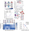
The organization of membrane proteins between and within membrane-bound compartments is critical to cellular function. Yet we lack approaches to regulate this organization in a range of membrane-based materials, such as engineered cells, exosomes, and liposomes. Uncovering and leveraging biophysical drivers of membrane protein organization to design membrane systems could greatly enhance the functionality of these materials. Towards this goal, we use de novo protein design, molecular dynamic simulations, and cell-free systems to explore how membrane-protein hydrophobic mismatch could be used to tune protein cotranslational integration and organization in synthetic lipid membranes. We find that membranes must deform to accommodate membrane-protein hydrophobic mismatch, which reduces the expression and co-translational insertion of membrane proteins into synthetic membranes. We use this principle to sort proteins both between and within membranes, thereby achieving one-pot assembly of vesicles with distinct functions and controlled split-protein assembly, respectively. Our results shed light on protein organization in biological membranes and provide a framework to design self-organizing membrane-based materials with applications such as artificial cells, biosensors, and therapeutic nanoparticles.
© 2024. The Author(s).
Conflict of interest statement
N.P.K., J.A.P. and J.S. are inventors on a U.S. provisional patent submitted by Northwestern University that covers organizing cell-free expressed membrane proteins in synthetic membranes. D.B. and P.L. are inventors on U.S. patents that cover the computational design of multipass transmembrane proteins and transmembrane pores submitted by the University of Washington. The remaining authors declare no competing interests.
Figures


Similar articles
-
Biopores/membrane proteins in synthetic polymer membranes.Biochim Biophys Acta Biomembr. 2017 Apr;1859(4):619-638. doi: 10.1016/j.bbamem.2016.10.015. Epub 2016 Oct 29. Biochim Biophys Acta Biomembr. 2017. PMID: 27984019 Review.
-
Controlled fusion of synthetic lipid membrane vesicles.Acc Chem Res. 2013 Dec 17;46(12):2988-97. doi: 10.1021/ar400065m. Epub 2013 Jul 23. Acc Chem Res. 2013. PMID: 23879805
-
The importance of membrane defects-lessons from simulations.Acc Chem Res. 2014 Aug 19;47(8):2244-51. doi: 10.1021/ar4002729. Epub 2014 Jun 3. Acc Chem Res. 2014. PMID: 24892900
-
DNA-Origami Line-Actants Control Domain Organization and Fission in Synthetic Membranes.J Am Chem Soc. 2023 May 24;145(20):11265-11275. doi: 10.1021/jacs.3c01493. Epub 2023 May 10. J Am Chem Soc. 2023. PMID: 37163977 Free PMC article.
-
Tethered bilayer lipid membranes (tBLMs): interest and applications for biological membrane investigations.Biochimie. 2014 Dec;107 Pt A:135-42. doi: 10.1016/j.biochi.2014.06.021. Epub 2014 Jul 4. Biochimie. 2014. PMID: 24998327 Review.
Cited by
-
X-Ray Crystal and Cryo-Electron Microscopy Structure Analysis Unravels How the Unique Thylakoid Lipid Composition Is Utilized by Cytochrome b6f for Driving Reversible Proteins' Reorganization During State Transitions.Membranes (Basel). 2025 May 8;15(5):143. doi: 10.3390/membranes15050143. Membranes (Basel). 2025. PMID: 40422753 Free PMC article.
-
Engineered membrane receptors with customizable input and output functions.Trends Biotechnol. 2023 Mar;41(3):276-277. doi: 10.1016/j.tibtech.2023.01.002. Epub 2023 Jan 14. Trends Biotechnol. 2023. PMID: 36646525 Free PMC article.
-
Computational electrostatic engineering of nanobodies for enhanced SARS-CoV-2 receptor binding domain recognition.Front Mol Biosci. 2025 Mar 10;12:1512788. doi: 10.3389/fmolb.2025.1512788. eCollection 2025. Front Mol Biosci. 2025. PMID: 40129869 Free PMC article.
-
CD24 Regulates the Formation of Ectosomes in B Lymphocytes.J Extracell Vesicles. 2025 May;14(5):e70093. doi: 10.1002/jev2.70093. J Extracell Vesicles. 2025. PMID: 40415253 Free PMC article.
-
Enhancing extracellular vesicle cargo loading and functional delivery by engineering protein-lipid interactions.Nat Commun. 2024 Jul 4;15(1):5618. doi: 10.1038/s41467-024-49678-z. Nat Commun. 2024. PMID: 38965227 Free PMC article.
References
-
- Guo, Y., Sirkis, D. W. & Schekman, R. Protein sorting at the trans-golgi network. Annu. Rev. Cell Dev. Biol.30, 169–206 (2014). - PubMed
MeSH terms
Substances
Grants and funding
LinkOut - more resources
Full Text Sources

