. 2024 Apr 11;14(1):8472.
doi: 10.1038/s41598-024-52810-0.

An RNA-hydrolyzing recombinant minibody prevents both influenza A virus and coronavirus in co-infection models

Affiliations

An RNA-hydrolyzing recombinant minibody prevents both influenza A virus and coronavirus in co-infection models

Quynh Xuan Thi Luong et al. Sci Rep. .

Abstract

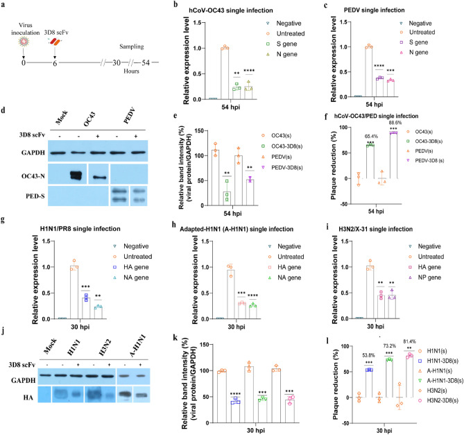
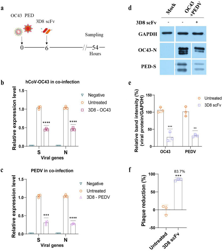
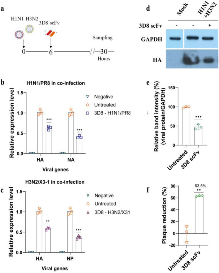
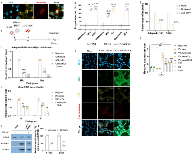
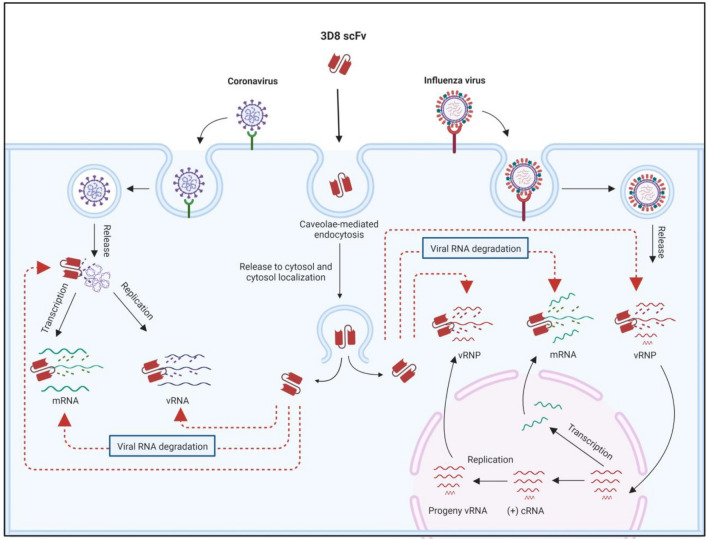
With the lifting of COVID-19 non-pharmaceutical interventions, the resurgence of common viral respiratory infections was recorded in several countries worldwide. It facilitates viral co-infection, further burdens the already over-stretched healthcare systems. Racing to find co-infection-associated efficacy therapeutic agents need to be rapidly established. However, it has encountered numerous challenges that necessitate careful investigation. Here, we introduce a potential recombinant minibody-associated treatment, 3D8 single chain variable fragment (scFv), which has been developed as a broad-spectrum antiviral drug that acts via its nucleic acid catalytic and cell penetration abilities. In this research, we demonstrated that 3D8 scFv exerted antiviral activity simultaneously against both influenza A viruses (IAVs) and coronaviruses in three established co-infection models comprising two types of coronaviruses [beta coronavirus-human coronavirus OC43 (hCoV-OC43) and alpha coronavirus-porcine epidemic diarrhea virus (PEDV)] in Vero E6 cells, two IAVs [A/Puerto Rico/8/1934 H1N1 (H1N1/PR8) and A/X-31 (H3N2/X-31)] in MDCK cells, and a combination of coronavirus and IAV (hCoV-OC43 and adapted-H1N1) in Vero E6 cells by a statistically significant reduction in viral gene expression, proteins level, and approximately around 85%, 65%, and 80% of the progeny of 'hCoV-OC43-PEDV', 'H1N1/PR8-H3N2/X-31', and 'hCoV-OC43-adapted-H1N1', respectively, were decimated in the presence of 3D8 scFv. Taken together, we propose that 3D8 scFv is a promising broad-spectrum drug for treatment against RNA viruses in co-infection.


Conflict of interest statement

The authors declare that the research was conducted in the absence of any commercial or financial relationships that could be construed as a potential conflict of interest.

Figures

Figure 1
Figure 1
3D8 scFv protected cells from single infection with cytoplasmic or nuclear RNA viruses. (a) Scheme of the experimental procedure. Coronaviruses or IAVs were inoculated to Vero E6 or MDCK cells for 6 h. The cells and supernatants were collected at 24 or 48 h after treatment with 10 µM 3D8 scFv. One-step RT-qPCR was performed to determine the expression of the S and N genes of (b) hCoV-OC43 and (c) PEDV at 54 hpi. Simultaneously, the cells were lysed using RIPA buffer in order to check (d) the protein level of nucleoprotein (N) of hCoV-OC43 and spike protein (S) of PEDV via western blotting assay, (e) the percentage of band intensity was calculated based on normalizing viral protein intensity to GAPDH intensity. The supernatants from the 3D8-treated cells were harvested, and a plaque reduction assay was performed to observe the infectious viral titer, and percent plaque reduction of (f) coronaviruses hCoV-OC43 and PEDV. Similarly, HA and NA genes of (g) H1N1/PR8 and (h) adapted-H1N1 at 30 hpi, (i) HA and NP genes of H3N2/X-31 at 30 hpi were indicated by RT-qPCR. (j) The hemagglutinin (HA) protein levels of IAVs were tested, and (k) the percentage of band intensity was calculated based on normalizing viral protein intensity to GAPDH intensity. The percent plaque reduction of (l) influenza viruses H1N1/PR8, H3N2/X-31, and adapted-H1N1 was observed. All assays were in triplicates. The significant difference was determined by unpaired t-test (*P < 0.05, **P < 0.01, ***P < 0.001, ****P < 0.0001). The original western blots are presented in Fig. S4.
Figure 2
Figure 2
Antiviral activity of 3D8 scFv against co-infection with coronaviruses. (a) Diagram of procedure of co-infection with coronaviruses. 3D8 scFv (10 µM) was treated to Vero E6 cells co-infected with coronaviruses hCoV-OC43 (MOI 0.1) and PEDV (MOI 0.02). At 54 h after coronaviruses infection, the cells and supernatants were harvested to check S and N gene expression level of (b) hCoV-OC43 and (c) PEDV using one-step RT-qPCR, (d) determine viral protein level using western blotting, (e) the relative band intensity of hCoV-OC43 N protein and PEDV S protein was normalized to GAPDH intensity. The total viral titer of the two coronaviruses was detected using plaque reduction assay, the plaques were counted and (f) the percentage of plaque reduction was calculated. All assays were conducted in triplicates. Significant differences were determined using unpaired t-test (*P < 0.05, **P < 0.01, ***P < 0.001, ****P < 0.0001). The original western blots are presented in Fig. S4.
Figure 3
Figure 3
Antiviral effects of 3D8 scFv against co-infection with IAVs. (a) Diagram of procedure of co-infection with IAVs. MDCK cells were simultaneously inoculated with H1N1/PR8 (MOI 1) and H3N2/X-31 (MOI 2) at 37 °C. At 6 hpi, the cells were treated with 10 µM 3D8 scFv in MEM supplemented with 1 µg/mL TPCK, after which the cells were incubated for a total duration of 30 h. (b) HA and NA genes of H1N1/PR8 as well as the (c) HA and NP genes of H3N2/X-31 expression level in 3D8 scFv-treated and untreated cells were assessed using one-step RT-qPCR. In addition, (d) 3D8-treated cells were collected for western blotting assay using influenza HA primary antibody; (e) the viral protein expression was normalized to GAPDH expression. (f) Plaque reduction assay was conducted; the total progeny of IAVs in the co-infection model was quantified, and the percentage reduction was calculated. All assays were conducted in triplicates. Significant differences were determined using unpaired t-test (*P < 0.05, **P < 0.01, ***P < 0.001, ****P < 0.0001). The original western blots are presented in Fig S4.
Figure 4
Figure 4
3D8 scFv suppressed co-infection with influenza virus and coronavirus. (a) Simultaneous infection of adapted-H1N1 (A-H1N1) and coronavirus (hCoV-OC43) was detected in infected Vero E6 cells using immunocytochemistry at 30 hpi. (b) Diagram of influenza virus (adapted-H1N1) and coronavirus (hCoV-OC43) co-infection procedure. Vero E6 cells were co-infected with adapted-H1N1 and hCoV-OC43 (MOI: 1 and 0.1, respectively). After 6 h of co-infection, the cells were treated with 10 µM of 3D8 scFv, oseltamivir (positive control), chloroquine (positive control), or DPBS (negative control) in DMEM supplemented with 1 µg/ml TPCK. (c) The HA and NA genes of adapted-H1N1 and (d) the S and N genes of hCoV-OC43 were quantified using one-step RT-qPCR. (e) The influenza HA protein and hCoV-OC43 nucleoprotein were examined via western blotting using the appropriate specific primary antibodies, in which (f) the relative band intensity of both viral proteins was normalized to GAPDH intensity. The supernatants were collected for progeny virus quantification using plaque reduction assay; plaques of adapted-H1N1 and hCoV-OC43 were separately counted, then (g) the plaque reductions for all treatments were combined in one graph to obtain the full view of plaque reduction under co-infection. (h) Antiviral activity of 3D8 scFv against adapted-H1N1 and hCoV-OC43 under co-infection in Vero E6 cells was measured using immunofluorescence, in which (i) the viral protein signal was converted to relative intensity percentages using CellProfiler 4.2.1, and the viral protein intensity was normalized to DAPI intensity. (j) TLR7 expression level in treated and untreated cells. All assays were conducted in triplicates. Significant differences were determined via unpaired t-test (*P < 0.05, **P < 0.01, ***P < 0.001, ****P < 0.0001). The original western blots are presented in Fig. S4.
Figure 5
Figure 5
Proposed mode of action of 3D8 scFv against co-infection with coronavirus and influenza virus. Upon cell entry, 3D8 scFv localizes in the cytosol, where it binds to the viral nucleic acid (regardless of coronavirus or IAV) and viral mRNA and then degrades them via nuclease activity. As 3D8 scFv can hydrolyze all types of viral RNA in both viruses at different periods of viral life cycles, 3D8 scFv can inhibit viral propagation. Created with BioRender.com.

Similar articles

Cited by

References

    1. Pawlowski, C., et al., SARS-CoV-2 and Influenza Co-infection Throughout the COVID-19 Pandemic: An Assessment of Co-infection Rates and Cohort Characterization. medRxiv, 2022: p. 2022.02.02.22270324.
    1. Chen N, et al. Epidemiological and clinical characteristics of 99 cases of 2019 novel coronavirus pneumonia in Wuhan, China: A descriptive study. The Lancet. 2020;395:1. doi: 10.1016/S0140-6736(20)30211-7. - DOI - PMC - PubMed
    1. Bai L, et al. Coinfection with influenza A virus enhances SARS-CoV-2 infectivity. Cell Research. 2021;31(4):395–403. doi: 10.1038/s41422-021-00473-1. - DOI - PMC - PubMed
    1. Bao L, et al. Sequential infection with H1N1 and SARS-CoV-2 aggravated COVID-19 pathogenesis in a mammalian model, and co-vaccination as an effective method of prevention of COVID-19 and influenza. Signal Transduct. Target. Therapy. 2021;6(1):200. doi: 10.1038/s41392-021-00618-z. - DOI - PMC - PubMed
    1. Ghildiyal R, Gabrani R. Computational approach to decipher cellular interactors and drug targets during co-infection of SARS-CoV-2, Dengue, and Chikungunya virus. VirusDisease. 2021;32(1):55–64. doi: 10.1007/s13337-021-00665-8. - DOI - PMC - PubMed