Review
. 2024 Oct;25(10):698-714.
doi: 10.1038/s41576-024-00715-z. Epub 2024 Apr 11.

Human embryonic genetic mosaicism and its effects on development and disease

Affiliations
Review

Human embryonic genetic mosaicism and its effects on development and disease

Sarah M Waldvogel et al. Nat Rev Genet. 2024 Oct.

Abstract

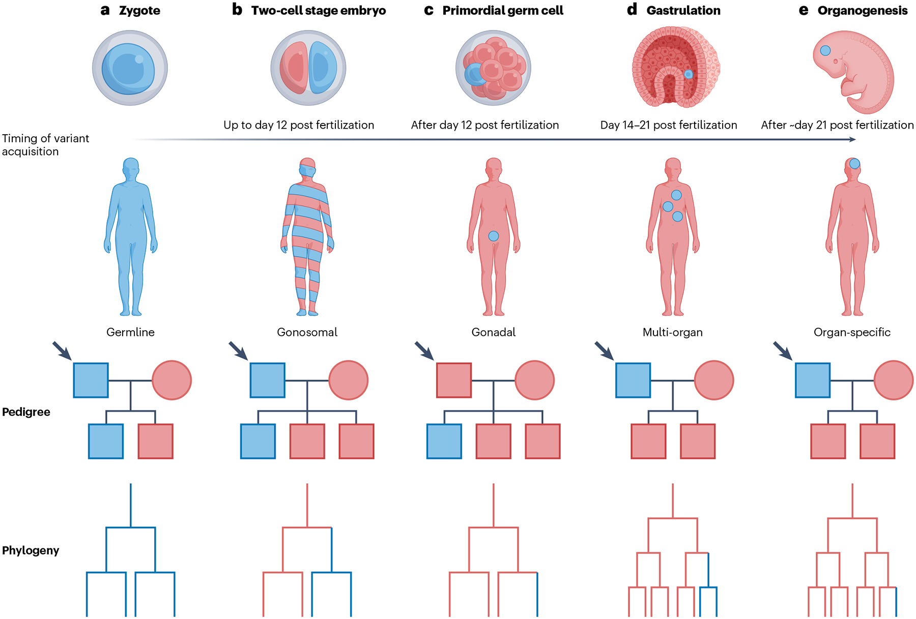
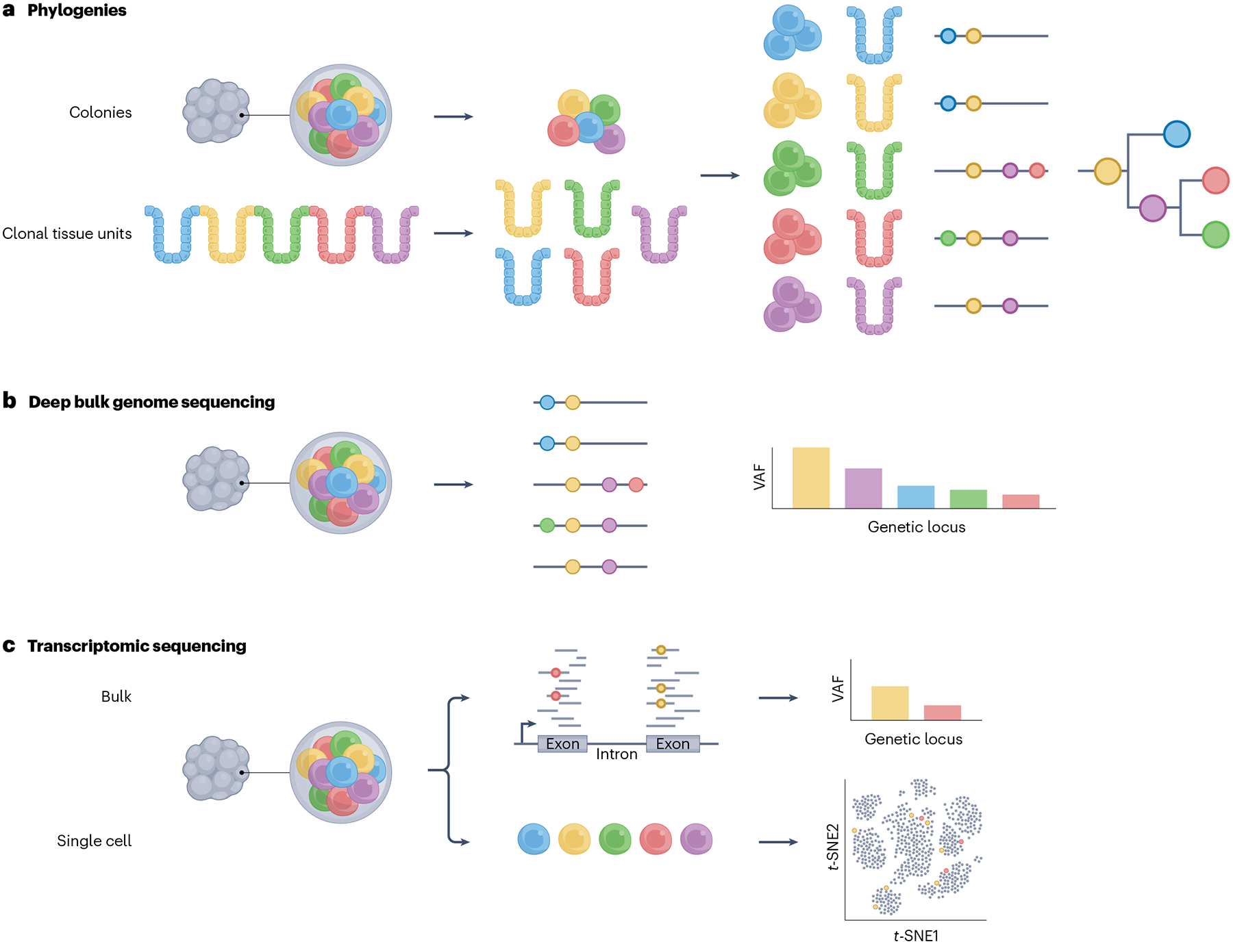
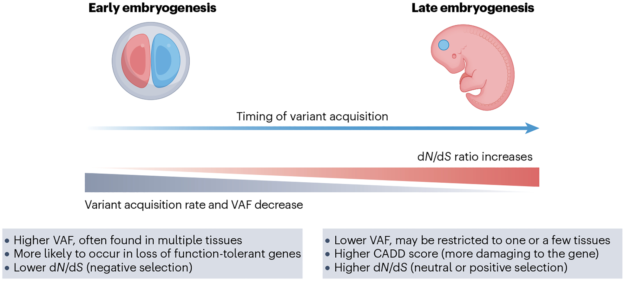
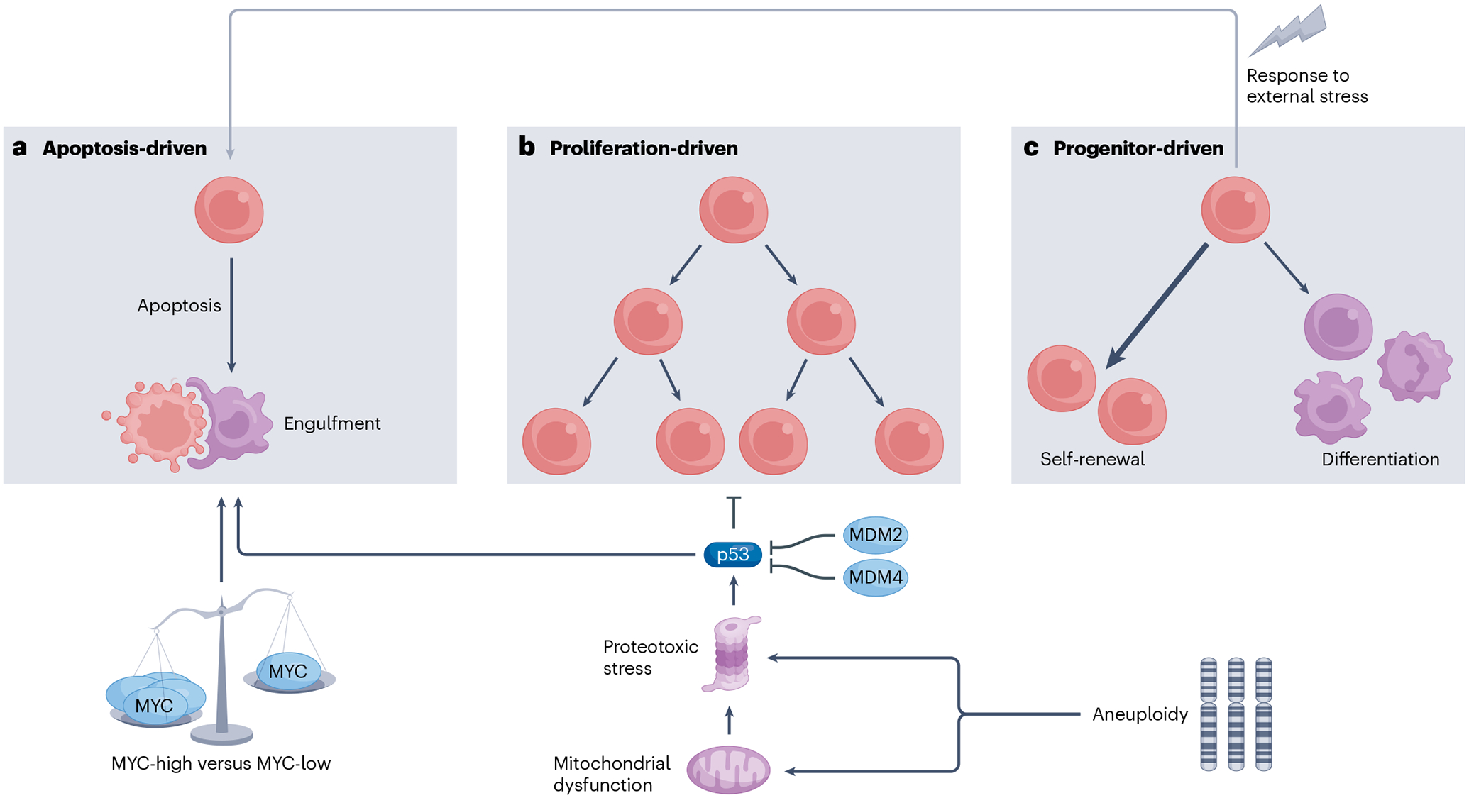
Nearly every mammalian cell division is accompanied by a mutational event that becomes fixed in a daughter cell. When carried forward to additional cell progeny, a clone of variant cells can emerge. As a result, mammals are complex mosaics of clones that are genetically distinct from one another. Recent high-throughput sequencing studies have revealed that mosaicism is common, clone sizes often increase with age and specific variants can affect tissue function and disease development. Variants that are acquired during early embryogenesis are shared by multiple cell types and can affect numerous tissues. Within tissues, variant clones compete, which can result in their expansion or elimination. Embryonic mosaicism has clinical implications for genetic disease severity and transmission but is likely an under-recognized phenomenon. To better understand its implications for mosaic individuals, it is essential to leverage research tools that can elucidate the mechanisms by which expanded embryonic variants influence development and disease.


Conflict of interest statement

Competing interests

The authors declare no competing interests.

Figures

Fig. 1 |
Fig. 1 |. Mosaicism is defined by the timing and lineage of variant acquisition.
Variants can be acquired at different stages of embryo development, and this timing is partially responsible for the ultimate distribution of the variant. This transmission of a heterozygous variant driving a dominant disease trait is shown, including cells carrying a variant or individuals potentially affected by the disease (blue). The proband represented by the human figure is the male at the top left of the pedigrees (indicated by the arrow). a, If the variant is acquired at or before fertilization, it will be present in all cells of the individual. Transmission to the next generation follows the rules of Mendelian genetics, and the phylogeny of cells in the individual shows the variant present at all branchpoints. Transmission rate is 50%. b, If the variant occurs in the very early embryo, before specification of the primordial germ cells, it will be present in both somatic and germ cell tissue. The individual may be phenotypically affected (depending on the impacted tissues and degree of mosaicism) and can pass the variant to his or her offspring. The variant occurs in a branchpoint of the phylogeny that represents a shared ancestor of somatic and germ cells. Transmission rate may be less than 50%. c, If the variant occurs in a primordial germ cell, it will be limited to the germ cells and can be transmitted to offspring, but the mosaic individual is unlikely to exhibit a phenotype. The last common phylogenetic cellular ancestor was already confined to the germ cell lineage. Transmission rate may be lower than 50%. d, If a variant arises during gastrulation, the variant will be confined to the tissues derived from that specific germ layer, but it may be seen in multiple cell types from that germ layer depending on the timing of acquisition. The last common phylogenetic ancestor is confined to the ectoderm, mesoderm or endoderm. The individual may be phenotypically affected, depending on the variant and the organ. e, If the variant arises later during embryogenesis, it is likely limited to one organ. The individual may be phenotypically affected, depending on the variant and the organ. The variant appears at a late branchpoint of the phylogenetic tree.
Fig. 2 |
Fig. 2 |. The timing of variant acquisition and the variant allele frequency can be estimated from adult or fetal tissue.
Numerous sequencing techniques can be used to reconstruct early cell lineages and determine the variant allele frequency (VAF) of mosaic variants. a, Genome sequencing is performed on individual single cell-derived colonies or clonal tissue units such as colonic crypts (obtained using a strategy that can capture a clean clone, such as laser capture microdissection). The shared variants between the clonal genomes are used to construct a phylogenetic tree of clone relationships. This can help infer timing of variant acquisition. For example, if the variant arose early in development of that tissue or region, the variant will be present in all or most clones. b, Genome sequencing is performed on bulk tissue samples. The percentage of sequencing reads that contain a particular variant is used to calculate the VAF. The VAF can be used to infer the proportion of cells within the bulk sample that carry the variant. Because most variants will be heterozygous, the proportion of cells harbouring the variant is calculated to be double that of the VAF. c, Bulk RNA sequencing (RNA-seq) data can be used to determine the VAF of variants that are present in the exons of expressed genes in a particular tissue. Because mRNAs can be expressed at high or low levels, deep sequencing is usually needed to identify variants. Furthermore, some variant-containing RNAs are degraded rapidly, making it difficult to identify the variant. If variants are at the 5′ end of the transcript, sensitivity may be reduced as RNA-seq is usually biased towards the 3′ end. Single-cell RNA sequencing (scRNA-seq) can also provide information about the identity of cells carrying a variant, although it has limited sensitivity due to the requirement for high coverage of transcripts.
Fig. 3 |
Fig. 3 |. The features of mosaic variants vary from early to late embryogenesis.
The first few cell divisions in the early embryo are the most mutagenic. Mosaic variants acquired during early embryogenesis are generally present at higher variant allele frequencies (VAFs) in the resulting individual. They are subjected to stronger negative selective pressures, with a lower ratio of non-synonymous to synonymous variants (dN/dS ratio), indicating negative selection against deleterious variants. By contrast, mosaic variants acquired later in embryogenesis are generally found at lower VAFs and are confined to a smaller number of tissues. Because of this, they may have a higher combined annotation-dependent deletion (CADD) score, suggesting a higher degree of damage to the gene, and thus deleteriousness at the population level. However, in aggregate, they exhibit a higher dN/dS ratio, suggesting positive selection, because genes that are deleterious at the population level or harmful if they occur in a very high proportion of embryonic cells may be favourable in certain tissues or embryonic contexts when present in a mosaic state at low VAF.
Fig. 4 |
Fig. 4 |. Cell competition in mosaicism occurs through separate but interrelated mechanisms.
Wild-type and variant cells in a mosaic individual compete via multiple pathways that converge on three main processes. a, Apoptosis-driven: aneuploid cells, cells with mitochondrial defects, cells with higher levels of proteotoxic stress, cells with higher levels of p53 or cells with low levels of MYC are more likely to undergo apoptosis when competed against their wild-type counterparts. Following initiation of apoptosis, ‘winner’ cells may engulf ‘loser’ cells. b, Proliferation-driven: cells with higher levels of p53 or lower levels of the p53 negative regulators MDM2 and MDM4 outcompete wild-type cells by increasing their proliferation rates. c, Progenitor-driven: variant progenitor cells may exhibit a self-renewal advantage, leading to their enrichment in the stem cell pool. They may exhibit decreased differentiation potential, or a bias towards certain cell types. They may also have a decreased tendency to undergo apoptosis when subjected to environmental stresses.
Fig. 5 |
Fig. 5 |. Differences in VAF across time, tissues and environmental exposures affect clinical detection of mosaicism.
Depending on which sample is collected (1 versus 2), a completely different clinical result may be identified and reported to a patient. a, Variant allele frequency (VAF) in a particular tissue, such as the blood, can vary over time due to selection for or against cells carrying that variant within the sampled tissue. b, VAF can be high in some tissues and low in others within the same individual, so sampling of one particular tissue can obscure the mosaic state. c, VAF can change with increasing exposure to an environmental insult, such as smoking, infection or chemotherapy. d, In sample 1, mosaicism is not detected because the VAF is low and is interpreted as background noise (arrow). e, In sample 2, mosaicism is misdiagnosed as a germline heterozygous variant because the VAF is high and is read as 50%, suggesting that the variant is present in all cells when it is actually mosaic. This scenario could occur if a variant-containing clone dominated the sampled tissue (even if it was rare in other tissues).

Similar articles

  • Somatic mutations reveal asymmetric cellular dynamics in the early human embryo.
    Ju YS, Martincorena I, Gerstung M, Petljak M, Alexandrov LB, Rahbari R, Wedge DC, Davies HR, Ramakrishna M, Fullam A, Martin S, Alder C, Patel N, Gamble S, O'Meara S, Giri DD, Sauer T, Pinder SE, Purdie CA, Borg Å, Stunnenberg H, van de Vijver M, Tan BK, Caldas C, Tutt A, Ueno NT, van 't Veer LJ, Martens JW, Sotiriou C, Knappskog S, Span PN, Lakhani SR, Eyfjörd JE, Børresen-Dale AL, Richardson A, Thompson AM, Viari A, Hurles ME, Nik-Zainal S, Campbell PJ, Stratton MR. Ju YS, et al. Nature. 2017 Mar 30;543(7647):714-718. doi: 10.1038/nature21703. Epub 2017 Mar 22. Nature. 2017. PMID: 28329761 Free PMC article.
  • The rate and spectrum of mosaic mutations during embryogenesis revealed by RNA sequencing of 49 tissues.
    Muyas F, Zapata L, Guigó R, Ossowski S. Muyas F, et al. Genome Med. 2020 May 27;12(1):49. doi: 10.1186/s13073-020-00746-1. Genome Med. 2020. PMID: 32460841 Free PMC article.
  • Mosaicism and clinical genetics.
    Spinner NB, Conlin LK. Spinner NB, et al. Am J Med Genet C Semin Med Genet. 2014 Dec;166C(4):397-405. doi: 10.1002/ajmg.c.31421. Epub 2014 Nov 25. Am J Med Genet C Semin Med Genet. 2014. PMID: 25424979 Review.
  • Somatic mosaicism: implications for disease and transmission genetics.
    Campbell IM, Shaw CA, Stankiewicz P, Lupski JR. Campbell IM, et al. Trends Genet. 2015 Jul;31(7):382-92. doi: 10.1016/j.tig.2015.03.013. Epub 2015 Apr 21. Trends Genet. 2015. PMID: 25910407 Free PMC article. Review.
  • Extensive phylogenies of human development inferred from somatic mutations.
    Coorens THH, Moore L, Robinson PS, Sanghvi R, Christopher J, Hewinson J, Przybilla MJ, Lawson ARJ, Spencer Chapman M, Cagan A, Oliver TRW, Neville MDC, Hooks Y, Noorani A, Mitchell TJ, Fitzgerald RC, Campbell PJ, Martincorena I, Rahbari R, Stratton MR. Coorens THH, et al. Nature. 2021 Sep;597(7876):387-392. doi: 10.1038/s41586-021-03790-y. Epub 2021 Aug 25. Nature. 2021. PMID: 34433963

Cited by

References

    1. Behjati S et al. Genome sequencing of normal cells reveals developmental lineages and mutational processes. Nature 513, 422–425 (2014). - PMC - PubMed
    2. This study is one of the first to use colony sequencing from a multicellular organism to reconstruct phylogenies based on shared variants.

    1. Ju YS et al. Somatic mutations reveal asymmetric cellular dynamics in the early human embryo. Nature 543, 714–718 (2017). - PMC - PubMed
    1. Ye AY et al. A model for postzygotic mosaicisms quantifies the allele fraction drift, mutation rate, and contribution to de novo mutations. Genome Res. 28, 943–951 (2018). - PMC - PubMed
    1. Spencer Chapman M et al. Lineage tracing of human development through somatic mutations. Nature 595, 85–90 (2021). - PubMed
    1. Coorens THH et al. Extensive phylogenies of human development inferred from somatic mutations. Nature 597, 387–392 (2021). - PubMed

LinkOut - more resources