. 2024 Apr 11;25(1):359.
doi: 10.1186/s12864-024-10289-z.

A murine model for the del(GJB6-D13S1830) deletion recapitulating the phenotype of human DFNB1 hearing impairment: generation and functional and histopathological study

Affiliations

A murine model for the del(GJB6-D13S1830) deletion recapitulating the phenotype of human DFNB1 hearing impairment: generation and functional and histopathological study

María Domínguez-Ruiz et al. BMC Genomics. .

Abstract

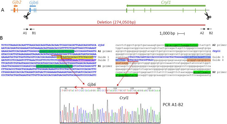
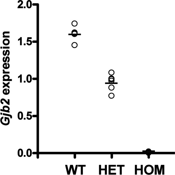
Inherited hearing impairment is a remarkably heterogeneous monogenic condition, involving hundreds of genes, most of them with very small (< 1%) epidemiological contributions. The exception is GJB2, the gene encoding connexin-26 and underlying DFNB1, which is the most frequent type of autosomal recessive non-syndromic hearing impairment (ARNSHI) in most populations (up to 40% of ARNSHI cases). DFNB1 is caused by different types of pathogenic variants in GJB2, but also by large deletions that keep the gene intact but remove an upstream regulatory element that is essential for its expression. Such large deletions, found in most populations, behave as complete loss-of-function variants, usually associated with a profound hearing impairment. By using CRISPR-Cas9 genetic edition, we have generated a murine model (Dfnb1em274) that reproduces the most frequent of those deletions, del(GJB6-D13S1830). Dfnb1em274 homozygous mice are viable, bypassing the embryonic lethality of the Gjb2 knockout, and present a phenotype of profound hearing loss (> 90 dB SPL) that correlates with specific structural abnormalities in the cochlea. We show that Gjb2 expression is nearly abolished and its protein product, Cx26, is nearly absent all throughout the cochlea, unlike previous conditional knockouts in which Gjb2 ablation was not obtained in all cell types. The Dfnb1em274 model recapitulates the clinical presentation of patients harbouring the del(GJB6-D13S1830) variant and thus it is a valuable tool to study the pathological mechanisms of DFNB1 and to assay therapies for this most frequent type of human ARNSHI.

Keywords: GJB2; Connnexin-26; DFNB1; Inherited hearing impairment; Murine models.


Conflict of interest statement

The authors declare no competing interests.

Figures

Fig. 1
Fig. 1
Deletion engineered at the murine Dfnb1 locus. A Map of the chromosome 14 region targeted in this study, including genes of interest. Scissors indicate the localization of CRISPR guides, and arrows show the primers used to test for the deletion (primer arrows not to scale). B Genomic sequences of Gjb6 exon 3 and Cryl1 exon 1 regions, showing the location of the CRISPR guides, deletion breakpoints and primers that were used. Exonic sequences appear in blue bold capital letters, primers are highlighted in green background and underlined, and CRISPR guides are marked with different background colors. The actual breakpoints in each region are indicated by a red zigzag line, as deduced from the Sanger sequence of A1-B2 breakpoint PCR (bottom)
Fig. 2
Fig. 2
Auditory phenotype of P30 Dfnb1em274 HOM mice. a ABR in response to click stimulus at different level from 90 to 30 dB SPL, in WT, HET and HOM Dfnb1em274 mice. Latin numbers (I-V) indicate the standard ABR peaks. b Scheme showing the main neural centers in the auditory pathway and their correspondence with the ABR wave peaks (AN (I): auditory nerve; CN (II), cochlear nucleus; SOC (III): superior olivary complex; LL (IV): lateral lemniscus; IC (V), inferior colliculus; NLL: Lateral lemniscus nuclei; MGB, medial geniculate body; SC, superior colliculus; TL, temporal lobe; AC, auditory cortex. c, d ABR thresholds (in dB SPL) in response to click (c) and 4–40 kHz pure tones (d) in WT (white), HET (grey) and HOM (black) Dfnb1em274 mice. Statistically significant differences in click and 4, 8 and 16 kHz thresholds were found between WT and HOM mutant mice. (e) Input–output curves (latency/intensity and amplitude/intensity) for ABR peak I and (f) wave interpeak latencies I-II, II-IV and I-IV in WT and HET mutant mice
Fig. 3
Fig. 3
Cx30 expression in the cochlea of P30 Dfnb1em274 HOM mice. Representative micrographs of cochlear paraffin sections stained with hematoxylin–eosin (A, B) or immunostained against Cx30 (C, D), showing the basal region of WT (A, C) and Dfnb1em274 HOM (B, D) mice. WT mice present a normal cochlear cytoarchitecture and Cx30 expression in the spiral limbus, organ of Corti (surrounding supporting cells, arrows in C) and lateral wall. Dfnb1em274 HOM mice show a complete absence of Cx30 expression (D) and gross alterations in the Reissner membrane, tectorial membrane, and organ of Corti, compared to WT (B). LW, lateral wall; RM, Reissner membrane; SG, spiral ganglion; SL, spiral limbus; SM, scala media; ST, scala tympani; sTV, stria vascularis; SV, scala vestibuli. Scale bar: 100 mm
Fig. 4
Fig. 4
Organ of Corti cytoarchitecture in P30 Dfnb1em274 HOM mice. Representative micrographs of historesin cochlear sections stained with cresyl violet, showing the organ of Corti at the basal (A, B) and apical (C, D) regions in WT (A, C) and Dfnb1em274 HOM (B, D) mice. WT mice present normal Corti cytoarchitecture, whereas Dfnb1em274 HOM mice show an absent spiral sulcus (SS) with presence of large inner sulcus cells, vacuolated interdental cells, and reduced or collapsed tunnel of Corti (TC, asterisks in B, D) due to the lack of separation between pillar cells (PC, arrows in B, D). These alterations are most severe in the basal turn of the cochlea. At apex region, the homozygous mouse shows spiral sulcus separated from the tectorial membrane, inner sulcus cells lower than in more basal regions (small arrows in D), and small tunnel of Corti (asterisk in D) with separate pillars. BC, border cells; DC, Deiter cells; IDC, interdental cells; IHC, inner hair cells; IPhC, inner phalangeal cells; ISC, inner sulcus cells; OHC, outer hair cell; PC, pillar cells; SL, spiral limbus; SS, spiral sulcus (inner sulcus); TC, tunnel of Corti; TM, tectorial membrane. Scale bar: 50 mm
Fig. 5
Fig. 5
Cx26 expression in the cochlea of P30 Dfnb1em274 HOM mice. Representative micrographs of cochlear paraffin sections stained with hematoxylin–eosin or immunostained against connexin-26 in WT (A-D) and Dfnb1em274 HOM mice (E–H). WT mice show normal cochlear cytoarchitecture (A) and connexin 26 expression (B) in the spiral limbus (C), organ of Corti (supporting cells, arrows in C) and lateral wall (D), both in basal (A-D) and apical (not shown) regions. In contrast, Dfnb1.em274 HOM mice present gross morphological alterations such as collapse of the tunnel of Corti (insets in E and G, asterisk in G) and a drastic reduction in connexin-26 expression, which is detected residually in just a few pillar cells (arrow in F) and supporting cells (arrows in H). These changes were most severe in the basal turn (E–F) compared to apical turn (G-H). LW, lateral wall; PC, pillar cells; SG, spiral ganglion; SL, spiral limbus; sTV, stria vascularis; TC, tunnel of Corti; TM, tectorial membrane. Scale bar: 100 μm (A-B) and 50 μm (C-H)
Fig. 6
Fig. 6
Gjb2 expression in the whole cochleae of wild-type (WT), heterozygous (HET), and homozygous (HOM) P30 Dfnb1em274 mice. Gjb2 expression was quantified by using real-time qPCR. The Y axis indicates the relative amounts of Gjb2 transcript with regard to the expression of the reference gene Hprt, as calculated according to the mathematical model of Pfaffl [34]. Points on the graph correspond to three different cDNA concentrations for one cochlea from each mouse (2 WT, 2 HET, 2 HOM), each point being the mean of two independent experiments. Horizontal lines show the mean of the expression level of Gjb2 for each genotype

Similar articles

Cited by

References

    1. Anselmi F, Hernandez VH, Crispino G, Seydel A, Ortolano S, Roper SD, Kessaris N, Richardson W, Rickheit G, Filippov MA, Monyer H, Mammano F. ATP release through connexin hemichannels and gap junction transfer of second messengers propagate Ca2+ signals across the inner ear. Proc Natl Acad Sci U S A. 2008;105(48):18770–18775. doi: 10.1073/pnas.0800793105. - DOI - PMC - PubMed
    1. Behringer R, Gertsenstein M, Vintersten Nagy K, Nagy A. Manipulating the mouse embryo - A laboratory manual. New York: Cold Spring Harbor Laboratory Press; 2014.
    1. Beltramello M, Piazza V, Bukauskas FF, Pozzan T, Mammano F. Impaired permeability to Ins(1,4,5)P3 in a mutant connexin underlies recessive hereditary deafness. Nat Cell Biol. 2005;7(1):63–69. doi: 10.1038/ncb1205. - DOI - PubMed
    1. Boulay AC, del Castillo FJ, Giraudet F, Hamard G, Giaume C, Petit C, Avan P, Cohen-Salmon M. Hearing is normal without connexin30. J Neurosci. 2013;33(2):430–434. doi: 10.1523/JNEUROSCI.4240-12.2013. - DOI - PMC - PubMed
    1. Cediel R, Riquelme R, Contreras J, Díaz A, Varela-Nieto I. Sensorineural hearing loss in insulin-like growth factor I-null mice: a new model of human deafness. Eur J Neurosci. 2006;23(2):587–590. doi: 10.1111/j.1460-9568.2005.04584.x. - DOI - PubMed

LinkOut - more resources