. 2024 Apr 11;22(1):346.
doi: 10.1186/s12967-024-05156-0.

The integration of single-cell and bulk RNA-seq atlas reveals ERS-mediated acinar cell damage in acute pancreatitis

Affiliations

The integration of single-cell and bulk RNA-seq atlas reveals ERS-mediated acinar cell damage in acute pancreatitis

Kaige Yang et al. J Transl Med. .

Abstract

Background: Acute pancreatitis (AP) is a clinically common acute abdominal disease, whose pathogenesis remains unclear. The severe patients usually have multiple complications and lack specific drugs, leading to a high mortality and poor outcome. Acinar cells are recognized as the initial site of AP. However, there are no precise single-cell transcriptomic profiles to decipher the landscape of acinar cells during AP, which are the missing pieces of jigsaw we aimed to complete in this study.

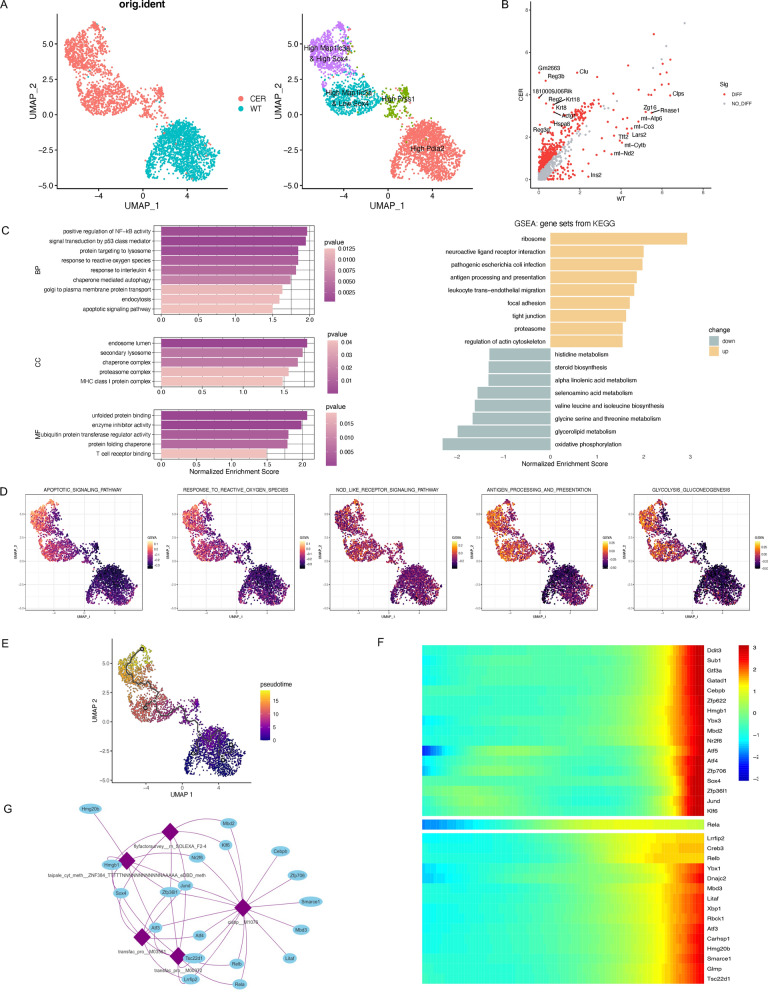
Methods: A single-cell sequencing dataset was used to identify the cell types in pancreas of AP mice and to depict the transcriptomic maps in acinar cells. The pathways' activities were evaluated by gene sets enrichment analysis (GSEA) and single-cell gene sets variation analysis (GSVA). Pseudotime analysis was performed to describe the development trajectories of acinar cells. We also constructed the protein-protein interaction (PPI) network and identified the hub genes. Another independent single-cell sequencing dataset of pancreas samples from AP mice and a bulk RNA sequencing dataset of peripheral blood samples from AP patients were also analyzed.

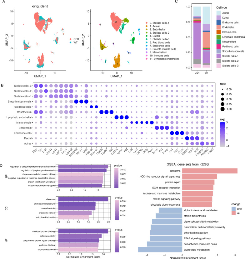
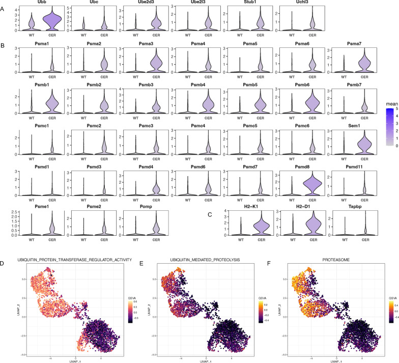
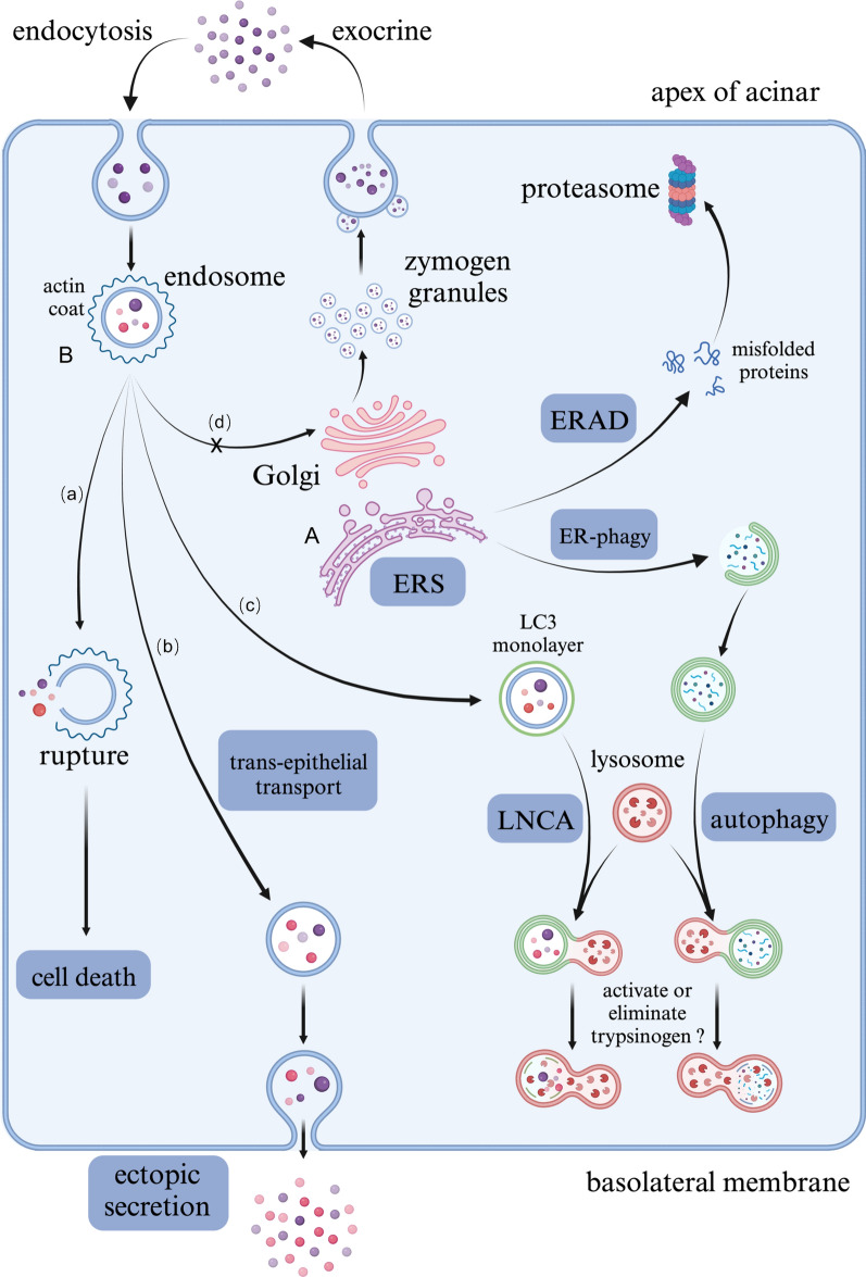
Results: In this study, we identified genetic markers of each cell type in the pancreas of AP mice based on single-cell sequencing datasets and analyzed the transcription changes in acinar cells. We found that acinar cells featured acinar-ductal metaplasia (ADM), as well as increased endocytosis and vesicle transport activity during AP. Notably, the endoplasmic reticulum stress (ERS) and ER-associated degradation (ERAD) pathways activated by accumulation of unfolded/misfolded proteins in acinar cells could be pivotal for the development of AP.

Conclusion: We deciphered the distinct roadmap of acinar cells in the early stage of AP at single-cell level. ERS and ERAD pathways are crucially important for acinar homeostasis and the pathogenesis of AP.

Keywords: Acute pancreatitis; Bulk RNA sequencing; ERAD; ERS; Single-cell RNA sequencing; Vesicle transport.


Conflict of interest statement

The authors declare that they have no competing interests.

Figures

Fig. 1
Fig. 1
Overview of single-cell transcriptional profiling of the pancreatic tissue. A UMAP plot of cells derived from WT/CER samples (left) and the 12 identified clusters (right). B Dotplot visualization of marker genes for each cluster. The size of the dots indicates the ratio of cells expressing this gene, and the shade of color represents the mean transcription levels in the corresponding clusters. C Relative proportion of each cell type in WT/CER samples. D GO (left) and KEGG (right) enrichment analyses of DEGs between the CER and WT samples based on GSEA
Fig. 2
Fig. 2
scRNA-seq analysis revealed the alteration of transcription programs in acinar cells. A UMAP plot of acinar derived from WT/CER samples (left) and subpopulations (right). B Scatter plot of DEGs (red points) in acinar cells showing the normalized expression level in CER (Y-axis) versus WT (X-axis) samples. The top 10 DEGs are highlighted by labels. C GO (left) and KEGG (right) enrichment analyses of DEGs in acinar cells based on GSEA. D UMAP plot visualizing the single-cell activity of specific pathways with GSVA scores. E Pseudotime trajectory of acinar cells in WT and CER samples. F Heatmap displaying the pseudotime changes of transcription factors (TFs) in acinar cells. G Binding motifs and expression regulatory network of the TFs in F
Fig. 3
Fig. 3
Acinar to ductal metaplasia (ADM) in AP. A Transcription levels of multiple enzymes and components of the zymogen granule membrane in acinar cells. B Transcription levels of ductal marker genes in acinar cells. C Transcription levels of regenerating family members in acinar cells
Fig. 4
Fig. 4
Endocytosis and endosomal recycling were promoted. A Transcription levels of endocytosis-, vesicular transportation- and endosomal recycling-associated genes. B Transcription levels of cytoskeleton-related genes. C UMAP plot depicting the single-cell activity of the “endocytosis” pathway with the GSVA score. D UMAP plot depicting the single-cell activity of the “trans-epithelial transport” pathway with the GSVA score
Fig. 5
Fig. 5
Endoplasmic reticulum stress was increased significantly. A Transcription levels of multiple genes involved in disulfide bond formation. B Transcription levels of genes involved in transportation across the ER membrane and the ubiquitin-mediated degradation of misfolded/unfolded proteins. C Transcription levels of ER stress marker genes. D Transcription levels of molecular chaperones that assist in protein folding. E UMAP plot depicting the single-cell activity of the “unfolded protein binding” pathway with the GSVA score. F UMAP plot depicting the single-cell activity of the “misfolded protein binding” pathway with the GSVA score. G Role of the unfolded protein response (UPR) in alleviating ERS
Fig. 6
Fig. 6
The ubiquitin–proteasome pathway was activated. A Transcription levels of ubiquitin and associated enzymes. B Transcription levels of proteasome components and Pomp. C Transcription levels of genes involved in the assembly of MHC-I and antigen peptide complexes. D UMAP plot depicting the single-cell activity of the “ubiquitin protein transferase regulator activity” pathway with the GSVA score. E UMAP plot depicting the single-cell activity of the “ubiquitin-mediated proteolysis” pathway with the GSVA score. F UMAP plot depicting the single-cell activity of the “proteasome” pathway with GSVA score
Fig. 7
Fig. 7
The transcription of autophagy–lysosome pathway associated proteins was increased. A Transcription levels of autophagy-related genes. B Transcription levels of lysosomal membrane components. C Transcription levels of lysosomal enzymes. D UMAP plot depicting the single-cell activity of the “chaperone-mediated autophagy” pathway with the GSVA score. E UMAP plot depicting the single-cell activity of the “secondary lysosome” pathway with the GSVA score. F UMAP plot depicting the single-cell activity of the “protein targeting to lysosome” pathway with the GSVA score. G UMAP plot depicting the single-cell activity of the “lysosome” pathway with GSVA score
Fig. 8
Fig. 8
Integrated analysis of another scRNA-seq and bulk RNA-seq data. A Venn diagram indicating the common DEGs in the scRNA-seq datasets GSE181276 and GSE188819. B GO enrichment of common DEGs in the GSE181276 and GSE188819 datasets. C Protein‒protein interaction (PPI) network showing the common DEGs in A. D Venn diagram indicating the common upregulated DEGs in the scRNA-seq dataset GSE181276 and the bulk RNA-seq dataset GSE194331. E KEGG enrichment of the common upregulated DEGs in the GSE181276 and GSE194331 datasets. F Chord diagram demonstrating the enriched KEGG pathways in E and relevant upregulated genes
Fig. 9
Fig. 9
Biological characteristics of acinar cells involved in ERS in AP. A ERS signaling was significantly activated in acinar cells due to excessive unfolded/misfolded proteins, which could be degraded by ERAD pathways including ubiquitin–proteasome system and autophagy-lysosome system. B The fate of endocytic vesicles containing mixed enzymes and zymogens: a ruptured when losing the protection by actin coat, which led to cell death; b underwent trans-epithelial transport of long distance then secreted outside of cells ectopically; c being wrapped by monolayer LC3 and degraded by LNCA; d recirculated into secretory compartments, which was blocked during AP

References

    1. Iannuzzi JP, King JA, Leong JH, Quan J, Windsor JW, Tanyingoh D, et al. Global incidence of acute pancreatitis is increasing over time: a systematic review and meta-analysis. Gastroenterology. 2022;162:122–134. doi: 10.1053/j.gastro.2021.09.043. - DOI - PubMed
    1. Mederos MA, Reber HA. Girgis MD acute pancreatitis: a review. JAMA. 2021;325:382–390. doi: 10.1001/jama.2020.20317. - DOI - PubMed
    1. Lee PJ. Papachristou GI new insights into acute pancreatitis. Nat Rev Gastroenterol Hepatol. 2019;16:479–496. doi: 10.1038/s41575-019-0158-2. - DOI - PubMed
    1. Boxhoorn L, Voermans RP, Bouwense SA, Bruno MJ, Verdonk RC, Boermeester MA, et al. Acute pancreatitis. Lancet. 2020;396:726–734. doi: 10.1016/S0140-6736(20)31310-6. - DOI - PubMed
    1. Del Poggetto E, Ho IL, Balestrieri C, Yen EY, Zhang S, Citron F, et al. Epithelial memory of inflammation limits tissue damage while promoting pancreatic tumorigenesis. Science. 2021 doi: 10.1126/science.abj0486. - DOI - PMC - PubMed

Publication types