. 2024 Mar 22;32(2):101242.
doi: 10.1016/j.omtm.2024.101242. eCollection 2024 Jun 13.

Subretinal AAV delivery of RNAi-therapeutics targeting VEGFA reduces choroidal neovascularization in a large animal model

Affiliations

Subretinal AAV delivery of RNAi-therapeutics targeting VEGFA reduces choroidal neovascularization in a large animal model

Silja Hansen Haldrup et al. Mol Ther Methods Clin Dev. .

Abstract

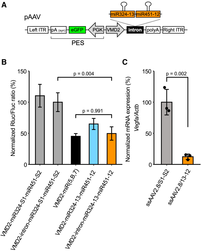
Neovascular age-related macular degeneration (nAMD) is a frequent cause of vision loss among the elderly in the Western world. Current disease management with repeated injections of anti-VEGF agents accumulates the risk for adverse events and constitutes a burden for society and the individual patient. Sustained suppression of VEGF using gene therapy is an attractive alternative, which we explored using adeno-associated virus (AAV)-based delivery of novel RNA interference (RNAi) effectors in a porcine model of choroidal neovascularization (CNV). The potency of VEGFA-targeting, Ago2-dependent short hairpin RNAs placed in pri-microRNA scaffolds (miR-agshRNA) was established in vitro and in vivo in mice. Subsequently, AAV serotype 8 (AAV2.8) vectors encoding VEGFA-targeting or irrelevant miR-agshRNAs under the control of a tissue-specific promotor were delivered to the porcine retina via subretinal injection before CNV induction by laser. Notably, VEGFA-targeting miR-agshRNAs resulted in a significant and sizable reduction of CNV compared with the non-targeting control. We also demonstrated that single-stranded and self-complementary AAV2.8 vectors efficiently transduce porcine retinal pigment epithelium cells but differ in their transduction characteristics and retinal safety. Collectively, our data demonstrated a robust anti-angiogenic effect of VEGFA-targeting miR-aghsRNAs in a large translational animal model, thereby suggesting AAV-based delivery of anti-VEGFA RNAi therapeutics as a valuable tool for the management of nAMD.

Keywords: AAV; AMD; CNV; RNAi therapeutics; anti-VEGF; large animal model; miR-agshRNA; pig; retinal gene therapy.


Conflict of interest statement

The authors declare no competing interests.

Figures

None
Graphical abstract
Figure 1
Figure 1
Design and knockdown efficacy of Vegfa targeting miR-agshRNA constructs (A) Schematic diagram of the miR-agshRNA units tested with or without intron embedment in pcDNA3.1-based plasmids. (B) Bar plot (mean ± SD) showing Vegfa knockdown activity of the different miR-agshRNA combinations with or without intron embedment using co-transfection of a dual-luciferase reporter plasmid in HEK293 cells. Renilla luciferase (Rluc) fused to the Vegfa sequence is related to a firefly luciferase (Fluc) serving as internal control. All miR-agshRNA constructs were tested against a previously published triple-targeting miRNA-based cluster, miR(5,B,7). Rluc/Fluc ratio is the mean of triplicates normalized to the pcDNA3.1-CMV-intron control. Statistical comparisons were performed using one-way ANOVA followed by Tukey’s post hoc test. CMV, cytomegalovirus promoter; poly(A), polyadenylation signal.
Figure 2
Figure 2
Design, in vitro, and in vivo Vegfa knockdown efficacy of VMD2-driven miR-agshRNA constructs (A) Schematic presentation of the AAV2-based vector (pAAV) intron-miR324-13-miR451-12-PES, which expresses two miR-agshRNA units from the VMD2 promotor and GFP from a back-to-back PGK promoter. (B) Bar plot (mean ± SD) showing Vegfa knockdown efficacy of the pAAV-VMD2-intron-324-13-451-12-PES tested against a corresponding construct without intron embedment, two controls with irrelevant miR-agshRNAs and a pAAV with a previously published triple-targeting miRNA-based cluster (VMD2-miR(5,B,7)) estimated by dual luciferase co-transfection assay in human melanoma cells. Rluc/Fluc ratio is the mean of triplicates normalized to the pAAV-VMD2-intron-324-S1-451-S2 control. Statistical comparisons were performed using one-way ANOVA followed by Tukey’s post hoc test. (C) Eight mice were injected in each group with 1 × 108 vg/eye of ssAAV2.8/13-12 or ssAAV2.8/S1-S2. From these eyes, three pools of GFP-positive cells were collected for RNA purification per group. RNA was purified from the GFP-positive pools, and Vegfa mRNA was quantified using RT-qPCR. Bar plot (mean ± SD) showing Vegfa mRNA expression relative to Actb and normalized to the ssAAV2.8/S1-S2-treated group. Each data point represents a pool of FACS-sorted GFP-positive murine RPE cells. Statistical comparison was performed using the Student’s t test. eGFP, enhanced green fluorescent protein; ITR, inverted terminal repeat; PES, PGK-eGFP-Syn-pA.; PGK, phosphoglycerate kinase 1 promotor; poly(A), polyadenylation signal; Syn-pA, synthetic polyadenylation signal; VMD2, vitelliform macular dystrophy 2 promoter.
Figure 3
Figure 3
Transduction characteristics of ssAAV2.8/GFP and scAAV2.8/GFP in the porcine retina (A) Schematic overview of ssAAV2.8 and scAAV2.8 encoding GFP driven by a CMV promotor. (B) Timeline of the delivery study showing the number (n) of animals injected with ssAAV2.8/GFP in one eye and scAAV2.8/GFP in the contralateral eye on D0 and at follow-ups on D14–D42. Black asterisks indicate clinical evaluation of ocular inflammation with slit lamp and fundus imaging. Circles indicate evaluation of GFP expression and retinal integrity using FI, FFI, and OCT in vivo and on RPE/choroidal flatmounts and retinal cross-sections. (C) Left and middle column: representative examples of RPE/choroidal flatmounts collected on D14 (left column) and D42 (middle column) after subretinal injection of ssAAV2.8/GFP (upper row) or scAAV2.8/GFP (lower row). GFP signal is presented in green. Scale bars, 50 μm. Right column: representative examples of retinal cross-sections from the injected area, obtained on D42 (right column) from eyes injected with ssAAV2.8/GFP (upper row) or scAAV2.8/GFP (lower row). DAPI staining (blue) and anti-GFP staining (green). White arrows point toward GFP expression. Scale bars, 100 μm. (D) Quantification of GFP expression on RPE/choroidal flatmounts obtained on D14, D28, and D42. Each dot represents one eye, n = 1–2. Data are plotted as mean ± SD. In two cases, only one eye was available for data analysis (scAAV2.8/GFP: D14; ssAVV2.8/GFP: D28). (E and F) Quantification of GFP signal by FFI on D14, D28, and D42. Each dot represents one eye, n = 3–7. Data are plotted as mean ± SD. In total, eight animals (P1–P8) were used in the delivery study. One eye (P3) was excluded. FFI settings: scAAV2.8/GFP (gain = 24 dB; flash level, 32 Ws), ssAAV2.8/GFP (gain = 24 dB; flash-level, 63 Ws). AAV, adeno-associated virus; AU, arbitrary units; BL, baseline; CMV, cytomegalovirus promoter; D0–D42, days 0–42; FI, fundus imaging; FFI, fundus fluorescence imaging; GFP, green fluorescent protein; INL, inner nuclear layer; IPL, inner plexiform layer; n, number of animals; OCT, optical coherence tomography; ONL, outer nuclear layer; PI, post-injection; PR, photoreceptor; RPE, retinal pigment epithelium; sc, self-complementary; ss, single-stranded.
Figure 4
Figure 4
In vivo examination of the porcine retina post-injection of ssAAV2.8/GFP and scAAV2.8/GFP Representative images of the porcine retina obtained with indicated imaging techniques on D0, D14, D28, and D42 after subretinal injection with ssAAV2.8/GFP (A) or scAAV2.8/GFP (B). FI (upper row) with black arrows indicating the border of the subretinal bleb on D0 and yellow arrows indicating the retinotomy and resulting scar tissue. Corresponding images obtained by FFI (middle row) showing GFP expression (white). Cross-sectional OCT scans (lower row) corresponding to the dashed line on FI. Light-blue arrowheads on OCT scans indicate hyperreflective irregularities adjacent to the RPE. FFI settings: scAAV2.8/GFP (gain = 24 dB; flash level = 32 Ws), ssAAV2.8/GFP (gain = 24 dB; flash level = 63 Ws). FI, fundus imaging; FFI, fundus fluorescence imaging; GFP, green fluorescent protein; OCT, optical coherence tomography; PI, post-injection.
Figure 5
Figure 5
Structural retinal changes after injection with scAAV2.8/GFP (A) Representative examples of FI (upper row) at baseline and on D42 after injection with ssAAV2.8/GFP or scAAV2.8/GFP. Light-pink arrows delimit the area of pigmentary changes. Cross-sectional OCT scan (lower row) corresponding to dashed white line on FI. White line indicates area outside the pigmentary changes, light-pink line indicates area with pigmentary changes. Light-blue arrow indicates hyperreflective irregularities adjacent to the RPE. Orange arrows point toward an area with loss of distinct layering of the RPE/PR complex. (B) Evaluation of structural retinal changes at different time points after subretinal injection of ssAAV2.8/GFP or scAAV2.8/GFP: Pigmentary changes (left), loss of distinct layering of the RPE/PR complex (middle), and hyperreflective irregularities adjacent to the RPE (right). Data are presented as percent of eyes available at the respective time point. ssAAV2.8/GFP (D14: n = 6; D28: n = 4; D42: n = 3); scAAV2.8/GFP (D14: n = 7, D28: n = 5; D42: n = 3). (C) H&E stains of retinal cross-section of the same eyes displayed in (A) injected with ssAAV2.8/GFP (GFP-positive area) or with scAAV2.8/GFP (GFP-positive and GFP-negative areas). Purple arrows point toward RPE cells with reduced pigment granules. Scale bars, 20 μm. BL, baseline; GFP, green fluorescent protein.
Figure 6
Figure 6
AAV-mediated subretinal delivery of VEGFA-targeting miR-agshRNA to the porcine retina (A) Timeline of the intervention study, showing the number of animals for subretinal injection of ssAAV2.8/13-12 and ssAAV2.8/S1-S2 on D0, laser-mediated CNV induction on D28 and follow-ups on D14 and D42 post-injection. Black asterisks indicate clinical evaluation of ocular inflammation using slit lamp and fundus imaging. Circles indicate evaluation of GFP expression, retinal integrity, and CNV formation with FI, FFI, and OCT in vivo and on RPE/choroidal flatmounts. (B) Representative examples of FI (upper row) and corresponding FFI (lower row) of an eye injected with ssAAV2.8/S1-S2 on D0 (PI), pre- and post-laser treatment on D28 and D42. Yellow arrows point toward the retinotomy and resulting scar tissue. Black arrows indicate the border of the subretinal bleb on D0. Blue arrowheads indicate laser lesions on D28 and D42. GFP expression is shown in white on the images obtained by FFI, white arrows indicate the border of the GFP-positive area. (C). Representative examples of cross-sectional OCT scans (inverted colors) obtained on D42 showing subretinal CNV lesions (marked with a white border) for eyes injected with ssAAV2.8/13-12 (upper row) and ssAAV2.8/S1-S2 (lower row). Scale bars, 450 μm. (D) Analysis of CNV area on cross-sectional OCT scans for eyes injected with ssAAV2.8/13-12 and ssAAV2.8/S1-S2. Dot plot and boxplot of individual CNV measurements are presented. Statistical comparison was performed on mean CNV area per eye using the Wilcoxon rank-sum test. P9–P18 represents individual animals, n = 9. Both eyes in P12 were excluded. AAV, adeno-associated virus; BL, baseline; CNV, choroidal neovascularization; D0–D42, days 0–42; n, number of animals; NA, not available; OCT, optical coherence tomography; PI, post-injection; PoL, post-laser; PrL, pre-laser; ss, single-stranded.
Figure 7
Figure 7
AAV-mediated subretinal delivery of VEGFA-targeting miR-agshRNA reduces CNV in a porcine model (A) Representative examples of RPE/choroidal flatmounts from eyes harvested on D42 after injection of ssAAV2.8/13-12 (upper row) or ssAAV2.8/S1-S2 (lower row). Overview of the RPE/choroidal flatmounts. GFP expression (green), CD31 (red), GS-IB4 (blue). Scale bar, 800 μm (first column). Magnified images of CNV lesions corresponds to the white box in the first column. CD31 staining (red), GS-IB4 staining (blue) and CD31/GS-IB4 staining (fourth column). Scale bar, 100 μm. (B) Quantification of CD31 CNV area and (C) GS-IB4 CNV area from ssAAV2.8/13-12 and ssAAV2.8/S1-S2-injected eyes. Dot plot and boxplot of individual CNV measurements are presented. Statistical comparisons were performed on mean CNV area per eye using the Student’s t test. P10–P18 represents individual animals, n = 8. Both eyes from P9 and P12 were excluded. AAV, adeno-associated virus; CD31, cluster of differentiation 31; CNV, choroidal neovascularization; GS-IB4, Griffonia simplicifolia type I lectin; ss, single-stranded.

References

    1. Coleman H.R., Chan C.C., Ferris F.L., 3rd, Chew E.Y. Age-related macular degeneration. Lancet. 2008;372:1835–1845. doi: 10.1016/s0140-6736(08)61759-6. - DOI - PMC - PubMed
    1. Rupert R.A.B., Jost B.J., Alain M.B., Maria Vittoria C., Aditi D., Seth R.F., David S.F., Jill E.K., John H.K., Janet L., et al. Prevalence and causes of vision loss in high-income countries and in Eastern and Central Europe in 2015: magnitude, temporal trends and projections. Br. J. Ophthalmol. 2018;102:575–585. doi: 10.1136/bjophthalmol-2017-311258. - DOI - PMC - PubMed
    1. Li J.Q., Welchowski T., Schmid M., Mauschitz M.M., Holz F.G., Finger R.P. Prevalence and incidence of age-related macular degeneration in Europe: a systematic review and meta-analysis. Br. J. Ophthalmol. 2020;104:1077–1084. doi: 10.1136/bjophthalmol-2019-314422. - DOI - PubMed
    1. Rein D.B., Wittenborn J.S., Burke-Conte Z., Gulia R., Robalik T., Ehrlich J.R., Lundeen E.A., Flaxman A.D. Prevalence of Age-Related Macular Degeneration in the US in 2019. JAMA Ophthalmol. 2022;140:1202–1208. doi: 10.1001/jamaophthalmol.2022.4401. - DOI - PMC - PubMed
    1. Fleckenstein M., Keenan T.D.L., Guymer R.H., Chakravarthy U., Schmitz-Valckenberg S., Klaver C.C., Wong W.T., Chew E.Y. Age-related macular degeneration. Nat. Rev. Dis. Primers. 2021;7:31. doi: 10.1038/s41572-021-00265-2. - DOI - PubMed

LinkOut - more resources