. 2024 Mar 26;32(2):101243.
doi: 10.1016/j.omtm.2024.101243. eCollection 2024 Jun 13.

Development of adenoviral vectors that transduce Purkinje cells and other cerebellar cell-types in the cerebellum of a humanized mouse model

Affiliations

Development of adenoviral vectors that transduce Purkinje cells and other cerebellar cell-types in the cerebellum of a humanized mouse model

Emre Kul et al. Mol Ther Methods Clin Dev. .

Abstract

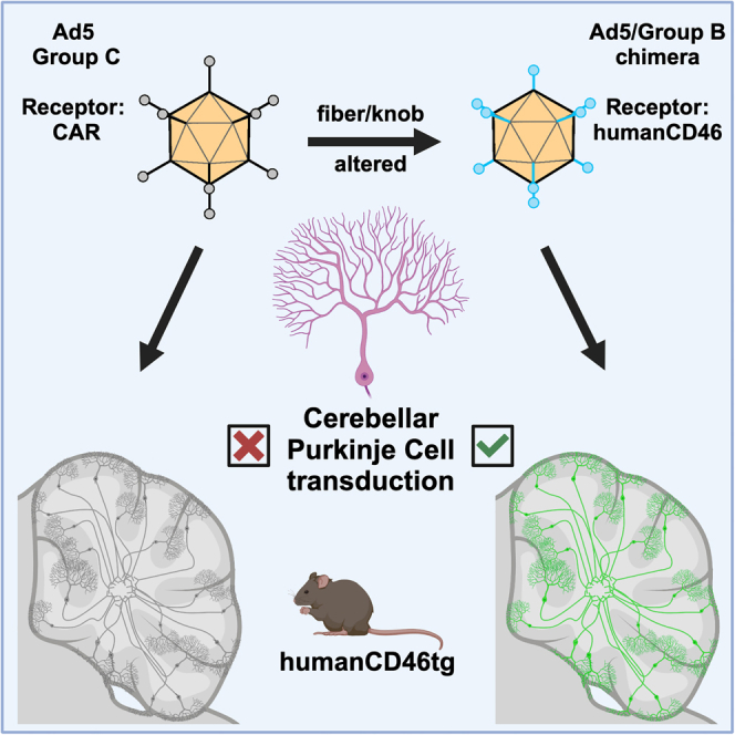
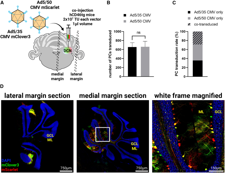
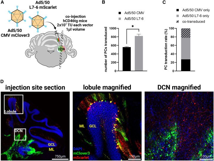
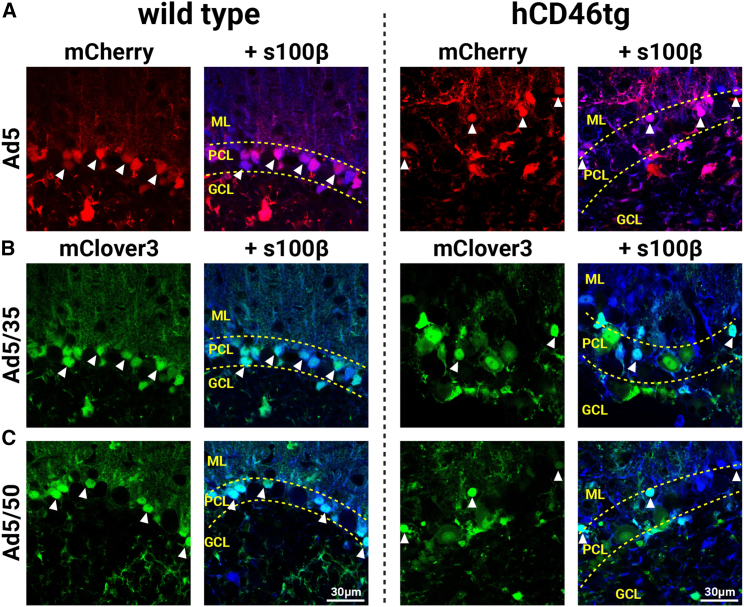
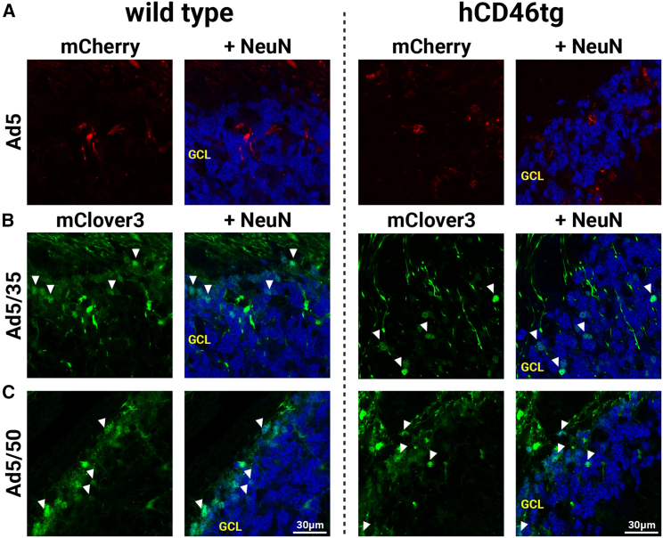
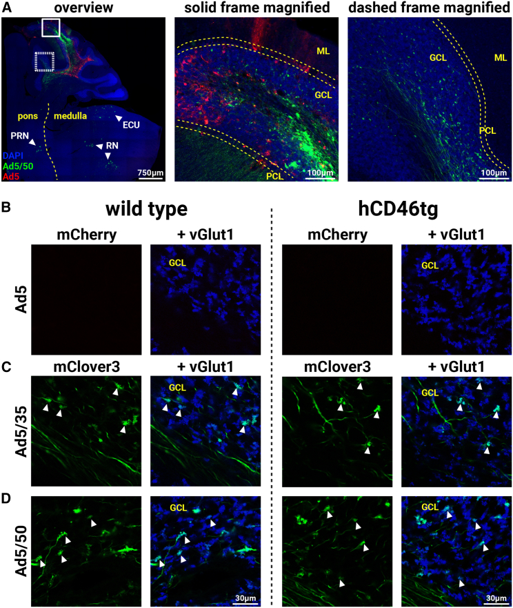
Viral vector gene therapy has immense promise for treating central nervous system (CNS) disorders. Although adeno-associated virus vectors (AAVs) have had success, their small packaging capacity limits their utility to treat the root cause of many CNS disorders. Adenoviral vectors (Ad) have tremendous potential for CNS gene therapy approaches. Currently, the most common vectors utilize the Group C Ad5 serotype capsid proteins, which rely on the Coxsackievirus-Adenovirus receptor (CAR) to infect cells. However, these Ad5 vectors are unable to transduce many neuronal cell types that are dysfunctional in many CNS disorders. The human CD46 (hCD46) receptor is widely expressed throughout the human CNS and is the primary attachment receptor for many Ad serotypes. Therefore, to overcome the current limitations of Ad vectors to treat CNS disorders, we created chimeric first generation Ad vectors that utilize the hCD46 receptor. Using a "humanized" hCD46 mouse model, we demonstrate these Ad vectors transduce cerebellar cell types, including Purkinje cells, that are refractory to Ad5 transduction. Since Ad vector transduction properties are dependent on their capsid proteins, these chimeric first generation Ad vectors open new avenues for high-capacity helper-dependent adenovirus (HdAd) gene therapy approaches for cerebellar disorders and multiple neurological disorders.

Keywords: CD46; Purkinje cell; cerebellum; chimeric adenoviral vector; helper-dependent adenoviral vector; retrograde transduction; tropism; viral vector gene therapy.


Conflict of interest statement

E.K. and S.M.Y. have a provisional patent filed related to the findings in the manuscript.

Figures

None
Graphical abstract
Figure 1
Figure 1
Mouse model and generation of viral vectors (A and B) Photomicrographs of RNA-ISH BaseScope Assay performed on the sagittal cerebellar sections of wild-type mouse (A) and hCD46tg mouse (B). N(wt/tg) = 3/3. Black arrowheads indicate Purkinje cells (PCs). Blue: hematoxylin, Red: chromogenic hCD46 probe signal, ML, molecular layer; PCL, Purkinje cell layer; GCL, granule cell layer. Dashed lines indicate PCL. (C and D) Immunofluorescence staining performed on cerebellar sections from wild-type (C) and hCD46tg (D) mice labeling PC-specific molecular marker, Pcp2 (green) and hCD46 (red). Blue: DAPI. N(wt/tg) = 1/1. (E and F) Confirmation of chimeric vector generation by distinct restriction enzyme digestion patterns of PCR-amplified fiber/knob fragments derived from purified vectors. Agarose gel image demonstrating AflIII digestion patterns, indicating successful incorporation of Ad21, Ad35, or Ad50 shaft and knob sequences into Ad5 genome. Expected band sizes were observed for Ad5 (lane 1), Ad5/21 (lane 2), Ad5/35 (lane 3), and Ad5/50 (lane 4). (G) Representative maps of viral genomes of E1/E3 deleted first generation Ad5 and Group B chimeric vectors, depicting the modifications made to the shaft and knob regions of the fiber domain.
Figure 2
Figure 2
hCD46-dependent transduction of Purkinje cells by Group B chimeric vectors (A) Illustration showcasing lobular administration of Ad5 individually co-mixed with Ad5/21, Ad5/35, or Ad5/50 into the mouse cerebellum and subsequent tissue processing steps. N(wt/tg) = 3/3 for each chimeric vector. VP, viral particle. (B) Fluorescent photomicrographs representing lack of Ad5 tropism to PCs in the cerebellar cortex of wild-type (left column) and hCD46tg (right column) mice. Red: Ad5 transduction signal (mCherry), Blue: Pcp2 immunofluorescence signal. ML, molecular layer; PCL, Purkinje cell layer; GCL, granule cell layer. Yellow arrowheads indicate non-transduced PCs, dashed lines indicate PCL. (C–E) Fluorescent photomicrographs representing Ad5/21 (C), Ad5/35 (D), and Ad5/50 (E) tropism to PCs in the cerebellar cortex of wild-type (left column) and hCD46tg (right column) mice. Green: chimeric vector transduction signal (mClover3). White arrowheads indicate transduced PCs. Data are derived from co-injections of each Ad5/Group B vector with the Ad5 vector, where 1 × 109 VP per vector was delivered in 1 μL volume into the cerebellar simple lobule.
Figure 3
Figure 3
Injection of chimeric vectors into the DCN transduces Purkinje cells via retrograde axonal transport (A) Illustration of sagittal mouse cerebellum section depicting DCN injection site targeting PC axon terminals. N(tg) = 3 for each chimeric vector. (B) Fluorescent photomicrographs representing lack of Ad5 tropism to PCs in the cerebellar cortex of hCD46tg mice upon DCN injection. Red: Ad5 transduction signal (mCherry), Blue: Pcp2 immunofluorescence signal. ML, molecular layer; PCL, Purkinje cell layer; GCL, granule cell layer. Dashed lines indicate PCL. (C and D) Fluorescent photomicrographs representing transduction of PCs by Ad5/35 (C) and Ad5/50 (D) in the cerebellar cortex of hCD46tg mice upon DCN injection. Green: chimeric vector transduction signal (mClover3). White arrowheads indicate transduced PCs. Data are derived from co-injections of each Ad5/Group B vector with the Ad5 vector, where 1 × 109 VP per vector was delivered in 1 μL volume into the DCN.
Figure 4
Figure 4
Ad5/35 and Ad5/50 transduce comparable number of Purkinje cells via DCN (A) Illustration of coronal mouse cerebellum section depicting DCN injection performed to compare Ad5/35 with Ad5/50 and margin sections obtained distant to the injection site (dashed lines). N(tg) = 3. TU, transducing units. (B) Quantification from sagittal serial sections of number of PCs transduced by either Ad5/35 or Ad5/50 under CMV promoter. ns, p = 0.9678, Student’s t test. Data are plotted as mean ± SEM. (C) Stacked bar graph indicating the percentage of PCs transduced by each vector and the percentage co-transduced by both vectors. (D) Tiled photomicrographs representing PC transduction at the lateral margin (left) and medial margin (middle) of the cerebellum. Magnified reproduction (right) of the white frame on the medial margin section. Green: Ad5/35 vector transduction signal (mClover3), Red: Ad5/50 vector transduction signal (mScarlet), Blue: DAPI, ML: molecular layer, GCL: granule cell layer. Data are derived from co-injections of the Ad5/35 vector with the Ad5/50 vector, where 2 × 107 TU per vector was delivered in 1 μL volume in to the DCN.
Figure 5
Figure 5
PC-specific L7-6 promoter increases transduction efficiency (A) Illustration of coronal mouse cerebellum section depicting DCN injection performed to compare CMV promoter to L7-6 promoter under the context of Ad5/50. N(tg) = 3. Dashed lines indicate section obtained from proximity to the injection site. TU: transducing units. (B) Quantification of number of PCs from sagittal serial sections transduced by Ad5/50 under either CMV or L7-6 promoters. ∗, p = 0.0173, Student’s t test. Data are plotted as mean ± SEM. (C) Stacked bar graph indicating the percentage of PCs transduced by each vector and the percentage co-transduced by both vectors. (D) Tiled photomicrograph representing Ad5/50 tropism at the injection site section (left) and magnified images corresponding to the lobule frame (middle) and DCN frame (right). Green: Ad5/50 CMV vector transduction signal (mClover3), Red: Ad5/50 L7-6 vector transduction signal (mScarlet), Blue: DAPI, ML, molecular layer; GCL, granule cell layer; DCN, deep cerebellar nuclei. Data are derived from co-injections of the Ad5/50 CMV vector with the Ad5/50 L7-6 vector, where 2 × 107 TU per vector was delivered in 1 μL volume into the DCN.
Figure 6
Figure 6
Ad5, Ad5/35, and Ad5/50 transduce Bergmann glia in a hCD46 independent manner (A) Fluorescent photomicrographs representing Ad5 tropism to Bergmann glia in wild-type (left column) and hCD46tg (right column) mice. Red: Ad5 transduction signal (mCherry), Blue: s100β immunofluorescence signal. ML, molecular layer; PCL, Purkinje cell layer; GCL, granule cell layer. (B and C) Fluorescent photomicrographs representing Ad5/35 (B) and Ad5/50 (C) tropism to Bergmann glia in wild-type (left column) and hCD46tg (right column) mice. White arrowheads indicate transduced Bergmann glia. Green: chimeric vector transduction signal (mClover3). N(wt/tg) = 3/3 for each chimeric vector. Data are derived from co-injections of each Ad5/Group B vector with the Ad5 vector, where 1 × 109 VP per vector was delivered in 1 μL volume into the cerebellar simple lobule.
Figure 7
Figure 7
Ad5/35 and Ad5/50 transduce granule cells in a hCD46 independent manner (A) Fluorescent photomicrographs of granule cell layer representing lack of Ad5 tropism to granule cells in wild-type (left column) and hCD46tg (right column) mice. Red: Ad5 transduction signal (mCherry), Blue: NeuN immunofluorescence signal. GCL, granule cell layer. (B and C) Fluorescent photomicrographs of granule cell layer representing Ad5/35 (B) and Ad5/50 (C) tropism to granule cells in wild-type (left column) and hCD46tg (right column) mice. White arrowheads indicate transduced granule cells. Green: chimeric vector transduction signal (mClover3). N(wt/tg) = 3/3 for each chimeric vector. Data are derived from co-injections of each Ad5/Group B vector with the Ad5 vector, where 1 × 109 VP per vector was delivered in 1 μL volume into the cerebellar simple lobule.
Figure 8
Figure 8
Ad5/35 and Ad5/50 transduce mossy fibers in a hCD46 independent manner (A) Tiled photomicrograph indicating retrograde transduction of mossy fibers leading to labeling of cell somata in various nuclei in pons and medulla. ECU, external cuneate nucleus; RN, reticular nuclei; PRN, pontine reticular nucleus. Dashed line traces the pontomedullary junction (left). Magnified image corresponding to the solid frame (middle) shows the transduction patterns at the injected simple lobule. Dashed frame indicates transduced mossy fiber terminals by Ad5/50 (green-mClover3) but not by Ad5 (red-mCherry) in a cerebellar lobule distant to the injected lobule (right). ML, molecular layer; PCL, Purkinje cell layer; GCL, granule cell layer. Blue: DAPI. Dashed lines in magnified reproductions indicate PCL. Representative image originates from a hCD46tg mouse. (B) Fluorescent photomicrographs of granule cell layer representing lack of Ad5 transduction of mossy fiber terminals in wild-type (left) and hCD46tg (right) mice. Red: Ad5 transduction signal (mCherry), Blue: vGlut1 immunofluorescence signal. (C and D) Fluorescent photomicrographs of granule cell layer representing Ad5/35 (C) and Ad5/50 (D) transduction of mossy fiber terminals in wild-type (left) and hCD46tg (right) mice. White arrowheads indicate transduced mossy fiber terminals. Green: chimeric vector transduction signal (mClover3). N(wt/tg) = 3/3 for each chimeric vector. Data are derived from co-injections of each Ad5/Group B vector with the Ad5 vector, where 1 × 109 VP per vector was delivered in 1 μL volume into the cerebellar simple lobule.

Similar articles

References

    1. Bulcha J.T., Wang Y., Ma H., Tai P.W.L., Gao G. Viral vector platforms within the gene therapy landscape. Signal Transduct. Targeted Ther. 2021;6:53. doi: 10.1038/s41392-021-00487-6. - DOI - PMC - PubMed
    1. Dunbar C.E., High K.A., Joung J.K., Kohn D.B., Ozawa K., Sadelain M. Gene therapy comes of age. Science. 2018;359 doi: 10.1126/science.aan4672. - DOI - PubMed
    1. Pupo A., Fernández A., Low S.H., François A., Suárez-Amarán L., Samulski R.J. AAV vectors: The Rubik's cube of human gene therapy. Mol. Ther. 2022;30:3515–3541. doi: 10.1016/j.ymthe.2022.09.015. - DOI - PMC - PubMed
    1. Li C., Samulski R.J. Engineering adeno-associated virus vectors for gene therapy. Nat. Rev. Genet. 2020;21:255–272. doi: 10.1038/s41576-019-0205-4. - DOI - PubMed
    1. Poletti V., Mavilio F. Designing Lentiviral Vectors for Gene Therapy of Genetic Diseases. Viruses. 2021;13 doi: 10.3390/v13081526. - DOI - PMC - PubMed

LinkOut - more resources