In vitro and in vivo degradation, biocompatibility and bone repair performance of strontium-doped montmorillonite coating on Mg-Ca alloy
- PMID: 38605854
- PMCID: PMC11007119
- DOI: 10.1093/rb/rbae027
In vitro and in vivo degradation, biocompatibility and bone repair performance of strontium-doped montmorillonite coating on Mg-Ca alloy
Abstract
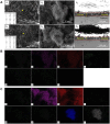
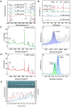
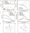
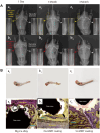
Poor bone growth remains a challenge for degradable bone implants. Montmorillonite and strontium were selected as the carrier and bone growth promoting elements to prepare strontium-doped montmorillonite coating on Mg-Ca alloy. The surface morphology and composition were characterized by SEM, EDS, XPS, FT-IR and XRD. The hydrogen evolution experiment and electrochemical test results showed that the Mg-Ca alloy coated with Sr-MMT coating possessed optimal corrosion resistance performance. Furthermore, in vitro studies on cell activity, ALP activity, and cell morphology confirmed that Sr-MMT coating had satisfactory biocompatibility, which can significantly avail the proliferation, differentiation, and adhesion of osteoblasts. Moreover, the results of the 90-day implantation experiment in rats indicated that, the preparation of Sr-MMT coating effectively advanced the biocompatibility and bone repair performance of Mg-Ca alloy. In addition, The Osteogenic ability of Sr-MMT coating may be due to the combined effect of the precipitation of Si4+ and Sr2+ in Sr-MMT coating and the dissolution of Mg2+ and Ca2+ during the degradation of Mg-Ca alloy. By using coating technology, this study provides a late-model strategy for biodegradable Mg alloys with good corrosion resistance, biocompatibility. This new material will bring more possibilities in bone repair.
Keywords: Mg–Ca alloy; biocompatibility; corrosion resistance; montmorillonite; osteogenesis; strontium.
© The Author(s) 2024. Published by Oxford University Press.
Figures


Similar articles
-
Corrosion resistance and antibacterial activity of zinc-loaded montmorillonite coatings on biodegradable magnesium alloy AZ31.Acta Biomater. 2019 Oct 15;98:196-214. doi: 10.1016/j.actbio.2019.05.069. Epub 2019 May 31. Acta Biomater. 2019. PMID: 31154057
-
The in vitro and in vivo performance of a strontium-containing coating on the low-modulus Ti35Nb2Ta3Zr alloy formed by micro-arc oxidation.J Mater Sci Mater Med. 2015 Jul;26(7):203. doi: 10.1007/s10856-015-5533-0. Epub 2015 Jul 8. J Mater Sci Mater Med. 2015. PMID: 26152510
-
Biomimetic Ca, Sr/P-Doped Silk Fibroin Films on Mg-1Ca Alloy with Dramatic Corrosion Resistance and Osteogenic Activities.ACS Biomater Sci Eng. 2018 Sep 10;4(9):3163-3176. doi: 10.1021/acsbiomaterials.8b00787. Epub 2018 Aug 27. ACS Biomater Sci Eng. 2018. PMID: 33435057
-
In-vivo bone remodeling potential of Sr-d-Ca-P /PLLA-HAp coated biodegradable ZK60 alloy bone plate.Mater Today Bio. 2022 Dec 27;18:100533. doi: 10.1016/j.mtbio.2022.100533. eCollection 2023 Feb. Mater Today Bio. 2022. PMID: 36619205 Free PMC article.
-
An in vivo study on the metabolism and osteogenic activity of bioabsorbable Mg-1Sr alloy.Acta Biomater. 2016 Jan;29:455-467. doi: 10.1016/j.actbio.2015.11.014. Epub 2015 Nov 11. Acta Biomater. 2016. PMID: 26577986
Cited by
-
Polydopamine/Montmorillonite-Based Brushite Calcium Phosphate Cements: Synergize Mechanical and Osteogenic Capacity for Bone Regeneration.Biomater Res. 2025 Jun 9;29:0213. doi: 10.34133/bmr.0213. eCollection 2025. Biomater Res. 2025. PMID: 40491507 Free PMC article.
-
Surface modification of polyetheretherketone for boosted osseointegration: A review.Biomater Transl. 2025 Jun 25;6(2):181-201. doi: 10.12336/bmt.24.00052. eCollection 2025. Biomater Transl. 2025. PMID: 40641993 Free PMC article. Review.
References
-
- Xie J, Wang L, Zhang J, Lu L, Zhang Z, He Y, Wu R.. Developing new Mg alloy as potential bone repair material via constructing weak anode nano-lamellar structure. J Magnes Alloys 2023;11:154–75.
-
- Hiromoto S, Nozoe E, Hanada K, Yoshimura T, Shima K, Kibe T, Nakamura N, Doi K.. In vivo degradation and bone formation behaviors of hydroxyapatite-coated Mg alloys in rat femur. Mater Sci Eng C Mater Biol Appl 2021;122:111942. - PubMed
-
- Kim SY, Kim YK, Kim KS, Lee KB, Lee MH.. Enhancement of bone formation on LBL-coated Mg alloy depending on the different concentration of BMP-2. Colloids Surf B Biointerfaces 2019;173:437–46. - PubMed
-
- Darbre PD. Aluminium and the human breast. Morphologie 2016;100:65–74. - PubMed
LinkOut - more resources
Full Text Sources
Research Materials
Miscellaneous

