Review
. 2024 Mar 28:17:1349123.
doi: 10.3389/fnmol.2024.1349123. eCollection 2024.

Cuproptosis in stroke: focusing on pathogenesis and treatment

Affiliations
Review

Cuproptosis in stroke: focusing on pathogenesis and treatment

Liwei Xing et al. Front Mol Neurosci. .

Abstract

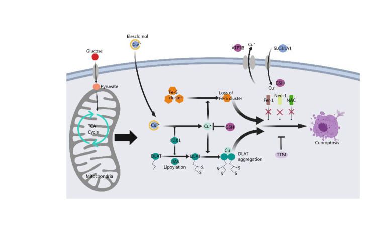
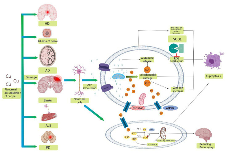
Annually, more than 15 million people worldwide suffer from stroke, a condition linked to high mortality and disability rates. This disease significantly affects daily life, impairing everyday functioning, executive function, and cognition. Moreover, stroke severely restricts patients' ability to perform daily activities, diminishing their overall quality of life. Recent scientific studies have identified cuproptosis, a newly discovered form of cell death, as a key factor in stroke development. However, the role of cuproptosis in stroke remains unclear to researchers. Therefore, it is crucial to investigate the mechanisms of cuproptosis in stroke's pathogenesis. This review examines the physiological role of copper, the characteristics and mechanisms of cuproptosis, the differences and similarities between cuproptosis and other cell death types, and the pathophysiology of cuproptosis in stroke, focusing on mitochondrial dysfunction and immune infiltration. Further research is necessary to understand the relationship between previous strokes and cuproptosis and to clarify the mechanisms behind these associations.

Keywords: cuproptosis; mechanism; pathogenesis; stroke; treatment.


Conflict of interest statement

The authors declare that the research was conducted in the absence of any commercial or financial relationships that could be construed as a potential conflict of interest.

Figures

Figure 1
Figure 1
Mechanism of cuproptosis. A novel cell death process called cuproptosis provides strong evidence for the concept that copper-induced cell death occurs through a well-established mechanism involving protein lipoacylation. Cuproptosis begins with copper directly interacting with the fatty acylated parts of the tricarboxylic acid (TCA) cycle. This interaction causes the accumulation of fatty acylated proteins and eventually disrupts iron–sulfur cluster proteins, leading to proteotoxic stress and resulting in cell death. Copper chelator TTM inhibited cuproptosis, while ferroptosis inhibitor (Fer-1), necrotizing apoptosis inhibitor (Nec-1) and oxidative stress inhibitor (NAC) had no effect on cuproptosis. FDX1 acts as a key regulator of protein lipoacylation, which is essential for controlling cuproptosis. Both FDX1 and protein lipoacylation are critical in managing this cell death process.
Figure 2
Figure 2
Schematic representation of possible mechanisms of cuproptosis in stroke. Abnormal accumulation of copper contributes to the development of a variety of neurodegenerative diseases, including stroke, Alzheimer's disease, amyotrophic lateral sclerosis, Parkinson's disease, glioma, and Huntington's disease. Mitochondrial oxidative stress injury is involved in the progression of stroke, providing conditions for the occurrence of cuproptosis. Therefore, reducing mitochondrial ROS production, excluding damaged mitochondria and refusing damaged mitochondria to reenter neuronal cells may be important measures to reduce neuronal oxidative stress injury and cuproptosis. Secondly, overexpression of LIAS reduced proinflammatory cytokines/chemokines and inhibited NF-κB activity. In addition, overexpression of LIAS can reduce oxidative stress and enhance antioxidant defense mechanisms, which may have some protective effect on mitochondria and reduce damage after stroke.

Similar articles

Cited by

References

    1. Acevedo K., Masaldan S., Opazo C. M., Bush A. I. (2019). Redox active metals in neurodegenerative diseases. J. Biol. Inorg. Chem. 24, 1141–1157. doi: 10.1007/s00775-019-01731-9 - DOI - PubMed
    1. Acilan C., Cevatemre B., Adiguzel Z., Karakas D., Ulukaya E., Ribeiro N., et al. . (2016). Validation data supporting the characterization of novel copper complexes as anticancer agents. Data Brief 9, 1160–1174. doi: 10.1016/j.dib.2016.11.063, PMID: - DOI - PMC - PubMed
    1. Agarwal P., Ayton S., Agrawal S., Dhana K., Bennett D. A., Barnes L. L., et al. . (2022). Brain copper may protect from cognitive decline and Alzheimer’s disease pathology: a community-based study. Mol. Psychiatry 27, 4307–4313. doi: 10.1038/s41380-022-01802-5, PMID: - DOI - PMC - PubMed
    1. Ayton S., Lei P., Bush A. I. (2013). Metallostasis in Alzheimer’s disease. Free Radic. Biol. Med. 62, 76–89. doi: 10.1016/j.freeradbiomed.2012.10.558, PMID: - DOI - PubMed
    1. Bica L., Liddell J. R., Donnelly P. S., Duncan C., Caragounis A., Volitakis I., et al. . (2014). Neuroprotective copper bis(thiosemicarbazonato) complexes promote neurite elongation. PLoS One 9:e90070. doi: 10.1371/journal.pone.0090070, PMID: - DOI - PMC - PubMed

LinkOut - more resources