. 2024 Mar 28:15:1394040.
doi: 10.3389/fphys.2024.1394040. eCollection 2024.

Motor properties of Myosin 5c are modulated by tropomyosin isoforms and inhibited by pentabromopseudilin

Affiliations

Motor properties of Myosin 5c are modulated by tropomyosin isoforms and inhibited by pentabromopseudilin

András Kengyel et al. Front Physiol. .

Abstract

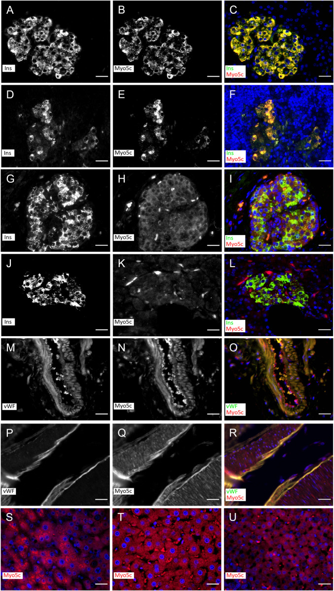
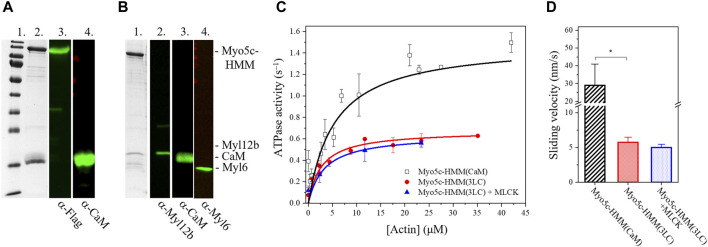
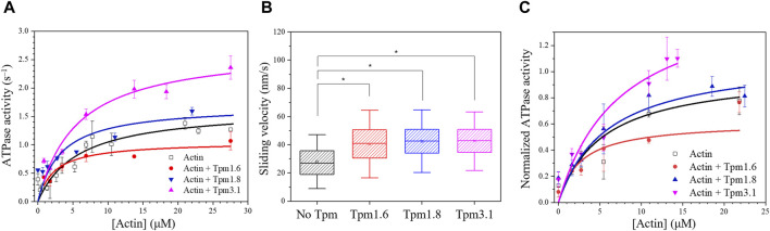
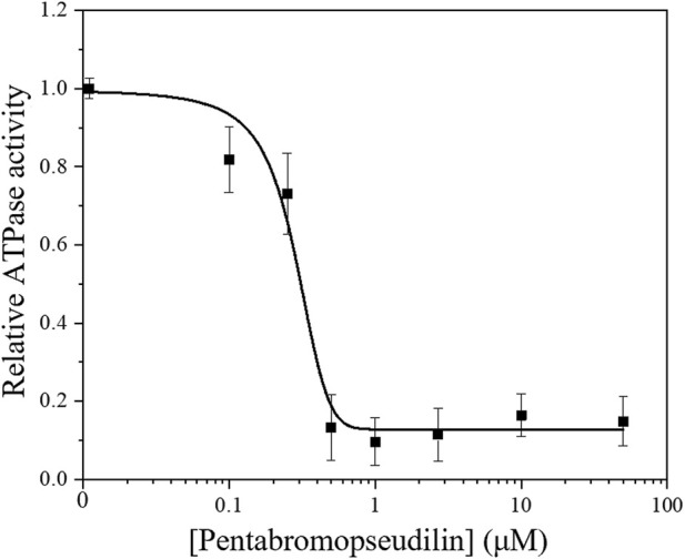
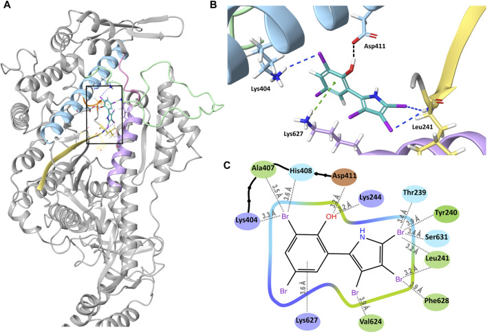
Myosin 5c (Myo5c) is a motor protein that is produced in epithelial and glandular tissues, where it plays an important role in secretory processes. Myo5c is composed of two heavy chains, each containing a generic motor domain, an elongated neck domain consisting of a single α-helix with six IQ motifs, each of which binds to a calmodulin (CaM) or a myosin light chain from the EF-hand protein family, a coiled-coil dimer-forming region and a carboxyl-terminal globular tail domain. Although Myo5c is a low duty cycle motor, when two or more Myo5c-heavy meromyosin (HMM) molecules are linked together, they move processively along actin filaments. We describe the purification and functional characterization of human Myo5c-HMM co-produced either with CaM alone or with CaM and the essential and regulatory light chains Myl6 and Myl12b. We describe the extent to which cofilaments of actin and Tpm1.6, Tpm1.8 or Tpm3.1 alter the maximum actin-activated ATPase and motile activity of the recombinant Myo5c constructs. The small allosteric effector pentabromopseudilin (PBP), which is predicted to bind in a groove close to the actin and nucleotide binding site with a calculated ΔG of -18.44 kcal/mol, inhibits the motor function of Myo5c with a half-maximal concentration of 280 nM. Using immunohistochemical staining, we determined the distribution and exact localization of Myo5c in endothelial and endocrine cells from rat and human tissue. Particular high levels of Myo5c were observed in insulin-producing β-cells located within the pancreatic islets of Langerhans.

Keywords: Myosin 5c; immunohistochemistry; molecular docking; pentabromopseudilin; tropomyosin.


Conflict of interest statement

The authors declare that the research was conducted in the absence of any commercial or financial relationships that could be construed as a potential conflict of interest.

Figures

FIGURE 1
FIGURE 1
Myo5c immunostaining in β–cells in islets of rat (A–F) and human (G–L) under non–diabetic and diabetic conditions and endothelial cells of vessels of rat (M–O) and human (P–R). Myo5c is colocalized with insulin (Ins) staining resulting in an orange–yellow overlay in the β–cells under non–diabetic (A–C,G–I) and diabetic (D–F,J–L) conditions. The endothelial layer of vessels shows a Myo5c expression in colocalization with the von Willebrand factor (vWF) (M–O,P–R). A moderate protein expression of Myo5c is found also in hepatocytes of human (S) and rat (T) as well as in endocrine cells of the rat adrenal cortical region (U). Nuclei are stained with DAPI. Scale Bar 25 µm.
FIGURE 2
FIGURE 2
Myo5c–HMM co–produced with CaM, RLC, and ELC light chains has lower motor activity as co–produced with CaM only. (A) Purified Myo5c–HMM co–produced with CaM light chain. (Lane 1) MW marker. (Lane 2) Coomassie gel showing the result of purification via Flag affinity chromatography. Blot of Myo5c–HMM stained with (Lane 3) anti–Flag or (Lane 4) anti–CaM. Marker bands from highest to lowest (180, 130,100, 70, 55, 40, 35, 25, 15, 10 kDa). (B) Purified Myo5c–HMM co–produced with CaM, RLC, and ELC light chains. (Lane 1) Coomassie gel showing the result of purification via Flag affinity chromatography. Blot of Myo5c–HMM stained with (Lane 2) anti–Myl12b, (Lane 3) anti–CaM or (Lane 4) anti–Myl6. (C) Steady–state actin–activated ATPase activity of human Myo5c coproduced with CaM only (black squares) or with CaM, RLC and ELC (red circles) at various F–actin concentration ranging between 0 and 45 µM. RLC was phosphorylated by MLCK (blue triangles). (D) Average sliding velocity of actin over the Myo5c–HMM–coated surface, obtained from in vitro motility assay, was determined from at least 300 individual trajectories (mean ± SD). RLC was phosphorylated by MLCK. Asterisk marks significance p < 0.01.
FIGURE 3
FIGURE 3
Tropomyosin isoforms affect the motor properties of Myo5c–HMM. (A) Steady-state actin-activated ATPase activity of human Myo5c–HMM (CaM) at various F-actin concentration ranging between 0 and 27 μM, in the presence of different Tpm isoforms. Tpm was added in a 1.3–fold molar excess, measurements were performed at 30°C (n = 2, mean ± SE). (B) Box chart showing the results of in vitro motility assay. Average sliding velocity of actin or actin–Tpm complex over the Myo5c-HMM (CaM)–coated surface was determined from at least 300 individual trajectories. 10 μM Tpm was added to all buffer solutions, assays were performed at 25°C. Box width shows percentile 25–75, whiskers represent ±SD. Asterisks mark significance p < 0.01. (C) Tpm isoforms have an impact on the motor function of Myo5c–HMM (3LC). Data were normalized to the kcat of the actin–activated ATPase activity of Myo5c–HMM (3LC) in the absence of Tpm, measurements were performed at 30°C (n = 3, mean ± SE).
FIGURE 4
FIGURE 4
Steady–state actin–activated ATPase activity of human Myo5c–HMM at 13 μM F–actin concentration, in the presence of 0–50 µM PBP. The semilogarithmic plot shows the concentration dependence of Myo5c–HMM inhibition. The data were fitted with a dose-response model, using the equation y = A1 + (A2−A1)/{1 + 10[(LOGx0−x)*P]}, where the coefficient P is an empirical parameter reflecting the allosteric nature of the mechanism of PBP-mediated inhibition. The concentrations of PBP required for half–maximal inhibition (IC50) of Myo5c–HMM was determined from the fit (n = 3, mean ± SE).
FIGURE 5
FIGURE 5
Binding of PBP to Myo5c. (A) Overview of PBP (cyan) bound into its binding groove (black square) in Myo5c. Helix 13 (Lys244–Val247) is shown in orange, Helix 21 (Arg391–Leu421) in blue, the strut loop (Asn566–Tyr571) in pink, loop 2 (Asn588–Thr623) in green, helix 29 (Val624–Asn640) in violet and the β–strand connecting β7 and helix 13 in yellow. (B) Close–up of the PBP binding site, shown from the direction of the back of the head rotated 90° to the right from the overview position. Hydrogen bond (black), halogen bonds (blue) and cation–pi interaction (green) between PBP and Myo5c are shown, structures of Myo5c in the same colors as in (A). (C) Ligand Interaction Diagram of PBP and Myo5c residues within 4 Å. Residues are colored by their properties: negative residues in orange, positive residues in violet, hydrophobic residues in green and polar residues in blue. Distances to the nearest atom are shown with grey dotted lines.

Similar articles

Cited by

References

    1. Agarwal S., Knölker H.-J. (2004). A novel pyrrole synthesis. Org. Biomol. Chem. 2 (21), 3060–3062. 10.1039/B412206B - DOI - PubMed
    1. Baboolal T. G., Sakamoto T., Forgacs E., White H. D., Jackson S. M., Takagi Y., et al. (2009). The SAH domain extends the functional length of the myosin lever. Proc. Natl. Acad. Sci. U. S. A. 106 (52), 22193–22198. 10.1073/pnas.0909851106 - DOI - PMC - PubMed
    1. Barua B., Nagy A., Sellers J. R., Hitchcock-DeGregori S. E. (2014). Regulation of nonmuscle myosin II by tropomyosin. Biochemistry 53 (24), 4015–4024. 10.1021/bi500162z - DOI - PMC - PubMed
    1. Barua B., Sckolnick M., White H. D., Trybus K. M., Hitchcock‐DeGregori S. E. (2018). Distinct sites in tropomyosin specify shared and isoform-specific regulation of myosins II and V. Cytoskeleton 75 (4), 150–163. 10.1002/cm.21440 - DOI - PMC - PubMed
    1. Behrens V. A., Münnich S., Adler-Gunzelmann G., Thiel C., Henn A., Latham S. L., et al. (2017). The conserved lysine-265 allosterically modulates nucleotide- and actin-binding site coupling in myosin-2. Sci. Rep. 7 (1), 7650. 10.1038/s41598-017-07933-y - DOI - PMC - PubMed