. 2024 Apr 11;14(16):11124-11140.
doi: 10.1039/d4ra01334d. eCollection 2024 Apr 3.

pH-responsive niosome-based nanocarriers of antineoplastic agents

Affiliations

pH-responsive niosome-based nanocarriers of antineoplastic agents

Viliana Gugleva et al. RSC Adv. .

Abstract

Differences in pH between the tumour interstitium and healthy tissues can be used to induce conformational changes in the nanocarrier structure, thereby triggering drug release at the desired site. In the present study, novel pH-responsive nanocarriers were developed by modifying conventional niosomes with hexadecyl-poly(acrylic acid)n copolymers (HD-PAAn). Niosomal vesicles were prepared by the thin film hydration method using Span 60, Span 60/Tween 60 and cholesterol as main constituents, and HD-PAA modifiers of different concentrations (0.5, 1, 2.5, 5 mol%). Next, two model substances, a water-soluble fluorescent dye (calcein) and a hydrophobic agent with pronounced antineoplastic activity (curcumin), were loaded in the aqueous core and hydrophobic membrane of the elaborated niosomes, respectively. Physicochemical properties of blank and loaded nanocarriers such as hydrodynamic diameter (Dh), size distribution, zeta potential, morphology and pH-responsiveness were investigated in detail. The cytotoxicity of niosomal curcumin was evaluated against human malignant cell lines of different origins (MJ, T-24, HUT-78), and the mechanistic aspects of proapoptotic effects were elucidated. The formulation composed of Span 60/Tween 60/cholesterol/2.5% HD-PAA17 exhibited optimal physicochemical characteristics (Dh 302 nm; ζ potential -22.1 mV; high curcumin entrapment 83%), pH-dependent drug release and improved cytotoxic and apoptogenic activity compared to free curcumin.


Conflict of interest statement

There are no conflicts to declare.

Figures
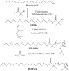
Fig. 1
Fig. 1. Synthesis of hexadecyl-poly(acrylic acid) polymers (HD-PAA) via ATRP of tert-butyl acrylate from hexadecyl-based macroinitiator (HD-Br), and subsequent hydrolysis of the poly(t-butyl acrylate) blocks with trifluoroacetic acid.
Fig. 2
Fig. 2. 1H-NMR spectra of hexadecanol (bottom), hexadecyl-based ATRP initiator (middle), and hexadecyl-poly(t-butyl acrylate)12 precursor (top) in CDCl3.
Fig. 3
Fig. 3. GPC chromatograms of HD-Br, HD-PtBA8 and HD-PtBA12 with THF as the eluent.
Fig. 4
Fig. 4. Cryo-TEM images of niosomes: (a) Sp60 : Chol, (b) Tw60 : Sp60 : Chol; (c) Sp60 : Chol : HD-PAA12 (2.5 mol%), (d) Tw60 : Sp60 : Chol : HD-PAA12 (2.5 mol%).
Fig. 5
Fig. 5. pH-dependent calcein release from HD-PAA modified niosomes after 10 min incubation at 37 °C in PBS pH 7.4/6.8/5.5/4.5. The selected pH values are representative for the pH in tumor interstitium (pH 7–6.8); early endosomes (pH 6.5); late endosomes (pH 5.5) and lysosomes (pH 4.5).
Fig. 6
Fig. 6. Calcein release in PBS pH 7.4 at 37 °C as a function of copolymer concentration.
Fig. 7
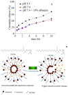
Fig. 7. Curcumin cumulative release from HD-PAA17 modified niosomes based on Sp60 : Tw60 : Chol as a function of pH and time (A) and schematic presentation of membrane rearrangements at acid pH (B).
Fig. 8
Fig. 8. (A) Representative microscopic images of cellular uptake of H1299 cells treated for 6 hours with pure curcumin (first line), non-modified niosomes (second line) formulation S4, drug encapsulated in S11 niosomes (third line), and drug encapsulated in S19 niosomes (fourth line). The natural fluorescence of curcumin is green, and nuclear contra staining with DAPI is blue. Arrows indicated cells with nuclear localization of curcumin-loaded T-17 niosomes. (B) Quantification of normalized integrated density in A.U. The integrated density was measured by Fiji quantification tool. For each condition, at least five images were measured. Multiple comparisons function of One-way ANOVA (Dunnett's multiple comparison test) was used to compare the mean of niosomes-treated cells with the mean of the control column of pure curcumin-treated cells. Probability values were considered significant at the *p < 0.05, **p < 0.01, and ****p < 0.0001. (C) Representative microscopic images showing the accumulation of curcumin encapsulated in T-17 niosomes in the nucleus of some cells. The natural fluorescence of curcumin is green, the cell membrane is stained with CellMask Orange in red, and nuclear contra staining with DAPI is blue.
Fig. 9
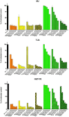
Fig. 9. Cytotoxic effect of free and loaded curcumin in conventional and pH-sensitive niosomes against a panel of human malignant cell lines determined by a MTT-dye reduction assay after 72 h continuous exposure. SD represents the mean values of 8 independent experiments.
Fig. 10
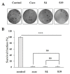
Fig. 10. Clonogenic assay of untreated H1299 cells (control) or cells treated with IC50 concentrations of curcumin loaded pH-sensitive Tw60 : Sp60 : Ch : HD-PAA17 niosomes (S19), curcumin-loaded plain niosomes (Tw60 : Sp60 : Ch) (S4) and pure curcumin. (A) A photograph of a representative experiment is shown. Graph quantifications of colony number (B). The mean values of two independent experiments performed in duplicates are presented with standard deviation (n = 2) ±SD. One-way ANOVA Tukey's multiple comparisons test was used to tests each experimental group against each control group. Probability values are ****p < 0.001.
Fig. 11
Fig. 11. Wound healing scratch assay of H1299 cells. (A) Wound closure over 24 hours for H1299 cells treated with IC50 concentrations of curcumin loaded pH-sensitive Tw60 : Sp60 : Ch : HD-PAA17 niosomes (S19), curcumin-loaded plain niosomes (Tw60 : Sp60 : Ch) (S4) and pure curcumin. Scale bar corresponds to 100 μM. (B) Bar graph presenting quantitative analyzes of wound closure. Results are representative of three independent experiments and displayed as mean + SEM for duplicate wells. ** = p < 0.01; **** = p < 0.001; statistical comparison of area was determined by ANOVA Tukey's multiple comparisons test.
Fig. 12
Fig. 12. Changes in expression levels of apoptosis-related proteins in T-24 cells following treatment with: curcumin (B); Tw60 : Sp60 : Chol – S4 (C); Tw60 : Sp60 : Chol : HD-PAA17 – S19 (D), as compared to untreated control (A). Cells were exposed to equieffective concentrations (IC50) of free curcumin and its niosome formulations for 24 h, following which a human proteome profiler assay was performed according to manufacturer's instructions. Further densitometric analysis of the array spots was conducted using ImageJ software and the most prominent changes in the proteome were ex-pressed graphically (E). Legend: 1 – bad; 2 – TRAILR1, 3 – TRAILR2; 4 – bcl-2; 5 – bcl-x; 6 – fas, CD95; 7 – pro-caspase 3; 8 – HIF-1α; 9 – HMOX1 (antiapoptotic); 10 – cIAP1; 11 – HSP60; 12 – survivin; 13 – claspin; 14 – HSP70; 15 – TNF RI; 16 – XIAP; 17 – HTRA/Omi.
Fig. 13
Fig. 13. Changes in expression levels of apoptosis-related proteins in MJ cells following treatment with: curcumin (B); Tw60 : Sp60 : Chol – S4 (C); Tw60 : Sp60 : Ch : HD-PAA17 – S19 (D), as compared to untreated control (A). Cells were exposed to equieffective concentrations (IC50) of free curcumin and its niosome formulations for 24 h, following which a human proteome profiler assay was performed according to manufacturer's instructions. Further densitometric analysis of the array spots was conducted using ImageJ software and the most prominent changes in the proteome were ex-pressed graphically (E). Legend: 1 – bcl-2; 2 – bcl-x; 3 – HIF-1α; 4 – phospho p53; 5 – cIAP1; 6 – cIAP2; 7 – survivin; 8 – claspin; 9 – HSP70; 10 – XIAP.
Fig. 14
Fig. 14. Changes in expression levels of inflammation-related proteins in T-24 cells following treatment with: curcumin (B); Tw60 : Sp60 : Chol – S4 (C); Tw60 : Sp60 : Ch : HD-PAA17 – S19 (D), as compared to untreated control (A). Cells were exposed to equieffective concentrations (IC50) of free curcumin and its niosome formulations for 24 h, following which a human cytokine profiler assay was performed according to manufacturer instructions. Further densitometric analysis of the array spots was conducted using ImageJ software and the most prominent changes in the proteome were expressed graphically (E). Legend: 1 – MIF; 2 – PAI1; 3 – G-CSF; 4 – ICAM-1; 5 – IL-6; 6 – IL-18; 7 – IL-8; 8 – CXCL1; 9 – IL-1α; 10 – IL-1β.
Fig. 15
Fig. 15. Changes in expression levels of inflammation-related proteins in MJ cells following treatment with: curcumin (B); Tw60 : Sp60 : Ch – S4 (C); Tw60 : Sp60 : Ch : PAA17 – S19 (D), as compared to untreated control (A). Cells were exposed to equieffective concentrations (IC50) of free curcumin and its niosome formulations for 24 h, following which a human cytokine profiler assay was performed according to manufacturer instructions. Further densitometric analysis of the array spots was conducted using ImageJ software and the most prominent changes in the proteome were expressed graphically (E). Legend: 1 – CCL2/MCP-1; 2 – CXCL11/I-TAC; 3 – CXCL12/SDF-1; 4 – IL-13; 5 – IL-16; 6 – MIF; 7 – SERPIN E1/PAI-1; 8 – CCL5/RANTES; 9 – GM-CSF; 10 – IL-5; 11 – ICAM1/CD54; 12 – IL-6; 13 – IL-18; 14 – IL-21; 15 – IL-1α; 16 – IL-32α.

Similar articles

Cited by

References

    1. Rizvi S. A. A. Saleh A. M. Applications of nanoparticle systems in drug delivery technology. Saudi Pharm. J. 2018;26(1):64–70. - PMC - PubMed
    1. Afzal O. Altamimi A. S. A. Nadeem M. S. Alzarea S. I. Almalki W. H. Tariq A. Mubeen B. Murtaza B. N. Iftikhar S. Riaz N. Kazmi I. Nanoparticles in drug delivery: from history to therapeutic applications. Nanomaterials. 2022;12(24):4494. - PMC - PubMed
    1. Yao Y. Zhou Y. Liu L. Xu Y. Chen Q. Wang Y. Wu S. Deng Y. Zhang J. Shao A. Nanoparticle-based drug de-livery in cancer therapy and its role in overcoming drug resistance. Front. Mol. Biosci. 2020;7:193. - PMC - PubMed
    1. Mundekkad D. Cho W. C. Nanoparticles in clinical translation for cancer therapy. Int. J. Mol. Sci. 2022;23(3):1685. - PMC - PubMed
    1. Saraf S. Jain A. Tiwari A. Verma A. Panda P. K. Jain S. K. Advances in liposomal drug delivery to cancer: An overview. J. Drug Delivery Sci. Technol. 2020;56:101549.