

Exploring the Utilization of Imaging Modalities in the Diagnosis of Basal Cell Carcinoma: A Scoping Review
- PMID: 38606243
- PMCID: PMC11008926
- DOI: 10.7759/cureus.56047
Exploring the Utilization of Imaging Modalities in the Diagnosis of Basal Cell Carcinoma: A Scoping Review
Abstract
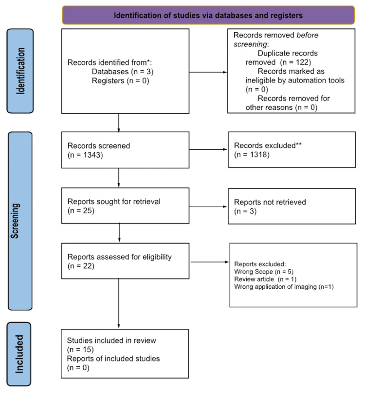
Basal cell carcinoma (BCC) is a common skin cancer that occurs due to various genetic and environmental factors. Diagnosis is made by a combination of clinical appearance, biopsy, imaging, and histopathological analysis. This review describes the current array of imaging modalities available to physicians to aid in the diagnosis of BCC. It is important to stay up-to-date with improvements in diagnostic screening, and knowledge of these options is instrumental in providing the best care to patients. Embase, Medline Industries, and PubMed were searched for articles within the past 10 years based on a search query that looked for imaging modalities used in the diagnosis and evaluation of a variety of dermatologic conditions. The search was further refined to focus on BCC and satisfy the inclusion/exclusion criteria determined by the authors. The research process was detailed in the Preferred Reporting Items for Systematic Reviews and Meta-Analyses diagram. Dermoscopy is a non-invasive in vivo microscopic technique used to evaluate skin lesions. Features of dermoscopy cannot be visualized with the naked eye, and studies found that dermoscopy increased diagnostic accuracy. Reflectance confocal microscopy (RCM) examines skin morphology, and recent studies found that 100% of patients with BCC had tumor-free margins when diagnosed with RCM. It allows for a one-stop-shop for diagnosis. Optical spectroscopy samples multiple sites without removing tissue. It helps detect subtle biophysical differences, allowing for earlier diagnosis. High-frequency ultrasound (HFUS) helps determine tumor size, structure, depth of invasion and spread. Studies found statistically significant positive correlations between depth of spread and HFUS readings. Optical coherence tomography takes cross-sectional images to analyze histopathology and morphology. It produces high-resolution images, confers slightly more accurate results than a biopsy, and expedites the treatment process through an earlier diagnosis without a biopsy.These results will advance the fields of dermatology and radiology as they describe unique uses for these imaging modalities. There are a variety of ways to use microscopy, and these techniques may be applied to many different lesions and help revolutionize the diagnosis and treatment of skin cancer and other lesions without the need for multiple, sometimes disfiguring surgical procedures. With the increase in diagnostic accuracy and decrease in diagnosis time, advanced imaging studies will become an integral part of dermatologic diagnosis and be included in future management and treatment plans, especially in the case of BCC.
Keywords: basal cell carcinoma; bcc; dermoscopy; optical spectroscopy; reflectance confocal microscopy.
Copyright © 2024, Basra et al.
Conflict of interest statement
The authors have declared that no competing interests exist.
Figures
Similar articles
-
Role of In Vivo Reflectance Confocal Microscopy in the Analysis of Melanocytic Lesions.Acta Dermatovenerol Croat. 2018 Apr;26(1):64-67. Acta Dermatovenerol Croat. 2018. PMID: 29782304 Review.
-
Dermoscopy and reflectance confocal microscopy-augmented characterization of pigmented micro-basal cell carcinoma (less than 2 mm diameter).Skin Res Technol. 2023 Jan;29(1):e13250. doi: 10.1111/srt.13250. Epub 2022 Dec 8. Skin Res Technol. 2023. PMID: 36482801 Free PMC article.
-
Novel imaging techniques for tumor margin detection in basal cell carcinoma: a systematic scoping review of FDA and EMA-approved imaging modalities.Int J Dermatol. 2025 Feb;64(2):287-301. doi: 10.1111/ijd.17496. Epub 2024 Oct 2. Int J Dermatol. 2025. PMID: 39358676 Free PMC article.
-
FDA and EMA-approved noninvasive imaging techniques for basal cell carcinoma subtyping: A systematic review.JAAD Int. 2025 Jun 10;21:73-86. doi: 10.1016/j.jdin.2025.05.006. eCollection 2025 Aug. JAAD Int. 2025. PMID: 40689050 Free PMC article. Review.
-
In vivo imaging characterization of basal cell carcinoma and cutaneous response to high-dose ionizing radiation therapy: A prospective study of reflectance confocal microscopy, dermoscopy, and ultrasonography.J Am Acad Dermatol. 2021 Jun;84(6):1575-1584. doi: 10.1016/j.jaad.2020.07.130. Epub 2020 Aug 20. J Am Acad Dermatol. 2021. PMID: 32827607 Free PMC article. Clinical Trial.
Cited by
-
Retrospective Analysis of Clinicopathological Characteristics of Surgically Treated Basal Cell Carcinomas of the Face: A Single-Centre Maxillofacial Surgery Experience.J Clin Med. 2024 Sep 14;13(18):5470. doi: 10.3390/jcm13185470. J Clin Med. 2024. PMID: 39336956 Free PMC article.
-
Ultrasound in Skin Cancer: Why, How, and When to Use It?Cancers (Basel). 2024 Sep 27;16(19):3301. doi: 10.3390/cancers16193301. Cancers (Basel). 2024. PMID: 39409920 Free PMC article. Review.
-
High-Frequency and Ultra-High-Frequency Ultrasound in Dermatologic Diseases and Aesthetic Medicine.Medicina (Kaunas). 2025 Jan 26;61(2):220. doi: 10.3390/medicina61020220. Medicina (Kaunas). 2025. PMID: 40005337 Free PMC article. Review.
References
-
- Basal cell carcinoma. Heath MS, Bar A. Dermatol Clin. 2023;41:13–21. - PubMed
-
- Optical coherence tomography for diagnosing recurrent or residual basal cell carcinoma after topical treatment: a diagnostic cohort study. Wolswijk T, Adan F, Nelemans PJ, Defauwes A, Mosterd K. J Am Acad Dermatol. 2023;89:728–733. - PubMed
Publication types
LinkOut - more resources
Full Text Sources
Miscellaneous