

doi: 10.1093/europace/euae094.
Targeting confluent areas of slow conduction and electrogram fragmentation for atrioventricular node re-entrant tachycardia ablation
Affiliations
- PMID: 38606815
- PMCID: PMC11094750
- DOI: 10.1093/europace/euae094
Item in Clipboard
Targeting confluent areas of slow conduction and electrogram fragmentation for atrioventricular node re-entrant tachycardia ablation
Europace.
.
No abstract available
Keywords: Conduction velocity; High-density mapping; Radiofrequency ablation; Slow pathway.
Conflict of interest statement
Conflict of interest: I have read the journal’s policy, and all the authors of this manuscript have declared that no competing interests exist.
Figures
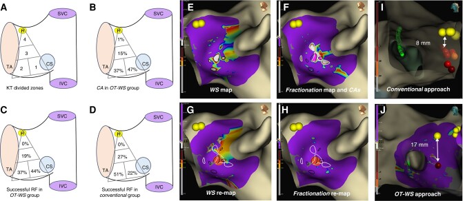
(A–H) Left lateral view of the right interatrial septum and the tricuspid valve. Panel A shows Koch’s triangle (KT), which was divided into four zones: Zones 1 and 2 were located at the base of the KT; Zone 1 at the septal isthmus, near the coronary sinus (CS) ostium and below its roof; and Zone 2 adjacent to Zone 1, towards the tricuspid annulus; Zone 3 and Zone 4 were located to the mid-septal and perihisian sites, respectively. Panels B and C display the percentage of confluent areas (CAs) and the percentage of successful RF applications in each zone, respectively, for the OT-WS group. Panel D shows the percentage of successful RF applications in each zone for the Conventional group. Panel E displays a WaveSpeed map, showing several zones with slow conduction located at KT’s base (slowest velocities in white, encircled in dark lines). Panel F shows a Fractionation Map, displaying also a few areas of fragmentation (most fragmented in white, encircled in white lines). One of these zones overlaps with two WaveSpeed slow conduction areas, delineating two small CAs (highlighted in pink colour). Panels G and H display post-ablation maps of WaveSpeed and Fractionation, respectively, showing a single (successful) RF application (dark orange dot, lesion index 4.2) in Zone 1, and the elimination of the previously encircled CAs. (I and J) Right lateral view focusing on the distance between the His signal (yellow dots) and the ablation site (red dots). A <10 mm distance is demonstrated from a patient belonging to the Conventional group (Panel I), while a much safer distance is noted from a patient belonging to the OT-WS group (Panel J). IVC, inferior vena cava; SVC, superior vena cava; H, His; TA, tricuspid annulus; CS, coronary sinus.
References
-
- Katritsis DG, Marine JE, Katritsis G, Latchamsetty R, Zografos T, Zimetbaum P, et al. Spatial characterization of the tachycardia circuit of atrioventricular nodal re-entrant tachycardia. Europace 2021;23:1596–602. - PubMed
-
- Pandozi C, Ficili S, Galeazzi M, Lavalle C, Russo M, Pandozi A, et al. Propagation of the sinus impulse into the Koch triangle and localization, timing, and origin of the multicomponent potentials recorded in this area. Circ Arrhythm Electrophysiol 2011;4:225–34. - PubMed
-
- Wakamatsu Y, Nagashima K, Kaneko Y, Mori H, Tsutsui K, Maegaki M, et al. Ablation strategy targeting the slow pathway visualized by ultrahigh-resolution mapping in typical slow-fast atrioventricular nodal reentrant tachycardia. Circ Arrhythm Electrophysiol 2023;16:e011497. - PubMed
-
- Gerontitis D, Pope MTB, Elmowafy M, Sadagopan S, Yue AM. High-density electroanatomic activation mapping to guide slow pathway modification in patients with persistent left superior vena cava. Heart Rhythm 2023;20:1018–25. - PubMed
-
- Katritsis DG, Anderson RH. New insights into the mechanisms of fast and slow conduction in the atrioventricular node. Heart Rhythm 2023;20:627–30. - PubMed
MeSH terms
LinkOut - more resources
Full Text Sources
