

Exploring Age-Related Variations in Carpal Bone Volume: Implications for Clinical Practice and Anatomical Understanding
- PMID: 38606949
- PMCID: PMC11571318
- DOI: 10.1177/15589447241242830
Exploring Age-Related Variations in Carpal Bone Volume: Implications for Clinical Practice and Anatomical Understanding
Abstract
Background: Clinically recognizing the changes in carpal bone volumes and understanding their implications in predicting osteoarthritis (OA) is crucial in clinical practice This study aimed to explore age-related differences in carpal bone volumes across genders, leveraging computed tomography (CT) wrist scans to create 3D surface models of these bones.
Methods: Carpal bone volumes were calculated using the 3D Slicer software from CT scans obtained from Frankston Hospital and additional datasets from Brown and Auckland Universities. The data were statistically processed using Stata V13. Double-sided P-values < .05 were considered statistically significant. The study was conducted in accordance with the ethical standards laid out in the Declaration of Helsinki.
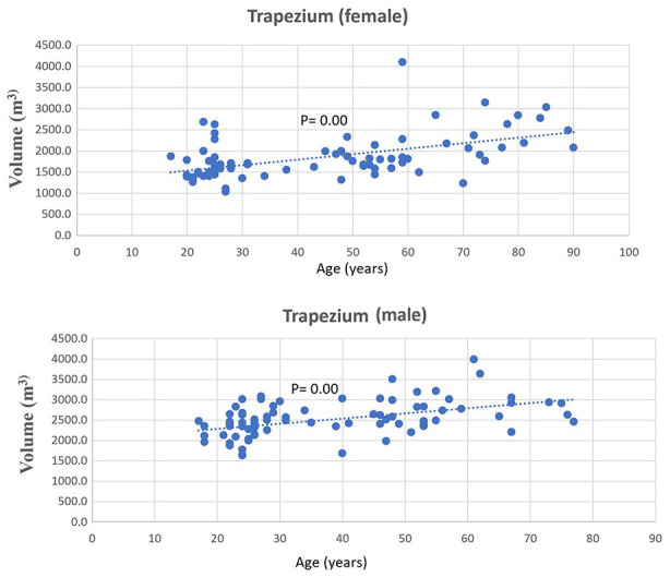
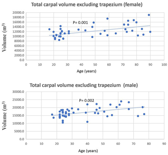
Results: A total of 181 patients were analyzed, and 48% of whom were female. A statistically significant positive Spearman correlation (rho = 0.37-0.611, P <.05) was observed between increasing age and the volume of all surveyed carpal bones (scaphoid, lunate, triquetrum, pisiform, hamate, capitate, and trapezium) across genders. Intrauser and interuser reliabilities for 3D Slicer-generated volumes of trapezium and pisiform bones were statistically significant, with Interclass Correlation Coefficient (ICC) values of 0.86 and 0.95, respectively.
Conclusion: Trapezial volumes increase with age, potentially due to the presence of OA and consequent osteophyte formation. This pattern is more prevalent among older individuals and women. However, the positive correlation between carpal bone volume and age was consistent across all carpal bones and both genders, regardless of OA presence. These findings suggest that carpal bone volume may naturally increase with age, independent of OA-related changes.
Level of evidence: III, cohort study.
Keywords: aging; anatomy; bones; carpus; morphology; wrist.
Conflict of interest statement
Declaration of Conflicting InterestsThe author(s) declared no potential conflicts of interest with respect to the research, authorship, and/or publication of this article.
Figures

References
-
- Taylor CL, Schwarz RJ. The anatomy and mechanics of the human hand. Artif Limbs. 1955;2(2):22-35. - PubMed
-
- Panchal-Kildare S, Malone K. Skeletal anatomy of the hand. Hand Clin. 2013;29(4):459-471. - PubMed
-
- Morimoto M, Utsumi M, Tohno Y, et al. Age-related changes of bone mineral density in human calcaneus, talus, and scaphoid bone. Biol Trace Elem Res. 2001;82(1-3):53-60. - PubMed
-
- Dalzell N, Kaptoge S, Morris N, et al. Bone micro-architecture and determinants of strength in the radius and tibia: age-related changes in a population-based study of normal adults measured with high-resolution pQCT. Osteoporos Int. 2009;20(10):1683-1694. - PubMed
MeSH terms
LinkOut - more resources
Full Text Sources
Research Materials
