

Localization and symbiotic status of probiotics in the coral holobiont
- PMID: 38606974
- PMCID: PMC11097643
- DOI: 10.1128/msystems.00261-24
Localization and symbiotic status of probiotics in the coral holobiont
Abstract
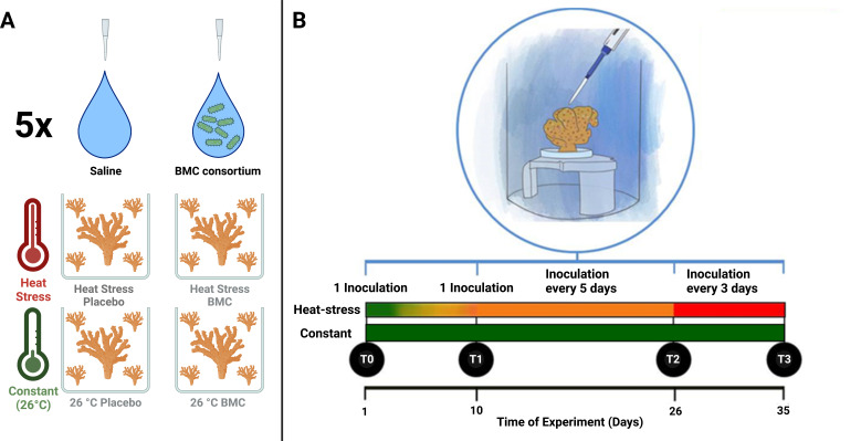
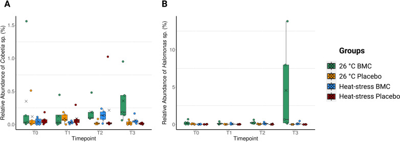
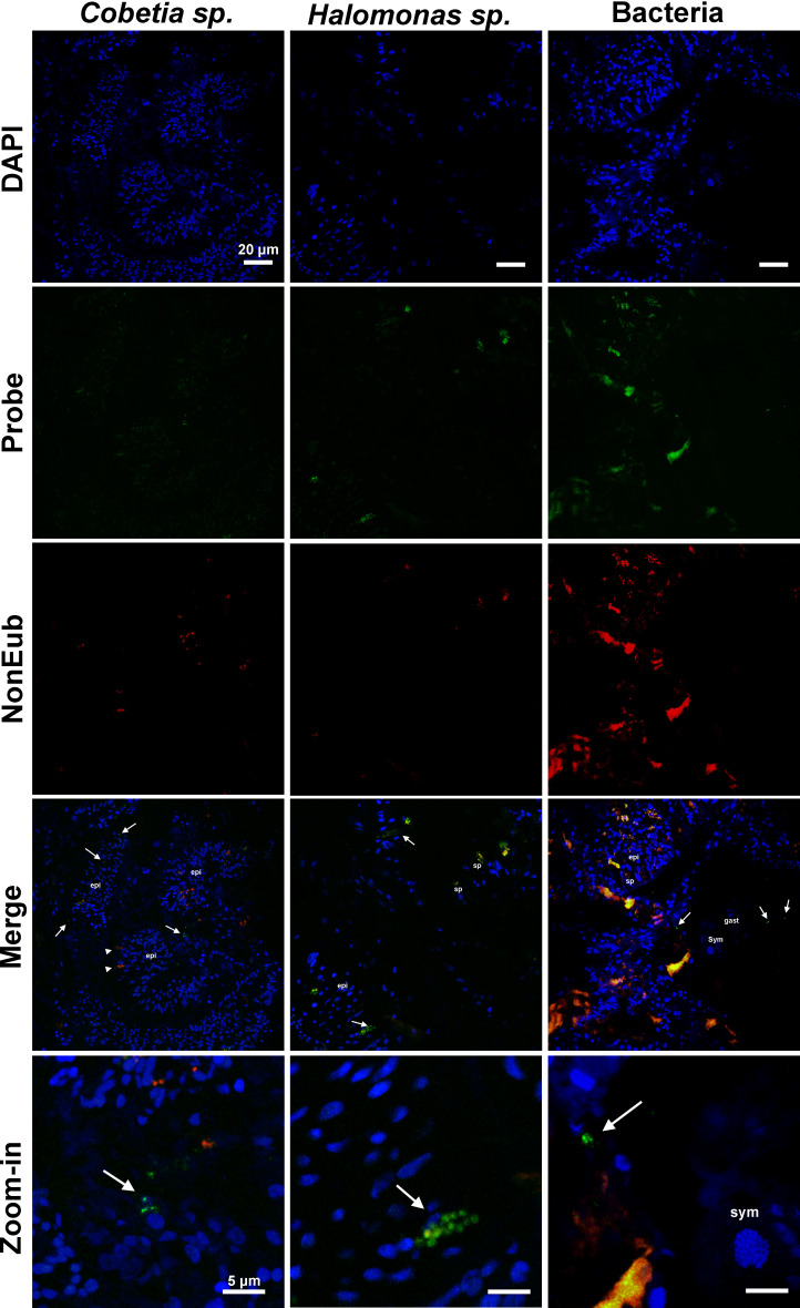
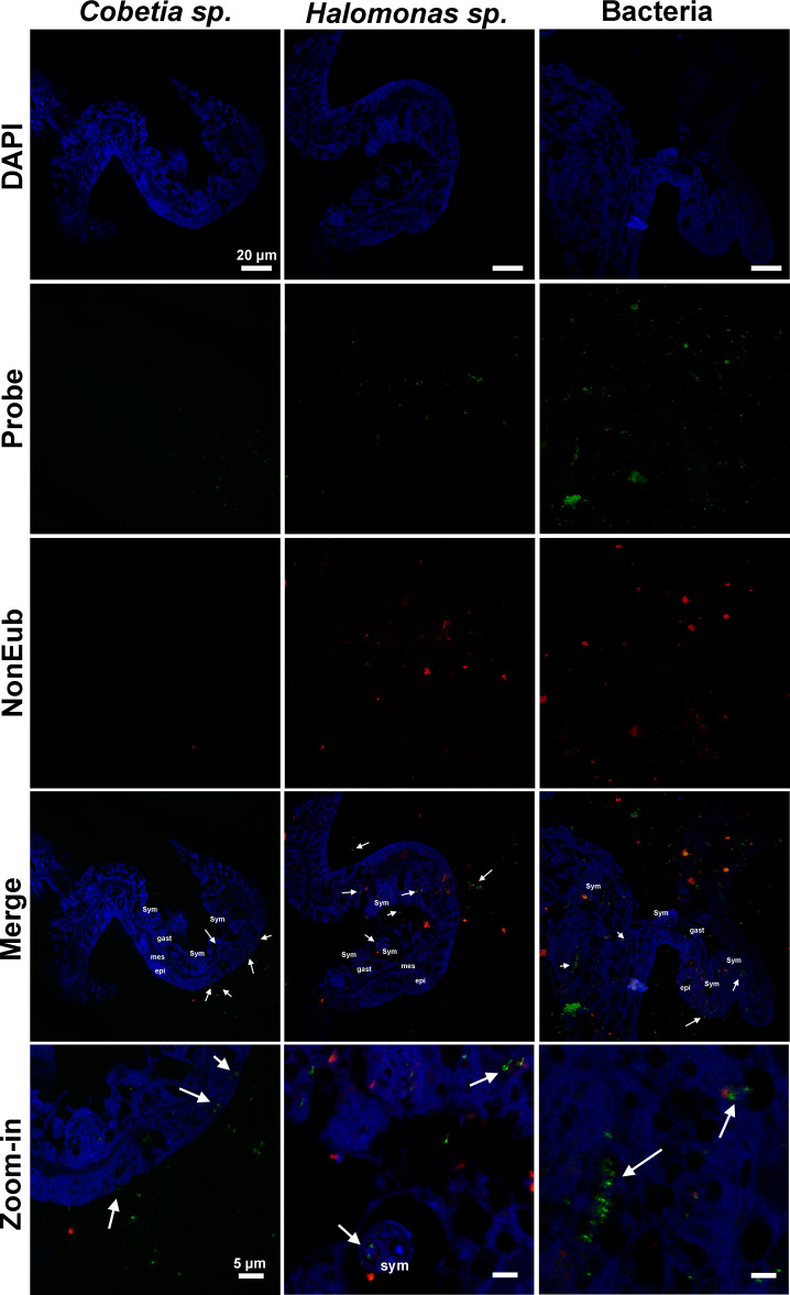
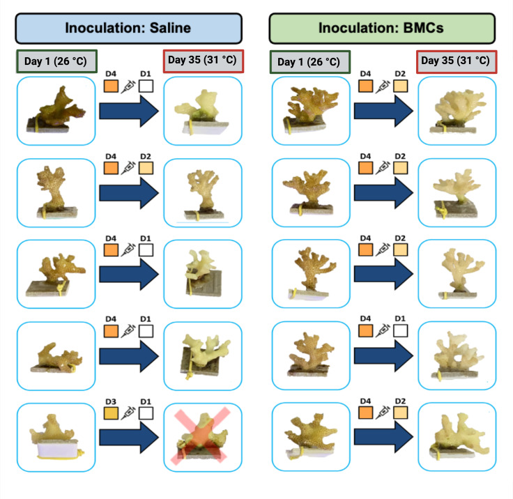
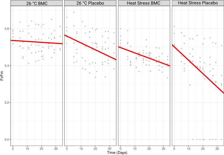
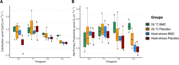
Corals establish symbiotic relationships with microorganisms, especially endosymbiotic photosynthetic algae. Although other microbes have been commonly detected in coral tissues, their identity and beneficial functions for their host are unclear. Here, we confirm the beneficial outcomes of the inoculation of bacteria selected as probiotics and use fluorescence in situ hybridization (FISH) to define their localization in the coral Pocillopora damicornis. Our results show the first evidence of the inherent presence of Halomonas sp. and Cobetia sp. in native coral tissues, even before their inoculation. Furthermore, the relative enrichment of these coral tissue-associated bacteria through their inoculation in corals correlates with health improvements, such as increases in photosynthetic potential, and productivity. Our study suggests the symbiotic status of Halomonas sp. and Cobetia sp. in corals by indicating their localization within coral gastrodermis and epidermis and correlating their increased relative abundance through active inoculation with beneficial outcomes for the holobiont. This knowledge is crucial to facilitate the screening and application of probiotics that may not be transient members of the coral microbiome.
Importance: Despite the promising results indicating the beneficial outcomes associated with the application of probiotics in corals and some scarce knowledge regarding the identity of bacterial cells found within the coral tissue, the correlation between these two aspects is still missing. This gap limits our understanding of the actual diversity of coral-associated bacteria and whether these symbionts are beneficial. Some researchers, for example, have been suggesting that probiotic screening should only focus on the very few known tissue-associated bacteria, such as Endozoicomonas sp., assuming that the currently tested probiotics are not tissue-associated. Here, we provide specific FISH probes for Halomonas sp. and Cobetia sp., expand our knowledge of the identity of coral-associated bacteria and confirm the probiotic status of the tested probiotics. The presence of these beneficial microorganisms for corals (BMCs) inside host tissues and gastric cavities also supports the notion that direct interactions with the host may underpin their probiotic role. This is a new breakthrough; these results argue against the possibility that the positive effects of BMCs are due to factors that are not related to a direct symbiotic interaction, for example, that the host simply feeds on inoculated bacteria or that the bacteria change the water quality.
Keywords: BMC; Cobetia sp.; FISH; Halomonas sp.; coral; coral-associated microbes; location; probiotics; tissue.
Conflict of interest statement
The authors declare no conflict of interest.
Figures
Similar articles
-
Coral Probiotics: Premise, Promise, Prospects.Annu Rev Anim Biosci. 2021 Feb 16;9:265-288. doi: 10.1146/annurev-animal-090120-115444. Epub 2020 Dec 15. Annu Rev Anim Biosci. 2021. PMID: 33321044 Review.
-
Marine probiotics: increasing coral resistance to bleaching through microbiome manipulation.ISME J. 2019 Apr;13(4):921-936. doi: 10.1038/s41396-018-0323-6. Epub 2018 Dec 5. ISME J. 2019. PMID: 30518818 Free PMC article.
-
Probiotics reshape the coral microbiome in situ without detectable off-target effects in the surrounding environment.Commun Biol. 2024 Apr 9;7(1):434. doi: 10.1038/s42003-024-06135-3. Commun Biol. 2024. PMID: 38594357 Free PMC article.
-
Exploring the Potential Molecular Mechanisms of Interactions between a Probiotic Consortium and Its Coral Host.mSystems. 2023 Feb 23;8(1):e0092122. doi: 10.1128/msystems.00921-22. Epub 2023 Jan 23. mSystems. 2023. PMID: 36688656 Free PMC article.
-
Beneficial Microorganisms for Corals (BMC): Proposed Mechanisms for Coral Health and Resilience.Front Microbiol. 2017 Mar 7;8:341. doi: 10.3389/fmicb.2017.00341. eCollection 2017. Front Microbiol. 2017. PMID: 28326066 Free PMC article. Review.
Cited by
-
Probiotics prevent mortality of thermal-sensitive corals exposed to short-term heat stress.ISME Commun. 2025 Mar 2;5(1):ycaf039. doi: 10.1093/ismeco/ycaf039. eCollection 2025 Jan. ISME Commun. 2025. PMID: 40151579 Free PMC article.
-
Phototrophic bacteria as potential probiotics for corals.NPJ Biodivers. 2025 Apr 29;4(1):16. doi: 10.1038/s44185-025-00085-7. NPJ Biodivers. 2025. PMID: 40301674 Free PMC article. Review.
-
The Coral Probiotics Village: An Underwater Laboratory to Tackle the Coral Reefs Crisis.Ecol Evol. 2025 Jul 4;15(7):e71558. doi: 10.1002/ece3.71558. eCollection 2025 Jul. Ecol Evol. 2025. PMID: 40625325 Free PMC article.
-
Bacterial Dynamics in Newly Settled Acropora kenti: Insights From Inoculations With Individual Probiotic Candidates.Environ Microbiol. 2025 Jul;27(7):e70143. doi: 10.1111/1462-2920.70143. Environ Microbiol. 2025. PMID: 40671239 Free PMC article.
References
-
- Oakley CA, Davy SK. 2018. Cell biology of coral bleaching, p 189–211. In van Oppen MJH, Lough JM (ed), Coral bleaching: patterns, processes, causes and consequences. Springer International Publishing, Cham.
-
- Wiedenmann J, D’Angelo C, Smith EG, Hunt AN, Legiret F-E, Postle AD, Achterberg EP. 2013. Nutrient enrichment can increase the susceptibility of reef corals to bleaching. Nat Clim Change 3:160–164. doi:10.1038/nclimate1661 - DOI
MeSH terms
Grants and funding
LinkOut - more resources
Full Text Sources
Miscellaneous
