. 2024 Mar 25;13(7):570.
doi: 10.3390/cells13070570.

Ubiquitinome Analysis Uncovers Alterations in Synaptic Proteins and Glucose Metabolism Enzymes in the Hippocampi of Adolescent Mice Following Cold Exposure

Affiliations

Ubiquitinome Analysis Uncovers Alterations in Synaptic Proteins and Glucose Metabolism Enzymes in the Hippocampi of Adolescent Mice Following Cold Exposure

Xin-Yue Li et al. Cells. .

Abstract

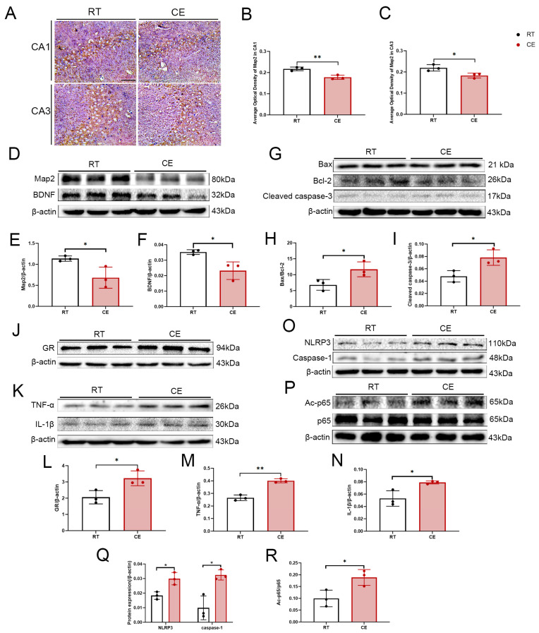
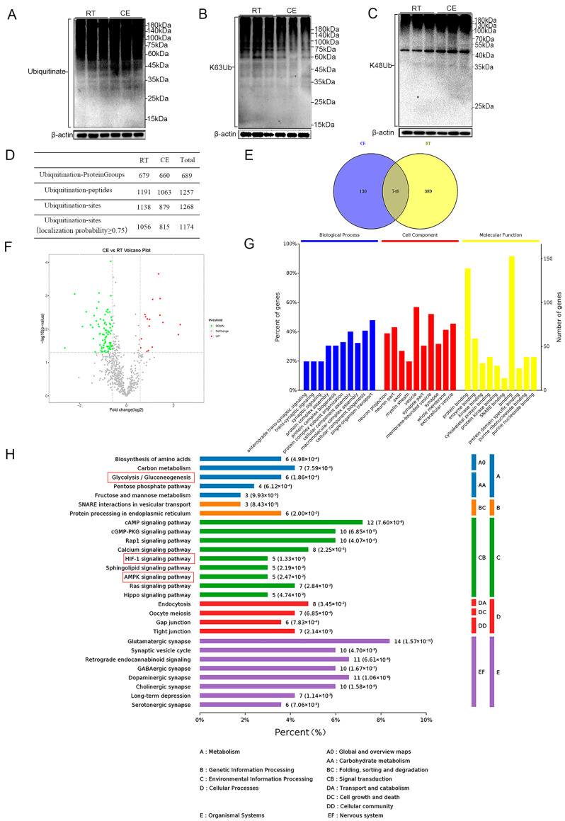
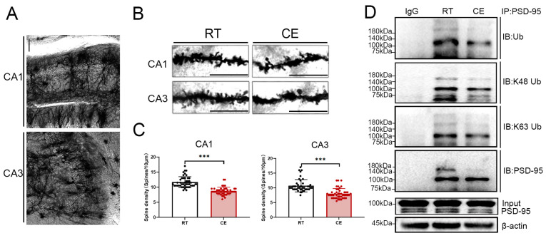
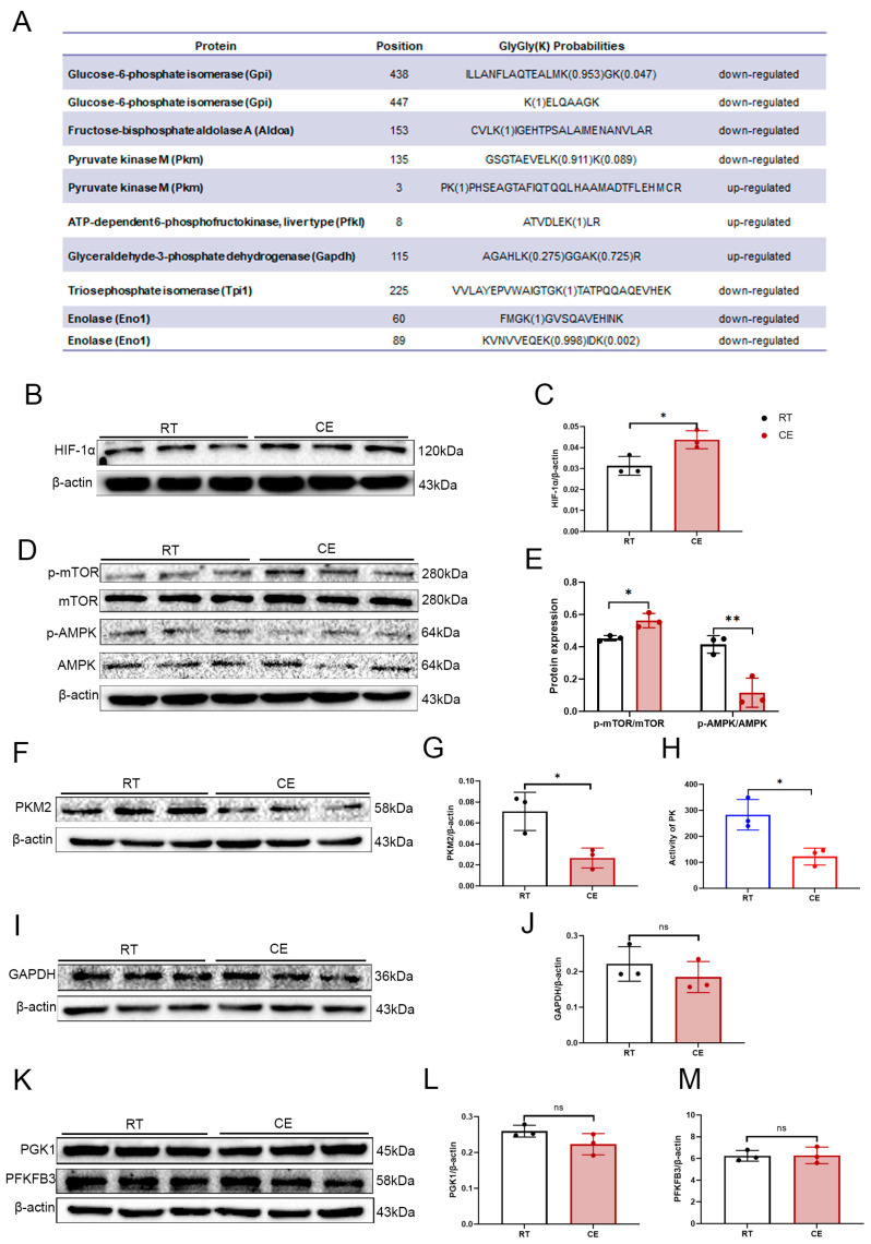
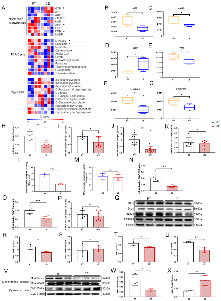
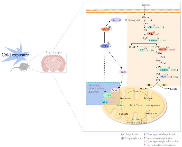
Cold exposure exerts negative effects on hippocampal nerve development in adolescent mice, but the underlying mechanisms are not fully understood. Given that ubiquitination is essential for neurodevelopmental processes, we attempted to investigate the effects of cold exposure on the hippocampus from the perspective of ubiquitination. By conducting a ubiquitinome analysis, we found that cold exposure caused changes in the ubiquitination levels of a variety of synaptic-associated proteins. We validated changes in postsynaptic density-95 (PSD-95) ubiquitination levels by immunoprecipitation, revealing reductions in both the K48 and K63 polyubiquitination levels of PSD-95. Golgi staining further demonstrated that cold exposure decreased the dendritic-spine density in the CA1 and CA3 regions of the hippocampus. Additionally, bioinformatics analysis revealed that differentially ubiquitinated proteins were enriched in the glycolytic, hypoxia-inducible factor-1 (HIF-1), and 5'-monophosphate (AMP)-activated protein kinase (AMPK) pathways. Protein expression analysis confirmed that cold exposure activated the mammalian target of rapamycin (mTOR)/HIF-1α pathway. We also observed suppression of pyruvate kinase M2 (PKM2) protein levels and the pyruvate kinase (PK) activity induced by cold exposure. Regarding oxidative phosphorylation, a dramatic decrease in mitochondrial respiratory-complex I activity was observed, along with reduced gene expression of the key subunits NADH: ubiquinone oxidoreductase core subunit V1 (Ndufv1) and Ndufv2. In summary, cold exposure negatively affects hippocampal neurodevelopment and causes abnormalities in energy homeostasis within the hippocampus.

Keywords: PSD-95; cold exposure; glucose metabolism; hippocampus; ubiquitinome.


Conflict of interest statement

The authors declare no conflict of interest.

Figures

Figure 1
Figure 1
Negative effects of cold exposure on hippocampal neurodevelopment in adolescent mice. (A) Representative images of MAP-2 immunohistochemistry of the hippocampi of control and cold-exposure mice. Bar = 50 μm. (B,C) Graphs depicting the average optical-density analysis of MAP-2 immunostaining of hippocampal sub-regions CA1 and CA3. (D) Western blot demonstrating the levels of MAP-2 and BDNF in the hippocampi of mice from the CE and RT groups. Graphs indicate the results of densitometric analyses of the expression ratio of (E) MAP-2/β-actin and (F) BDNF/β-actin. (G) Western blot demonstrating the levels of Bax, Bcl-2, and cleaved-caspase-3 in the hippocampi of mice from the CE and RT groups. The graph indicates the results of densitometric analyses of the expression ratio of (H) Bax/Bcl-2 and (I) cleaved-caspase-3/β-actin. (J) Expression of GR was assessed by Western blotting. (K) Expression of the inflammatory cytokines TNF-α and IL-1β in the hippocampus after cold exposure were assessed by Western blotting. (O) Expression of NLRP3, caspase-1 in the hippocampus after cold exposure were assessed by Western blotting. (P) Expression of Ac-p65 and p65 in the hippocampus after cold exposure was assessed by Western blotting. The graphs indicate the results of densitometric analyses of the expression ratios of (L) GR/β-actin, (M) TNF-α/β-actin, (N) IL-1β/β-actin, (Q) NLRP3/β-actin and Caspase-1/β-actin, and (R) Ac-p65/p65. Results are expressed as means ± SD; n = 3. * p < 0.05; ** p < 0.01.
Figure 2
Figure 2
Ubiquitinome profiling of the hippocampi of RT and CE mice. (A) Western blot analysis of Ub species in hippocampal tissue. (B) Western blot analysis of K63-polyUb species in hippocampal tissue lysates. (C) Western blot analysis of K48-polyUb species in hippocampal tissue lysates. (D) The table shows the statistical analysis results of ubiquitination proteomics. (E) The Venn diagram shows the comparison of ubiquitination sites identified in CE vs. RT. (F) The volcano plot shows the log2-transformed fold change and log10-transformed p-value of the identified ubiquitination sites, highlighting significantly regulated lysine (K) sites. Significantly upregulated ubiquitination sites are displayed as red dots, and downregulated ubiquitination sites are displayed as green dots. (G) GO annotation of the ubiquitinome. (H) KEGG pathway analysis of the ubiquitinome.
Figure 3
Figure 3
Cold exposure downregulates the density of dendritic spines and PSD-95 ubiquitination levels within the hippocampus. (A) A representative low magnification image of Golgi–Cox staining in the hippocampus. Bar = 100 μm. (B) Representative images of dendritic spines in the hippocampal CA1 and CA3 subregions from RT and CE mice. Bar = 10 μm. (C) Quantification of dendritic spines per 10 μm of the hippocampal CA1 and CA3 subregions from RT and CE mice. n = 45 dendritic segments from 3 animals per group. (D) Immunoprecipitation of PSD-95 from the hippocampi from RT and CE mice. Values are presented as the mean ± SD (n = 3). Statistically significant differences are indicated: *** p < 0.0001.
Figure 4
Figure 4
The effect of cold exposure on a variety of glycolytic enzymes. (A) Glucose-metabolizing enzymes with differential ubiquitination sites. (B) The expression of HIF-1α was analyzed by Western blotting. (C) The graph indicates the expression ratios of HIF-1α/β-actin obtained from the densitometric analyses. (D) The expression of p-mTOR, mTOR, p-AMPK, and AMPK in the hippocampal homogenates. (E) The graphs indicate the expression ratios of p-mTOR/mTOR and p-AMPK/AMPK based on the densitometric analyses. (F) The expression of PKM2 was analyzed by Western blotting. (G) The graph indicates the expression ratios of PKM2/β-actin based on the densitometric analyses. (H) Enzyme activity of PK within the hippocampi of RT and CE mice. (I) The expression of GAPDH was analyzed by Western blotting. (J) The graph indicates the expression ratios of GAPDH/β-actin based on the densitometric analyses. (K) The expression of PGK1 and PFKFB3 in the hippocampal homogenates. The graphs indicate the expression ratios of (L) PGK1/β-actin and (M) PFKFB3/β-actin based on the densitometric analyses. Values are presented as the mean ± SD (n = 3). Statistically significant differences are indicated: * p < 0.05, ** p < 0.01; ns: not significant.
Figure 5
Figure 5
Cold exposure affects the tricarboxylic acid cycle and mitochondrial function within the hippocampus. (A) Heat map showing the energy metabolomics within the hippocampi of RT and CE mice. Box diagrams depicting the content of (B) AMP, (C) cAMP, (D) GTP, (E) FMN, (F) L-malate, and (G) succinate in energy metabolomics. Values are presented as the mean ± SD (n = 4). Statistically significant differences are indicated: * p < 0.05, ** p < 0.01. The mRNA levels of Fh1 (H), Suclg1 (I), Suclg2 (J), Sucla2 (K) in the hippocampi of RT and CE mice (n = 6). (L) Activity of mitochondrial complex I within the hippocampi of RT and CE mice (n = 3). (M) Activity of mitochondrial complex IV within the hippocampi of RT and CE mice (n = 3). (N) The mRNA levels of Ndufv1 in the hippocampi of RT and CE mice (n = 6). (O) The mRNA levels of Ndufv2 in the hippocampi of RT and CE mice (n = 6). (P) The mRNA levels of Ndufs1 in the hippocampi of RT and CE mice (n = 6). (Q) Western blot showing the levels of Mfn1, Drp1, PINK1, PARKIN in the hippocampi of control and cold-exposure mice (n = 3). The graphs indicate the expression ratio of (R) PINK1/β-actin, (S) PARKIN/β-actin (n = 3), (T) Mfn/β-actin, and (U) Drp1/β-actin based on densitometric analyses. (V) Western blot showing the levels of PARKIN in the mitochondrial component and in the cytoplasmic component of the hippocampus for control and cold exposure mice (n = 3). The graphs indicate the expression ratios of (W) Mito Parkin/VDAC, (X) Cyto Parkin/β-actin (n = 3) based on densitometric analyses. Values are presented as the mean ± SD. Statistically significant differences are indicated: * p < 0.05, ** p < 0.01, *** p < 0.0001; ns: not significant.
Figure 6
Figure 6
A schematic diagram of the effect of chronic cold exposure on hippocampal energy metabolism. A series of glucose metabolic enzymes were found to have differentially ubiquitinated sites, including GPI (K438, K447), ALDOA (K153), PKM (K3, K135), PFKL (K8), GAPDH (K115), TPI (K225), and ENO 1 (K60, K89). In the TCA cycle, cold exposure selectively impairs the generation of L-malate and succinate. Cold exposure also inhibited the activity of PK. The hippocampus image in Figure 6 was created with BioRender.com (accessed on 1 June 2022).

Similar articles

Cited by

References

    1. Mäkinen T.M., Palinkas L.A., Reeves D.L., Pääkkönen T., Rintamäki H., Leppäluoto J., Hassi J. Effect of repeated exposures to cold on cognitive performance in humans. Physiol. Behav. 2006;87:166–176. doi: 10.1016/j.physbeh.2005.09.015. - DOI - PubMed
    1. Yang L., Wu J., Hu Z., Gao F., Hu X. Effects of workload on human cognitive performance of exposure to extremely cold environment. Physiol. Behav. 2021;230:113296. doi: 10.1016/j.physbeh.2020.113296. - DOI - PubMed
    1. Muller M.D., Gunstad J., Alosco M.L., Miller L.A., Updegraff J., Spitznagel M.B., Glickman E.L. Acute cold exposure and cognitive function: Evidence for sustained impairment. Ergonomics. 2012;55:792–798. doi: 10.1080/00140139.2012.665497. - DOI - PMC - PubMed
    1. Falla M., Micarelli A., Hüfner K., Strapazzon G. The Effect of Cold Exposure on Cognitive Performance in Healthy Adults: A Systematic Review. Int. J. Environ. Res. Public Health. 2021;18:9725. doi: 10.3390/ijerph18189725. - DOI - PMC - PubMed
    1. El Marzouki H., Aboussaleh Y., Najimi M., Chigr F., Ahami A. Effect of Cold Stress on Neurobehavioral and Physiological Parameters in Rats. Front. Physiol. 2021;12:660124. doi: 10.3389/fphys.2021.660124. - DOI - PMC - PubMed

Publication types

LinkOut - more resources