Review
. 2024 Mar 28;13(7):587.
doi: 10.3390/cells13070587.

An OX-Tra'Ordinary Tale: The Role of OX40 and OX40L in Atopic Dermatitis

Affiliations
Review

An OX-Tra'Ordinary Tale: The Role of OX40 and OX40L in Atopic Dermatitis

Kaviyon Sadrolashrafi et al. Cells. .

Abstract

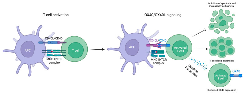
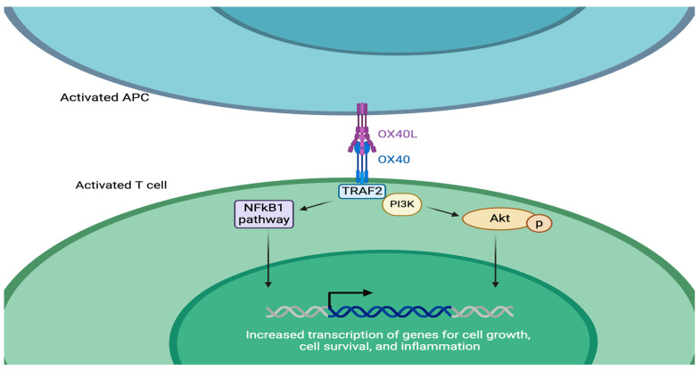
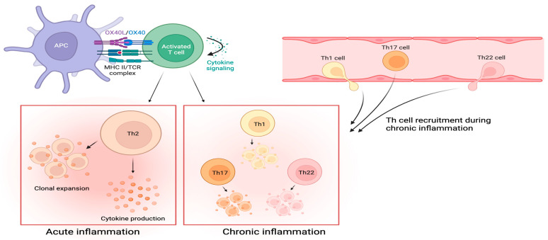
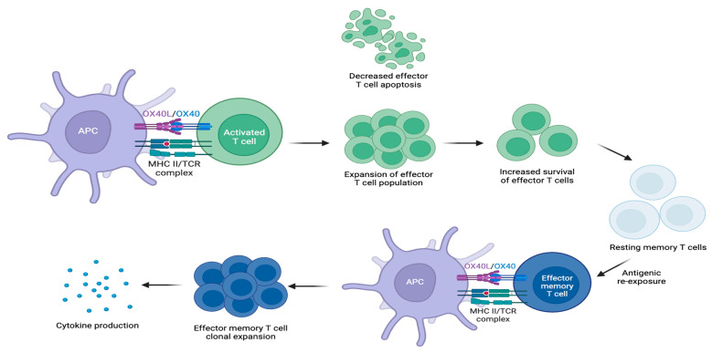
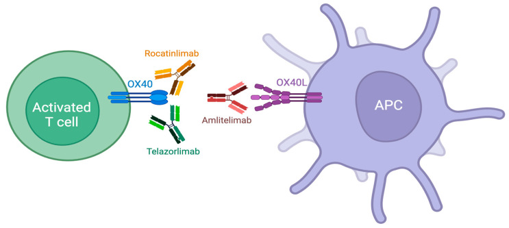
The transmembrane glycoprotein OX40 receptor (OX40) and its ligand, OX40L, are instrumental modulators of the adaptive immune response in humans. OX40 functions as a costimulatory molecule that promotes T cell activation, differentiation, and survival through ligation with OX40L. T cells play an integral role in the pathogenesis of several inflammatory skin conditions, including atopic dermatitis (AD). In particular, T helper 2 (TH2) cells strongly contribute to AD pathogenesis via the production of cytokines associated with type 2 inflammation (e.g., IL-4, IL-5, IL-13, and IL-31) that lead to skin barrier dysfunction and pruritus. The OX40-OX40L interaction also promotes the activation and proliferation of other T helper cell populations (e.g., TH1, TH22, and TH17), and AD patients have demonstrated higher levels of OX40 expression on peripheral blood mononuclear cells than healthy controls. As such, the OX40-OX40L pathway is a potential target for AD treatment. Novel therapies targeting the OX40 pathway are currently in development, several of which have demonstrated promising safety and efficacy results in patients with moderate-to-severe AD. Herein, we review the function of OX40 and the OX40-OX40L signaling pathway, their role in AD pathogenesis, and emerging therapies targeting OX40-OX40L that may offer insights into the future of AD management.

Keywords: AD; AD pathogenesis; OX40; OX40L; T cells; atopic dermatitis; cytokines; eczema; inflammation; monoclonal antibodies.


Conflict of interest statement

K.S., L.G., R.K., A.H., R.K.Y., H.C.T., S.N.B. and D.K.Y. declare no conflicts of interest. A.W.A. has served as a research investigator, scientific advisor, or speaker for AbbVie, Amgen, Almirall, Arcutis, ASLAN, Beiersdorf, BI, BMS, EPI, Incyte, Leo, UCB, Janssen, Lilly, Mindera, Nimbus, Novartis, Ortho, Sun, Dermavant, Dermira, Sanofi, Takeda, Organon, Regeneron, Pfizer, and Ventyx.

Figures

Figure 1
Figure 1
OX40 expression is induced on activated T cells, and OX40L expression is induced on APCs. OX40-OX40L ligation stimulates the clonal expansion of effector CD4+ and CD8+ T cell populations, upregulates the expression of proinflammatory cytokines, and promotes the survival of T cells.
Figure 2
Figure 2
OX40-OX40L ligation activates the PI3K-PKB/Akt and NF-κB1 pathways. These intracellular pathways regulate cellular division and promote cellular growth and T cell survival, which can contribute to inflammation.
Figure 3
Figure 3
Downstream effects of OX40-OX40L signaling. OX40-OX40L signaling expands effector CD4+ T cell populations when there is a surrounding inflammatory cytokine milieu. OX40-OX40L signaling also decreases the induction of Treg populations through FoxP3 suppression. Additionally, OX40-OX40L signaling promotes the clonal expansion of antigen-specific T cell populations.
Figure 4
Figure 4
Two phases of inflammation characterize AD. During the acute phase, OX40-OX40L ligation on activated T cells facilitates TH2-predominant signaling and differentiation, which produces type 2 inflammatory cytokines. The shift to the chronic phase is characterized by the recruitment of TH1, TH17, and TH22 cells expressing OX40. OX40-OX40L ligation on these activated T helper cell populations leads to effector cell proliferation and cytokine production that maintain the inflammatory response.
Figure 5
Figure 5
OX40-OX40L signaling causes effector T cell proliferation, which generates a primary immune response. Effector T cells undergo apoptosis following a primary immune response; however, OX40-OX40L signaling mediates the transition of some into resting memory T cells. Re-exposure to the same antigen converts resting memory T cells into effector memory T cells expressing OX40. OX40-OX40L ligation on effector memory T cells causes effector memory T cell proliferation, leading to a secondary immune response.
Figure 6
Figure 6
Novel therapies targeting the interaction between OX40 and OX40L include monoclonal antibodies against OX40 (rocatinlimab and telazorlimab) and OX40L (amlitelimab).

References

    1. Weidinger S., Beck L.A., Bieber T., Kabashima K., Irvine A.D. Atopic dermatitis. Nat. Rev. Dis. Primers. 2018;4:1. doi: 10.1038/s41572-018-0001-z. - DOI - PubMed
    1. Nutten S. Atopic dermatitis: Global epidemiology and risk factors. Ann. Nutr. Metab. 2015;66:8–16. doi: 10.1159/000370220. - DOI - PubMed
    1. Guttman-Yassky E., Krueger J.G., Lebwohl M.G. Systemic immune mechanisms in atopic dermatitis and psoriasis with implications for treatment. Exp. Dermatol. 2018;27:409–417. doi: 10.1111/exd.13336. - DOI - PubMed
    1. Ständer S. Atopic Dermatitis. N. Engl. J. Med. 2021;384:1136–1143. doi: 10.1056/NEJMra2023911. - DOI - PubMed
    1. Itamura M., Sawada Y. Involvement of Atopic Dermatitis in the Development of Systemic Inflammatory Diseases. Int. J. Mol. Sci. 2022;23:13445. doi: 10.3390/ijms232113445. - DOI - PMC - PubMed

LinkOut - more resources