Review
. 2024 Apr 3;13(7):621.
doi: 10.3390/cells13070621.

From Vessels to Neurons-The Role of Hypoxia Pathway Proteins in Embryonic Neurogenesis

Affiliations
Review

From Vessels to Neurons-The Role of Hypoxia Pathway Proteins in Embryonic Neurogenesis

Barbara K Stepien et al. Cells. .

Abstract

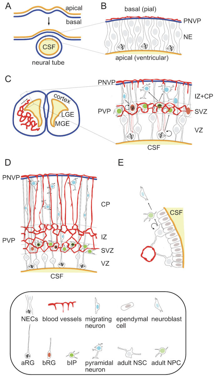
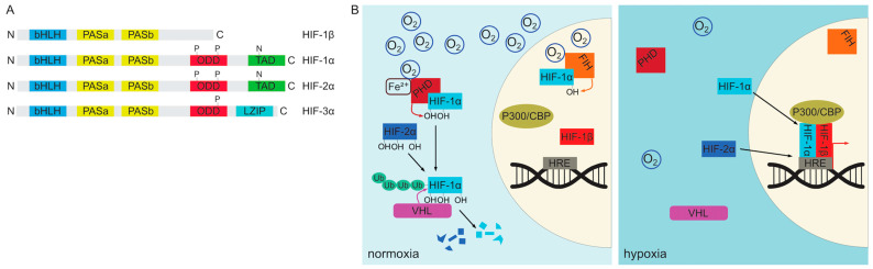
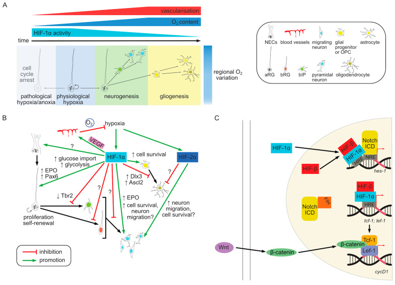
Embryonic neurogenesis can be defined as a period of prenatal development during which divisions of neural stem and progenitor cells give rise to neurons. In the central nervous system of most mammals, including humans, the majority of neocortical neurogenesis occurs before birth. It is a highly spatiotemporally organized process whose perturbations lead to cortical malformations and dysfunctions underlying neurological and psychiatric pathologies, and in which oxygen availability plays a critical role. In case of deprived oxygen conditions, known as hypoxia, the hypoxia-inducible factor (HIF) signaling pathway is activated, resulting in the selective expression of a group of genes that regulate homeostatic adaptations, including cell differentiation and survival, metabolism and angiogenesis. While a physiological degree of hypoxia is essential for proper brain development, imbalanced oxygen levels can adversely affect this process, as observed in common obstetrical pathologies such as prematurity. This review comprehensively explores and discusses the current body of knowledge regarding the role of hypoxia and the HIF pathway in embryonic neurogenesis of the mammalian cortex. Additionally, it highlights existing gaps in our understanding, presents unanswered questions, and provides avenues for future research.

Keywords: HIF; NSC; embryonic neurogenesis; hypoxia; neocortex; neural progenitor cells; vascularization.


Conflict of interest statement

The authors declare no conflicts of interest.

Figures

Figure 1
Figure 1
Neurogenesis in the developing neocortex follows a precisely regulated spatiotemporal order of events. During this process progenitor proliferation, neuronal generation and migration coincide with the ingrowth of new blood vessels from two independent sources. (A) Neurogenesis is preceded by the formation of a neural tube by the closure of neural folds. This leads to the reversal of the apicobasal polarity with the apical side of the neuroepithelium now facing the ventricular lumen filled with cerebrospinal fluid. The neocortex forms from the dorsal region of the telencephalic vesicle. (B) At this stage the neural stem cells are organized in a single layer avascular neuroepithelium. NECs divide symmetrically to expand its surface. Blood vessels are restricted to a pial perineural vascular plexus (PNVP) and do not yet penetrate the brain parenchyma. (C) At the onset of cortical neurogenesis NECs transform into aRG, which start dividing asymmetrically to produce more committed basal progenitors and neurons. These migrate basally to form a secondary germinal zone (SVZ) followed by the formation of a cortical plate (CP) by neurons migrating through the intermediate zone (IZ). While the aRG-containing VZ remains largely avascular, angiogenesis occurs in the more basal zones. The PNVP starts sprouting radial vessels through the CP towards the ventricular surface. In the same time a second vascular plexus, the periventricular vascular plexus (PVP) is formed at the border between VZ and SVZ due to the ingrowth of vessels from the ventral part of the brain. This plexus is organized in a honeycomb pattern, with bIPs located preferentially at vessel branch points. (D) Both plexuses form connecting branches leading to a singular vascular network as neurogenesis progresses and CP thickens. (E) In the germinal zones of an adult neocortex the NSCs reside close to the ventricle and maintain contact with nearby blood vessels. They remain largely quiescent, but can be activated to produce more committed NPCs and neuroblasts. Physiologically, in mice the newly generated neurons in the SVZ migrate along the rostral migratory stream to replenish the neurons of the olfactory bulbs.
Figure 2
Figure 2
Schematic representation of the HIF protein family members and the molecular mechanism of oxygen sensing via the HIF signaling pathway. (A) All HIF family proteins contain an N-terminal DNA-binding basic helix-loop-helix (bHLH) domain and two PAS dimerization regions. The α-subunits additionally contain an oxygen-dependent degradation (ODD) domain. While HIF-1α and HIF-2α have a C-terminal transactivation domain (TAD), HIF-3α has a leucine zipper. Alternative truncated versions of these proteins can also be expressed. (B) α subunits of the HIF complex are subject to oxygen-dependent ubiquitination and proteasomal degradation. In the presence of oxygen PHD family enzymes hydroxylate conserved Pro residues in the ODD, which directs HIF proteins to polyubiquitination by VHL. HIF-1α can also be additionally inhibited by asparagine hydroxylation by FIH-1 under normoxia. In contrast, under hypoxic conditions PHD and FIH-1 are inactive, leading to the stabilization and nuclear translocation of HIF-1α and HIF-2α proteins. In the nucleus they can bind the constitutively expressed HIF-1β subunit at the promoters of regulated genes via conserved hypoxia-responsive element (HRE) sequences. The complex can then recruit other transcriptional activators to drive the transcription of target genes.
Figure 3
Figure 3
Mechanism of oxygen sensing and HIF pathway activation in embryonic neurogenesis. (A) During early development, oxygen delivery to the embryonic neocortex is limited by the lack of vasculature. As a result, HIF-1α signaling is high in aRG cells, which promotes self-renewal. As neurogenesis progresses the ingrowth of blood vessels basally from VZ creates more oxygenated zones where more committed progenitor cells reside. These progenitors divide to produce postmitotic progeny, although its ultimate fate (neuronal or glial) appears to be regulated primarily by the developmental timing. The oxygen levels are also not uniform along the apicobasal axis, which adds another layer to the niche complexity. Although aRG cells reside in physiologically hypoxic regions, very low oxygen levels or anoxia can have adverse effects on progenitor proliferation and survival. (B) The effects of HIF-1α activity on different cell types in the developing neocortex. Activation of the HIF pathway generally maintains stemness and promotes survival and proliferation of aRG cells. This occurs at the expense of differentiative divisions and the production of more committed basal progenitors. This phenotype is caused at least in part, by increased glycolysis and HIF-dependent expression of trophic proteins such as EPO. Additionally, HIF promotes the expression of transcription factors such as Pax6 while being associated with the repression of differentiation markers such as Tbr2. However, the degree to which these genes are directly regulated by HIF is largely unknown. Moreover, the role of HIF in regulating basal progenitor subtypes is so far not understood. In newly generated neurons HIF-1α is critical for survival and may affect migration. HIF-1α also induces VEGF production by NPCs which is necessary for cortical vascularization. In glial progenitors HIF activity is particularly important for survival and proliferation of OPCs, while putting the brakes on oligodendrocyte maturation via Dlx3 and Ascl2 regulation. While HIF-2α is unlikely to be required for embryonic NPCs it plays a role in perinatal neuron survival, myelination and likely in ECs. (C) HIF signaling cross talks with other signaling pathways during neurogenesis. HIF-1α co-activates the transcription of Notch target genes such as hes-1 by binding to HRE elements directly as well as interacting with the Notch ICD. In addition, Notch can potentiate HIF-1α activity by sequestering FIH-1. HIF-1α also enhances Wnt signaling by inducing the expression of Tcf-1 and Lef-1 transcription factors, which bind β-catenin to activated Wnt-dependent transcription.

Similar articles

Cited by

References

    1. Lodato S., Arlotta P. Generating Neuronal Diversity in the Mammalian Cerebral Cortex. Annu. Rev. Cell Dev. Biol. 2015;31:699–720. doi: 10.1146/annurev-cellbio-100814-125353. - DOI - PMC - PubMed
    1. Rakic P. Evolution of the neocortex: A perspective from developmental biology. Nat. Rev. Neurosci. 2009;10:724–735. doi: 10.1038/nrn2719. - DOI - PMC - PubMed
    1. Sanides F. Comparative Architectonics of the Neocortex Of Mammals snd Their Evolutionary Interpretation. Ann. N. Y. Acad. Sci. 1969;167:404–423. doi: 10.1111/j.1749-6632.1969.tb20459.x. - DOI
    1. Krubitzer L.A., Prescott T.J. The Combinatorial Creature: Cortical Phenotypes within and across Lifetimes. Trends Neurosci. 2018;41:744–762. doi: 10.1016/j.tins.2018.08.002. - DOI - PubMed
    1. Striedter G.F. Principles of Brain Evolution. Sinauer Associates Inc.; Sunderland, MA, USA: 2005.

Publication types

LinkOut - more resources