. 2024 Apr 3;13(7):620.
doi: 10.3390/cells13070620.

The Natural Product Secoemestrin C Inhibits Colorectal Cancer Stem Cells via p38-S100A8 Feed-Forward Regulatory Loop

Affiliations

The Natural Product Secoemestrin C Inhibits Colorectal Cancer Stem Cells via p38-S100A8 Feed-Forward Regulatory Loop

Huimin Zhou et al. Cells. .

Abstract

Cancer stem cells (CSCs) are closely associated with tumor initiation, metastasis, chemoresistance, and recurrence, which represent some of the primary obstacles to cancer treatment. Targeting CSCs has become an important therapeutic approach to cancer care. Secoemestrin C (Sec C) is a natural compound with strong anti-tumor activity and low toxicity. Here, we report that Sec C effectively inhibited colorectal CSCs and non-CSCs concurrently, mainly by inhibiting proliferation, self-renewal, metastasis, and drug resistance. Mechanistically, RNA-seq analysis showed that the pro-inflammation pathway of the IL17 axis was enriched, and its effector S100A8 was dramatically decreased in Sec C-treated cells, whose roles in the stemness of CSCs have not been fully clarified. We found that the overexpression of S100A8 hindered the anti-CSCs effect of Sec C, and S100A8 deficiency attenuated the stemness traits of CSCs to enhance the Sec C killing activity on them. Meanwhile, the p38 signal pathway, belonging to the IL17 downstream axis, can also mediate CSCs and counter with Sec C. Notably, we found that S100A8 upregulation increased the p38 protein level, and p38, in turn, promoted S100A8 expression. This indicated that p38 may have a mutual feedback loop with S100A8. Our study discovered that Sec C was a powerful anti-colorectal CSC agent, and that the positive feedback loop of p38-S100A8 mediated Sec C activity. This showed that Sec C could act as a promising clinical candidate in colorectal cancer treatment, and S100A8 could be a prospective drug target.

Keywords: CSCs; S100A8; colorectal cancer; p38; secoemestrin C; stemness.


Conflict of interest statement

The authors declare that the research was conducted in the absence of any commercial or financial relationships that could be construed as a potential conflict of interest.

Figures
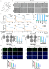
Figure 1
Figure 1
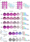
Sec C exhibits strong anti-colorectal tumor activity dose-dependently. (A) The chemical structure of Sec C. (B) The cell morphology of HCT8 and HT29 cells was observed after the treatment with 2.5 μmol/L Sec C for the indicated time points. Scale bar, 100 μm. (C) Different colorectal tumor cells were treated with the specific concentrations of Sec C or OXA for 48 h, and cell survival was detected by MTT assay. The dose-dependent curves were drawn via GraphPad Prism (Version 6.01). (D) IC50 of Sec C and OXA in different colorectal tumor cell lines. (E) The vitality of HCT8 and HT29 cells was determined via MTT assay after treatment with 2.5 μmol/L Sec C for the indicated time periods. A time-dependent curve was plotted by GraphPad Prism (Version 6.01). (F) HCT8 and HCT116 cells were seeded in 6-well plates and Sec C was added for incubation. Then, visible colonies containing more than 50 cells were counted. (G) HCT8 and HT29 cells were treated with Sec C for 24 h, and cell proliferation was detected with an EdU assay. Scale bar, 300 μm. The data are shown as mean ± SD (n = 3). ** p < 0.01, and **** p < 0.0001.
Figure 2
Figure 2
Sec C is effective at killing OXA-resistant cells. (A) HCT8 and HCT116 cells were treated with the Sec C or OXA for 48 h, and cell viability was detected with SRB assay. (B) HCT8, HCT116, HT29, and HCT15 cells were constantly stressed by OXA, and then the viability of OXA-R cells after being treated with Sec C was detected with SRB assay. (C) HCT8/L cells were treated with the indicated concentrations of OXA or Sec C for 48 h, and cell viability was detected by MTT assay. The dose-dependent curves were drawn using GraphPad Prism (Version 6.01). (D) HCT8/L cells were treated with Sec C for 48 h, and cell viability was detected with SRB assay. (E) HCT8/L cells were seeded in 6-well plates and Sec C was added for co-incubation. Then, visible colonies containing more than 50 cells were counted. The data are shown as mean ± SD (n = 3). * p < 0.05, ** p < 0.01, *** p < 0.001, and **** p < 0.0001. NS indicated that no significance was noted.
Figure 3
Figure 3
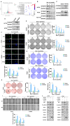
Sec C significantly inhibits colorectal CSCs and their stemness traits. (A) The visible colonies of HCT8 and HT29 cells cultivated in soft agar. Circular images, naked-eye observation. Square images, microscopic observation. Scale bar, 400 μm. (B) The grown spheres (diameter ≥ 60 μm) of HT29 cells were observed under a microscope. Scale bar, 150 μm. (C) The HT29 sphere cells (diameter ≥ 60 μm) were counted after being treated with Sec C under a microscope. Scale bar, 200 μm. (D) Representative images of the wound from HCT8 and RKO cells were recorded by microscope. Scale bar, 200 μm. (E) Western blotting for detecting the stemness and EMT markers in HCT116 and HT29 cells. (F) The changes in the proportion of CSCs from HCT8 cells were measured by FCM. (G) The stemness proteins decreased due to Sec C treatment according to RNA-seq analysis. The data are shown as mean ± SD (n = 3). * p < 0.05, ** p < 0.01, *** p < 0.001, and **** p < 0.0001. NS indicated that no significance was noted.
Figure 4
Figure 4
S100A8 reverses the effect of Sec C on colorectal tumor cells. (A) The genes had changed and the activated transduction pathways of Sec C-treated HT29 sphere cells. (B) HCT8, HT29, and HCT116 were treated by Sec C to detect the changes in S100A8 by Western blotting. (C) The expression of S100A8 in HCT8/L and sphere cells. (D) The expression of S100A8 in HCT8 and HT29 cells after being stably transfected by S100A8 plasmid. (E) The stable S100A8-overexpressing HCT8 and HT29 cells were seeded in 6-well plates and treated with Sec C. The visible colonies were counted as shown. (F) The S100A8-overexpressing HCT8 cells were treated with Sec C, and the DNA replication level was examined by EdU assay. Scale bar, 150 μm. (G) The visible colonies of the stable HCT8 and HT29 cells in soft agar were counted under a microscope. (H) The stable HCT8 cells were treated with Sec C and cultured with serum-free medium, and then the spheres (diameter ≥ 60 μm) were counted. Scale bar, 500 μm. (I) Representative images of the wound from S100A8-overexpressing HCT8 cells were recorded by microscope. Scale bar, 200 μm. (J) The Western blotting of S100A8-overexpressing HCT8 and HT29 cells for stemness and EMT markers. Vector, the plasmid empty vector. S100A8, the overexpressed plasmid group. The data are shown as mean ± SD (n = 3). * p < 0.05, ** p < 0.01, *** p < 0.001, and **** p < 0.0001. NS indicated that no significance was noted.
Figure 5
Figure 5
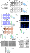
Downregulated S100A8 increases the sensitivity of colorectal tumor cells to Sec C. (A) The expression of S100A8 in HCT8 and HT29 cells after being stably transfected with Sh S100A8 plasmid. (B) The visible colonies of the stable S100A8-knockdown HCT8 and HT29 cells were counted as shown. (C) The stable HCT8 and HT29 cells were treated with Sec C and then seeded in soft agar. The visible colonies were counted after being stained. (D) The S100A8-knockdown HCT8 cells were treated with Sec C, and cell proliferation was detected with EdU assay. Scale bar, 75 μm. (E) The stable S100A8-knockdown HCT8 cells were treated with Sec C and cultured in 6-well low-adhesion culture plates, and then the spheres (diameter ≥ 60 μm) were counted. Scale bar, 500 μm. (F) Representative images of the wound from S100A8-knockdown HCT8 cells were recorded by microscope. Scale bar, 200 μm. (G) The Western blotting of S100A8-knockdown HCT8 and HT29 cells for stemness and EMT markers. Ctrl, the plasmid empty control. Sh or Si S100A8, the downregulated plasmid group. The data are shown as mean ± SD (n = 3). * p < 0.05, ** p < 0.01, *** p < 0.001, and **** p < 0.0001. NS indicated that no significance was noted.
Figure 6
Figure 6
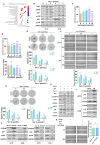
p38 weakens the effect of Sec C and interacts with S100A8. (A) The activated signaling pathways after S100A8 overexpression by RNA-seq analysis. (B) The changes of ERK, JNK, and p38 in Sec C-treated HCT8 and HT29 cells by Western blotting. (C) Anisomycin was mixed with Sec C to treat tumor cells in 96-well plates for 48 h, and the vitality of HCT8 cells were detected by MTT assay. (D) The survival rate of HCT8 cells treated by Sec C and SP600125 or FR180204 were detected by MTT assay. (E) The p38-overexpressing HCT8 cells were treated with Sec C for 24 h. Then, they were cultured in 6-well low-adhesion culture plates, and the spheres (diameter ≥ 60 μm) were counted. Scale bar, 200 μm. (F) The transfected HCT8 cells were seeded in 6-well plates and treated with Sec C. Representative images of the wound were recorded by microscope. Scale bar, 200 μm. (G) HCT8 cells with high p38 expression were cultured in serum-free medium, and then treated with Sec C for 24 h. The spheres (diameter ≥ 60 μm) were counted. Scale bar, 200 μm. (H) The proteins were extracted from transfected HCT8 cells after being treated with Sec C and immunoblotting was performed for stemness and EMT markers. (I) After p38 was overexpressed, S100A8 protein was detected in HCT8 and HT29 cells by Western blotting. (J) S100A8 was up- or downregulated in HCT8 and HT29 cells, respectively, and p38 was detected by Western blotting. (K) Representative images of the wound from HCT8 cells transfected by S100A8-downregulated and p38-upregulated plasmids were recorded by microscope. Scale bar, 200 μm. Vector, the plasmid empty vector. p38, the overexpressed plasmid group. The data are shown as mean ± SD (n = 3). * p < 0.05, ** p < 0.01, and *** p < 0.001. NS indicated that no significance was noted.
Figure 7
Figure 7
Sec C inhibits tumorigenesis in vivo. BALB/c nude mice were used to evaluate the anti-tumor activity of Sec C. After the mice were sacrificed, the variation curve of mouse weight (A), the variation curve of tumor volume (B), the weight (C), and the shapes (D) of tumors were all analyzed. The expression of S100A8 (E), p38 (F), and Ki67 (G) in the tumor tissue of BALB/c nude mice was detected by immunohistochemistry, and the IHC score was analyzed by ImageJ (Version 1.53). Scale bar, 100 μm. * p < 0.05, ** p < 0.01, and **** p < 0.0001. NS indicated that no significance was noted.

Similar articles

References

    1. Ahmad R., Singh J.K., Wunnava A., Al-Obeed O., Abdulla M., Srivastava S.K. Emerging trends in colorectal cancer: Dysregulated signaling pathways (Review) Int. J. Mol. Med. 2021;47:14. doi: 10.3892/ijmm.2021.4847. - DOI - PMC - PubMed
    1. Dekker E., Tanis P.J., Vleugels J.L.A., Kasi P.M., Wallace M.B. Colorectal cancer. Lancet. 2019;394:1467–1480. doi: 10.1016/S0140-6736(19)32319-0. - DOI - PubMed
    1. Yang I.P., Tsai H.L., Huang C.W., Huang M.Y., Hou M.F., Juo S.H., Wang J.Y. The functional significance of microRNA-29c in patients with colorectal cancer: A potential circulating biomarker for predicting early relapse. PLoS ONE. 2013;8:e66842. doi: 10.1371/journal.pone.0066842. - DOI - PMC - PubMed
    1. Zhao W., Dai S., Yue L., Xu F., Gu J., Dai X., Qian X. Emerging mechanisms progress of colorectal cancer liver metastasis. Front. Endocrinol. 2022;13:1081585. doi: 10.3389/fendo.2022.1081585. - DOI - PMC - PubMed
    1. Li J., Ma X., Chakravarti D., Shalapour S., DePinho R.A. Genetic and biological hallmarks of colorectal cancer. Genes Dev. 2021;35:787–820. doi: 10.1101/gad.348226.120. - DOI - PMC - PubMed

Publication types

MeSH terms