. 2024 Apr 4;13(7):623.
doi: 10.3390/cells13070623.

Enrichment, Characterization, and Proteomic Profiling of Small Extracellular Vesicles Derived from Human Limbal Mesenchymal Stromal Cells and Melanocytes

Affiliations

Enrichment, Characterization, and Proteomic Profiling of Small Extracellular Vesicles Derived from Human Limbal Mesenchymal Stromal Cells and Melanocytes

Sebastian Kistenmacher et al. Cells. .

Abstract

Limbal epithelial progenitor cells (LEPC) rely on their niche environment for proper functionality and self-renewal. While extracellular vesicles (EV), specifically small EVs (sEV), have been proposed to support LEPC homeostasis, data on sEV derived from limbal niche cells like limbal mesenchymal stromal cells (LMSC) remain limited, and there are no studies on sEVs from limbal melanocytes (LM). In this study, we isolated sEV from conditioned media of LMSC and LM using a combination of tangential flow filtration and size exclusion chromatography and characterized them by nanoparticle tracking analysis, transmission electron microscopy, Western blot, multiplex bead arrays, and quantitative mass spectrometry. The internalization of sEV by LEPC was studied using flow cytometry and confocal microscopy. The isolated sEVs exhibited typical EV characteristics, including cell-specific markers such as CD90 for LMSC-sEV and Melan-A for LM-sEV. Bioinformatics analysis of the proteomic data suggested a significant role of sEVs in extracellular matrix deposition, with LMSC-derived sEV containing proteins involved in collagen remodeling and cell matrix adhesion, whereas LM-sEV proteins were implicated in other cellular bioprocesses such as cellular pigmentation and development. Moreover, fluorescently labeled LMSC-sEV and LM-sEV were taken up by LEPC and localized to their perinuclear compartment. These findings provide valuable insights into the complex role of sEV from niche cells in regulating the human limbal stem cell niche.

Keywords: exosomes; extracellular vesicles; limbal epithelial progenitor cells; limbal melanocytes; limbal mesenchymal stromal cells; limbal stem cell niche; proteomics.


Conflict of interest statement

The authors declare no conflicts of interest.

Figures
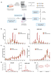
Figure 1
Figure 1
Enrichment and quantification of small extracellular vesicles (sEV): (A) Workflow used for enrichment of sEVs from conditioned media (~40 mL) of limbal mesenchymal stromal cells (LMSC) and limbal melanocytes (LM). (B) Nanoparticle tracking analysis showing particle counts in LM-sEV and LMSC-sEV fractions (F1–F8), void volume (V), and flow-through (FT) of conditioned and unconditioned media. Data presented as min-to-max whisker box plots (n = 10/cell type). (C) Protein concentration analysis by micro-BCA assay showing higher protein levels in later fractions (F7, F8) and flow-through (FT) compared to earlier fractions for both LM-sEVs and LMSC-sEVs (n = 10/cell type sEVs). Data presented as min-to-max whisker box plots (n = 10/cell type). (D) Particle-to-protein ratio analysis indicating highest purity in fraction 3 (F3) for both LM-sEVs and LMSC-sEVs (n = 10/cell type). Data presented as min-to-max whisker box plots (n = 10/cell type). (E) Size distribution of LM-sEV and LMSC-sEV particles in fraction 3 (F3) measured by nanoparticle tracking analysis. Data presented as min-to-max whisker box plots (n = 10/cell type).
Figure 2
Figure 2
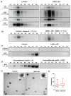
Characterization of small extracellular vesicles (sEV): (A) Western blot analysis of EV-specific markers in fractions F1–F8 from LM- and LMSC-sEVs. Enrichment of EV markers was predominantly observed in F3. Uncropped versions of the Western blot are shown in Supplementary Files S2 and S3. (B) Cell-specific markers CD90 (LMSC) and Melan-A (LM) were detected in F2–F4, with highest levels in F3. Uncropped versions of the Western blot are shown in Supplementary Files S2 and S3. (C) Absence of the endoplasmic reticulum marker calnexin (CANX) across all fractions indicates lack of cellular contamination. Residual bovine serum albumin (BSA) was detected in LM-sEV fractions. Uncropped versions of the Western blot are shown in Supplementary File S4. (D) BSA was present in unconditioned media controls for LM-sEVs but not LMSC-sEVs. Uncropped versions of the Western blot are shown in Supplementary File S4. (E) Transmission electron microscopy images of F3 fractions showing classical EV morphology with intact membranes. (F) Size distribution box plots of LM-sEVs and LMSC-sEVs in F3 based on TEM analysis. Data presented as min-to-max whisker box plots (n = 3/cell type).
Figure 3
Figure 3
Multiplex bead-based flow cytometry assay: (A) Column graphs showing normalized median fluorescence intensity (nMFI) values for various EV surface antigens detected by multiplex bead array analysis of fraction 3 (F3) from limbal melanocyte-derived sEVs (LM-sEVs) and limbal mesenchymal stromal cell-derived sEVs (LMSC-sEVs). Data presented as mean ± standard deviation (SD), n = 5/cell type. (B) Western blot validation confirming predominant expression of CD146 and MCSP in LM-sEVs (F3) compared to LMSC-sEVs. Cell lysate of both LMSC and LM showing the expression of CD146 and MCSP. Uncropped versions of the Western blot are shown in Supplementary File S5. (C) Immunohistochemical staining of organ-cultured limbal tissue sections showing absence of CD146 (green) and MSCP (green) in melanocytes (arrows, red, gp100+) and stromal cells (cyan, vimentin+). Vessels in the stroma stained for CD146 (arrow heads, green) and MCSP (arrow heads, green). Dashed line represent the basement membrane. Nuclei are counterstained with DAPI (blue).
Figure 4
Figure 4
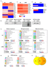
Proteomic characterization of small extracellular vesicles (sEV): (A) Heatmaps showing protein intensity profiles (based on iBAQ mean intensity, n = 5) of (i) plasma membrane and endosomal proteins, (ii) cytosolic proteins, (iii) apolipoproteins, and (iv) cellular contaminants across LMSC-sEV and LM-sEV samples. We employed relative quantification (iBAQ) to compare the abundance levels of known EV markers and potential contaminants (such as serum components or cytosolic proteins) within the sample. (B) Western blot validation confirming the presence of CD90 (LMSC), Melan-A and TYRP1 (LM), and β-actin in fraction 3 (F3) of respective sEV populations. Uncropped versions of the Western blot are shown in Supplementary File S7. (C,D) Top 10 enriched Gene Ontology (GO) terms for (C) Biological Process, Molecular Function, Cellular Component, and (D) KEGG pathways in LM-sEV and LMSC-sEVs. (E) Venn diagram showing the number of unique and shared proteins identified in LM-sEVs and LMSC-sEV.
Figure 5
Figure 5
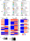
Distinct protein signatures in limbal melanocyte (LM)-derived small extracellular vesicles (sEV) and limbal mesenchymal stromal cell (LMSC)-derived small extracellular vesicles (sEV). (A) Gene ontology analysis highlighting the unique biological processes, cellular components, molecular functions, and KEGG pathways enriched in proteins found exclusively in LM-sEVs versus LMSC-sEVs. (B) Heatmaps showing selected proteins (based on LFQ mean intensity, n = 5) uniquely present in LM-sEVs versus LMSC-sEVs, categorized based on their roles in melanogenesis, extracellular matrix composition and remodeling, angiogenesis, and cell migration. (C) Western blot validation confirming the presence of fibronectin (FN1) and SPARC in LMSC sEV populations. Uncropped versions of the Western blot are shown in Supplementary File S8.
Figure 6
Figure 6
Uptake of isolated small extracellular vesicles (sEVs) by recipient cells: (A) Flow cytometry analysis demonstrating increased fluorescence intensity in limbal epithelial progenitor cells (LEPC) treated with limbal mesenchymal stromal cell derived small extracellular vesicles (LMSC-sEVs) or limbal melanocyte derived small extracellular vesicles (LM-sEVs) for 3 or 6 h compared to untreated LEPC or LEPC treated with sEVs derived from unconditioned media. (B) Confocal microscopy images showing the localization of fluorescently labeled LMSC-sEVs and LM-sEVs (green) in the perinuclear region of LEPC after 6 h of incubation. Nuclei are counterstained with DAPI (blue). (C) LEPC co-stained with Phalloidin-TRITC (red) to visualize F-actin. Nuclei are counterstained with DAPI (blue).

Similar articles

Cited by

References

    1. Cotsarelis G., Cheng S.Z., Dong G., Sun T.T., Lavker R.M. Existence of Slow-Cycling Limbal Epithelial Basal Cells That Can Be Preferentially Stimulated to Proliferate: Implications on Epithelial Stem Cells. Cell. 1989;57:201–209. doi: 10.1016/0092-8674(89)90958-6. - DOI - PubMed
    1. Li W., Hayashida Y., Chen Y.-T., Tseng S.C.G. Niche Regulation of Corneal Epithelial Stem Cells at the Limbus. Cell Res. 2007;17:26–36. doi: 10.1038/sj.cr.7310137. - DOI - PMC - PubMed
    1. Ordonez P., Di Girolamo N. Limbal Epithelial Stem Cells: Role of the Niche Microenvironment. Stem Cells. 2012;30:100–107. doi: 10.1002/stem.794. - DOI - PubMed
    1. Mei H., Gonzalez S., Deng S.X. Extracellular Matrix Is an Important Component of Limbal Stem Cell Niche. J. Funct. Biomater. 2012;3:879–894. doi: 10.3390/jfb3040879. - DOI - PMC - PubMed
    1. Dziasko M.A., Tuft S.J., Daniels J.T. Limbal Melanocytes Support Limbal Epithelial Stem Cells in 2D and 3D Microenvironments. Exp. Eye Res. 2015;138:70–79. doi: 10.1016/j.exer.2015.06.026. - DOI - PubMed

Publication types

LinkOut - more resources