

Prognostic value of radiological T category using conventional MRI in patients with oral tongue cancer: comparison with pathological T category
- PMID: 38607437
- PMCID: PMC11133020
- DOI: 10.1007/s00234-024-03345-8
Prognostic value of radiological T category using conventional MRI in patients with oral tongue cancer: comparison with pathological T category
Abstract
Purpose: This study aimed to compare the radiological tumor (T)-category using multiparametric MRI with the pathological T category in patients with oral tongue squamous cell carcinoma (OTSCC) and to examine which is a better predictor of prognosis.
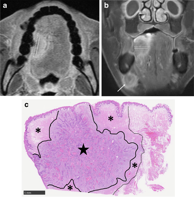
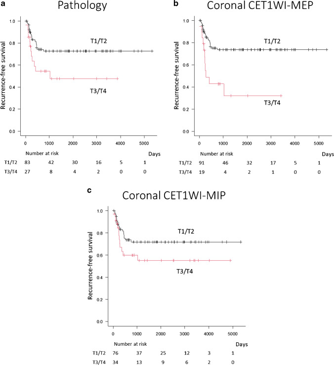
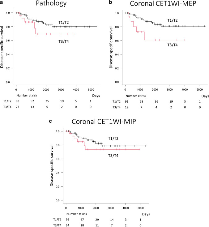
Methods: This retrospective study included 110 consecutive patients with surgically resected primary OTSCC who underwent preoperative contrast-enhanced MRI. T categories determined by maximum diameter and depth of invasion were retrospectively assessed based on the pathological specimen and multiparametric MRI. The MRI assessment included the axial and coronal T1-weighted image (T1WI), axial T2-weighted image (T2WI), coronal fat-suppressed T2WI, and axial and coronal fat-suppressed contrast-enhanced T1WI (CET1WI). Axial and coronal CET1WI measurements were divided into two groups: measurements excluding peritumoral enhancement (MEP) and measurements including peritumoral enhancement. The prognostic values for recurrence and disease-specific survival after radiological and pathological T categorization of cases into T1/T2 and T3/T4 groups were compared.
Results: The T category of MEP on coronal CET1WI was the most relevant prognostic factor for recurrence [hazard ratio (HR) = 3.30, p = 0.001] and the HR was higher than the HR for pathological assessment (HR = 2.26, p = 0.026). The T category determined by MEP on coronal CET1WI was also the most relevant prognostic factor for disease-specific survival (HR = 3.12, p = 0.03), and the HR was higher than the HR for pathological assessment (HR = 2.02, p = 0.20).
Conclusion: The T category determined by MEP on the coronal CET1WI was the best prognostic factor among all radiological and pathological T category measurements.
Keywords: MRI; Oral tongue squamous cell carcinoma; Peritumoral enhancement; Prognosis; Tumor staging.
© 2024. The Author(s).
Conflict of interest statement
The authors declare no competing interests.
Figures
Similar articles
-
Value of radiological depth of invasion in non-pT4 Oral tongue squamous cell carcinoma: implication for preoperative MR T-staging.Eur Radiol. 2024 Sep;34(9):6047-6059. doi: 10.1007/s00330-024-10598-7. Epub 2024 Feb 3. Eur Radiol. 2024. PMID: 38308013 Free PMC article.
-
Assessment of tumor depth in oral tongue squamous cell carcinoma with multiparametric MRI: correlation with pathology.Eur Radiol. 2022 Jan;32(1):254-261. doi: 10.1007/s00330-021-08148-6. Epub 2021 Jul 13. Eur Radiol. 2022. PMID: 34255162 Free PMC article.
-
Utility of Diffusion-weighted MR Imaging for Evaluating the Depth of Invasion in Oral Tongue Squamous Cell Carcinoma.Magn Reson Med Sci. 2025 Apr 1;24(2):210-219. doi: 10.2463/mrms.mp.2023-0137. Epub 2024 Mar 7. Magn Reson Med Sci. 2025. PMID: 38447989 Free PMC article.
-
Correlation between clinical and MRI assessment of depth of invasion in oral tongue squamous cell carcinoma.J Otolaryngol Head Neck Surg. 2016 Nov 22;45(1):61. doi: 10.1186/s40463-016-0172-0. J Otolaryngol Head Neck Surg. 2016. PMID: 27876067 Free PMC article.
-
Radiological approach for the newly incorporated T staging factor, depth of invasion (DOI), of the oral tongue cancer in the 8th edition of American Joint Committee on Cancer (AJCC) staging manual: assessment of the necessity for elective neck dissection.Jpn J Radiol. 2020 Sep;38(9):821-832. doi: 10.1007/s11604-020-00982-w. Epub 2020 Apr 30. Jpn J Radiol. 2020. PMID: 32356237 Review.
References
-
- Zittel S, Moratin J, Horn D, Metzger K, Ristow O, Engel M, Mrosek J, Freier K, Hoffmann J, Freudlsperger C. Clinical outcome and prognostic factors in recurrent oral squamous cell carcinoma after primary surgical treatment: a retrospective study. Clin Oral Invest. 2022;26(2):2055–2064. doi: 10.1007/s00784-021-04186-y. - DOI - PMC - PubMed
-
- Takamura M, Kobayashi T, Nikkuni Y, Katsura K, Yamazaki M, Maruyama S, Tanuma JI, Hayashi T. A comparative study between CT, MRI, and intraoral US for the evaluation of the depth of invasion in early stage (T1/T2) tongue squamous cell carcinoma. Oral Radiol. 2022;38(1):114–125. doi: 10.1007/s11282-021-00533-7. - DOI - PMC - PubMed
Publication types
MeSH terms
Substances
LinkOut - more resources
Full Text Sources
Medical
