. 2024 Apr 1;151(7):dev201102.
doi: 10.1242/dev.201102. Epub 2024 Apr 12.

The journey of a generation: advances and promises in the study of primordial germ cell migration

Affiliations

The journey of a generation: advances and promises in the study of primordial germ cell migration

Lacy J Barton et al. Development. .

Erratum in

Abstract

The germline provides the genetic and non-genetic information that passes from one generation to the next. Given this important role in species propagation, egg and sperm precursors, called primordial germ cells (PGCs), are one of the first cell types specified during embryogenesis. In fact, PGCs form well before the bipotential somatic gonad is specified. This common feature of germline development necessitates that PGCs migrate through many tissues to reach the somatic gonad. During their journey, PGCs must respond to select environmental cues while ignoring others in a dynamically developing embryo. The complex multi-tissue, combinatorial nature of PGC migration is an excellent model for understanding how cells navigate complex environments in vivo. Here, we discuss recent findings on the migratory path, the somatic cells that shepherd PGCs, the guidance cues somatic cells provide, and the PGC response to these cues to reach the gonad and establish the germline pool for future generations. We end by discussing the fate of wayward PGCs that fail to reach the gonad in diverse species. Collectively, this field is poised to yield important insights into emerging reproductive technologies.

Keywords: ECM; Gonad; Migration; Primordial germ cells; Signaling.


Conflict of interest statement

Competing interests The authors declare no competing or financial interests.

Figures
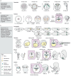
Fig. 1.
Fig. 1.
Routes PGCs migrate along to reach the somatic gonad. Three broad pathways are followed by PGCs among studied model organisms. (A) One path is exemplified by the zebrafish model Danio rerio, in which PGCs are specified at multiple locations in the early embryo, often at cleavage furrows. Then, PGCs actively migrate dorsally during the shield stage, converging from four populations into two bilateral groups. Then, at the somite stages, PGCs migrate laterally and anteriorly as they colonize the gonad. (B) A second PGC route is found in chicken (Gallus gallus domesticus). Like zebrafish, chicken PGCs are specified near cleavage furrows. In contrast to zebrafish, chicken PGCs reach the gonad through both passive translocation and active migration. First, PGCs move anteriorly toward the germinal crescent, where they enter blood vessels. After circulating, PGCs exit the vascular endothelia and actively migrate through the dorsal mesentery toward bilateral developing gonads. (C) A third, common PGC route is found in several model organisms, including Drosophila melanogaster, Xenopus laevis and mouse (Mus musculus). This is also the route that PGCs likely follow in humans, although the origin of human PGCs is as yet unclear. In Drosophila, Xenopus and mouse, PGCs are specified at the border of the embryo and extra-embryonic tissue. From this peripheral location, PGCs are passively internalized into the endoderm during gastrulation. Mouse PGCs actively move to the endoderm, but once associated with the hindgut epithelium they move passively with the gastrulating hindgut. From the gut, PGCs traverse the endodermal epithelium, entering the mesoderm (commonly the dorsal mesentery) where they actively migrate and sort bilaterally to reach the two gonadal ridges. In Drosophila, the germ-band, which contains the somatic gonadal precursors (green), retracts during PGC migration, depicted by the black arrows. Cell movements are indicated by black arrows. For all species, developmental progression is shown from left to right. Each embryonic stage is shown underneath the corresponding embryo schematic. A, anterior; D, dorsal; E, embryonic day; hpf, hours post-fertilization; HH, Hamburger and Hamilton stage; P, posterior; St., stage; V, ventral; W(PF), weeks post fertilization.
Fig. 2.
Fig. 2.
Adhesion properties during common phases in PGC migration. Schematic of the five stages of PGC migration in fruit flies and frogs. Mice follow the same stages, but actively migrate to enter the endoderm at Stage 2 and then continue to migrate within it. (A) Stage 1: PGCs are specified at the periphery of embryos or extra-embryonic regions early in embryogenesis. Stage 2: PGCs are passively translocated by gastrulation into the interior of the embryo, often with the future endoderm. Stage 3: PGCs transmigrate through an epithelial barrier to access the mesoderm. Stage 4: PGCs directionally migrate through mesoderm containing cells and extracellular matrix. Stage 5: PGCs are ensconced by the somatic component of the gonad. (B,C) Common cell–cell and cell–ECM adhesion molecules that PGCs use to reach the gonad. During passive translocation (B), PGC–PGC and PGC–somatic cell contacts are commonly mediated by E-cadherin (E-cad). During active migration (C), PGCs migrate upon other cells and ECM in the mesoderm. PGCs express E-cadherin early in development whereas mesoderm cells express N-cadherin (N-cad). This suggests PGCs may use heterotypic cadherin contacts (E-cadherin on PGC to N-cadherin on mesoderm) for cell-on-cell migration. PGCs also express integrin receptors to migrate on ECM. BM, basement membrane; ECM, extracellular matrix; EMT, epithelial-to-mesenchymal transition; SGP, somatic gonadal precursor.
Fig. 3.
Fig. 3.
PGC migration strategies and signaling logic. Migrating PGCs interpret chemical guidance cues using different classes of receptors and lipid phosphate phosphatases. Rho-GTPases commonly establish cell polarity, but their upstream linkage to guidance receptors is largely unclear. Different receptor–cue pairs that affect PGC guidance (classified by a role in early or late migration if known, following the convention described in Table 1) are shown on the right of each species-specific PGC schematic. Lipid phosphate phosphatases (LPPs) dephosphorylate unidentified phospholipids to establish permissive migration zones. (A) Signaling pathways at the front and rear of zebrafish PGCs. In a typical front bleb cycle, Rac1 signaling first activates Arp2/3 to create an F-actin template. RhoA and calcium-MLCK signaling then drive actomyosin contractility to form a pressurized bleb, a region of plasma membrane detached from the actin cortex. Retrograde F-actin flow occurs from front to back, moving F-actin-linked proteins, such as Ezrin and septins, to the back to inhibit bleb formation and enhance front–back polarity. (B) Signaling pathways at the front and rear of Drosophila PGCs. Net actin polymerization at the cell front is required to maintain front-to-rear cortical actin flows, but the molecular details are unclear. A proposed front F-actin polymerization pathway includes the generation of PIP2 leading to Arp2/3-driven branched actin polymerization. However, inhibition of the Arp2/3 complex only mildly perturbs cortical flow, suggesting that a formin is involved. At the rear of PGCs, AMPK phosphorylates RhoGEF2, mediating its release from inhibitory microtubule interactions. This allows RhoGEF2 to activate RhoA and generate actomyosin contractility to direct cortical flow. (C) Signaling pathways at the front and rear of Xenopus PGCs. In culture, motile Xenopus PGCs are elongated with a leading bleb. Similar to Drosophila PGCs, RhoA signaling appears to be more crucial than Rac1 and is likely active at the rear. PIP3 has been shown to be enriched in blebs in a Kif13b-dependent manner and likely regulates actomyosin contractility to generate blebs. Retrograde F-actin flow likely occurs, but it is unclear whether it is local or global. (D) Signaling pathways at the front and rear of mouse PGCs. The importance of PI3K and Src are inferred from in vitro studies with inhibitors, whereas the importance of Rac1 has been shown in vivo. No functional data exists for signaling or cytoskeletal function at the rear of mouse PGCs. AMPK, AMP-activated protein kinase; Cxcl12, C-X-C motif chemokine ligand 12; Cxcr4, C-X-C motif chemokine receptor 4; FGF, fibroblast growth factor; FGFR, fibroblast growth factor receptor; GPCR, G protein-coupled receptor; HH, hedgehog; Jak, Janus kinases; JH, Juvenile hormone; KIF, kinesin family member 13B; LPP, lipid phosphate phosphatase; MT, microtubule; PI3K, phosphoinositide 3-kinase; PIP5K, phosphatidylinositol-4-phosphate 5-kinase; PIP2, phosphatidylinositol 4,5-bisphosphate [PtdIns(4,5)P2]; PIP3, phosphatidylinositol (3,4,5)-trisphosphate [PtdIns(3,4,5)P3]; Ptc, Patched; Ror2, receptor tyrosine kinase like orphan receptor 2; RTK, receptor tyrosine kinase; Src, Rous sarcoma oncogene; Stat, signal transducer and activator of transcription; TGFβ, transforming growth factor β; TGFβR, transforming growth factor β receptor.
Fig. 4.
Fig. 4.
PGC traction forces. PGCs generate traction forces to migrate on other cells and ECM by coupling rearward F-actin flow to various molecular clutches, which transmit this force to the extracellular environment. Molecular clutches are adaptors that bind F-actin and are linked to transmembrane proteins either directly or indirectly. (A) Cadherin clutches. α-Catenin couples F-actin to cadherins and allows PGCs to migrate on other cells expressing the cognate cadherin or possibly another cadherin family member or a transmembrane protein that can transiently interact with the PGC cadherin. (B) Integrin clutches. Integrin clutches are well-described and allow cells to migrate on ECM. Talin serves as the molecular clutch between F-actin and ECM-binding integrin receptors. (C) Alternative clutches. Presumably, any protein that can interact with F-actin and a transmembrane protein can serve as a molecular clutch. These transmembrane proteins might generate traction by interacting with other transmembrane proteins or possibly allow migration via multiple non-specific environmental interactions. ECM, extracellular matrix.

Similar articles

Cited by

References

    1. Aeckerle, N., Drummer, C., Debowski, K., Viebahn, C. and Behr, R. (2015). Primordial germ cell development in the marmoset monkey as revealed by pluripotency factor expression: suggestion of a novel model of embryonic germ cell translocation. Mol. Hum. Reprod. 21, 552. 10.1093/molehr/gav016 - DOI - PMC - PubMed
    1. Alvarez-Buylla, A. and Merchant-Larios, H. (1986). Mouse primordial germ cells use fibronectin as a substrate for migration. Exp. Cell. Res. 165, 362-368. 10.1016/0014-4827(86)90590-2 - DOI - PubMed
    1. Alves-Lopes, J. P., Wong, F. C. K., Tang, W. W. C., Gruhn, W. H., Ramakrishna, N. B., Jowett, G. M., Jahnukainen, K. and Surani, M. A. (2023). Specification of human germ cell fate with enhanced progression capability supported by hindgut organoids. Cell Rep. 42, 111907. 10.1016/j.celrep.2022.111907 - DOI - PubMed
    1. Amadei, G., Handford, C. E., Qiu, C., De Jonghe, J., Greenfeld, H., Tran, M., Martin, B. K., Chen, D. Y., Aguilera-Castrejon, A., Hanna, J. H.et al. (2022). Embryo model completes gastrulation to neurulation and organogenesis. Nature 610, 143-153. 10.1038/s41586-022-05246-3 - DOI - PMC - PubMed
    1. Anderson, R., Copeland, T. K., Scholer, H., Heasman, J. and Wylie, C. (2000). The onset of germ cell migration in the mouse embryo. Mech. Dev. 91, 61-68. 10.1016/S0925-4773(99)00271-3 - DOI - PubMed