. 2024 Apr 12;15(4):259.
doi: 10.1038/s41419-024-06651-4.

RUVBL1 ubiquitination by DTL promotes RUVBL1/2-β-catenin-mediated transcriptional regulation of NHEJ pathway and enhances radiation resistance in breast cancer

Affiliations

RUVBL1 ubiquitination by DTL promotes RUVBL1/2-β-catenin-mediated transcriptional regulation of NHEJ pathway and enhances radiation resistance in breast cancer

Jie Tian et al. Cell Death Dis. .

Abstract

Radiotherapy effectiveness in breast cancer is limited by radioresistance. Nevertheless, the mechanisms behind radioresistance are not yet fully understood. RUVBL1 and RUVBL2, referred to as RUVBL1/2, are crucial AAA+ ATPases that act as co-chaperones and are connected to cancer. Our research revealed that RUVBL1, also known as pontin/TIP49, is excessively expressed in MMTV-PyMT mouse models undergoing radiotherapy, which is considered a murine spontaneous breast-tumor model. Our findings suggest that RUVBL1 enhances DNA damage repair and radioresistance in breast cancer cells both in vitro and in vivo. Mechanistically, we discovered that DTL, also known as CDT2 or DCAF2, which is a substrate adapter protein of CRL4, promotes the ubiquitination of RUVBL1 and facilitates its binding to RUVBL2 and transcription cofactor β-catenin. This interaction, in turn, attenuates its binding to acetyltransferase Tat-interacting protein 60 (TIP60), a comodulator of nuclear receptors. Subsequently, ubiquitinated RUVBL1 promotes the transcriptional regulation of RUVBL1/2-β-catenin on genes associated with the non-homologous end-joining (NHEJ) repair pathway. This process also attenuates TIP60-mediated H4K16 acetylation and the homologous recombination (HR) repair process. Expanding upon the prior study's discoveries, we exhibited that the ubiquitination of RUVBL1 by DTL advances the interosculation of RUVBL1/2-β-catenin. And, it then regulates the transcription of NHEJ repair pathway protein. Resulting in an elevated resistance of breast cancer cells to radiation therapy. From the aforementioned, it is evident that targeting DTL-RUVBL1/2-β-catenin provides a potential radiosensitization approach when treating breast cancer.


Conflict of interest statement

The authors declare no competing interests.

Figures

Fig. 1
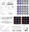
Fig. 1. RUVBL1 expression is comparatively high in the radiation breast-tumor tissue.
A FVB-TgN (MMTV-PyMT) transgenic female mice were irradiated when their tumors reached 200 mm3. Every 2 days, 3 Gy each time. B The irradiated mice were dissected and the tumors were photographed and compared at the end of the experiment. C Tumor growth was monitored every 2 days for 5 times, and the volume was recorded till the experiment ended (n = 7 mice). D Heatmaps showing the 348 differentially expressed proteins in irradiated and non-irradiated tumor tissue. E Volcano plot comparing control and irradiated tumor tissue. F Enrichment of differentially expressed proteins in signaling pathways is portrayed as a bar chart. Y-axis represents pathways, and the X-axis represents rich factor. G Heatmap of DNA repair pathway. H Immunohistochemical (IHC) for RUVBL1 in tumor tissues (×10). Scale bars: 100 μm. I Western blot for RUVBL1 and β-actin in tumor tissues. Data presented as mean ± SD, *p < 0.05, statistical differences were assessed using two-tailed unpaired Student’s t-test in (C). (**p < 0.01).
Fig. 2
Fig. 2. RUVBL1 regulates radiation resistance of breast cancer cells.
A Western blot for RUVBL1 and β-actin in MDA-MB-231 RUVBL1 cell line. B Western blot for RUVBL1 and β-actin in MDA-MB-231 shRUVBL1 cell line. C, D The stable cell lines underwent a series of radiation treatments at 0, 1, 2, 4, 6 and 8 Gy, followed by an MTT test (n = 3). EH The same cell lines were subjected to radiation at 0, 1, 2, 4, and 6 Gy, prior to being cloned and evaluated for survival curve (n = 3). I Western blot for γ-H2AX and β-actin in MDA-MB-231 RUVBL1 cells with radiation (5 Gy). J Immunofluorescence for γ-H2AX in MDA-MB-231 RUVBL1 cells with radiation (5 Gy) (×10). Scale bars: 100 μm. K 2 × 10⁵ control or RUVBL1 MDA-MB-231 cells were subcutaneously injected into nude mice (n = 5). Since the 12th day, each group of nude mice were treated with radiation (Every 3 days, 3 Gy each time). Tumor growth curves were shown. Tumors (L) and tumor size (M) of mice were shown. Data presented as mean ± SD, *p < 0.05, statistical differences were assessed using two-tailed unpaired Student’s t-test (C, E, F, H, K, M). (*p < 0.05, **p < 0.01, ***p < 0.001).
Fig. 3
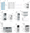
Fig. 3. RUVBL1 enhances DNA damage repair and is ubiquitinated by DTL.
A, B HEK 293T cells transfected with control vector or Myc-RUVBL1 were immunoprecipitated with Myc antibody and subjected to mass spectrometry analysis. DTL was identified as a novel binding partner for RUVBL1. C HEK293T cells transfected with Myc-RUVBL1 were harvested for immunoprecipitation with Myc antibody, followed by western blot. D HEK293T cells transfected with Myc-RUVBL1, DTL or HA-Ub were immunoprecipitated with Myc antibody and western blot analysis. E MDA-MB-231-RUVBL1 cells were harvested for immunoprecipitation with RUVBL1 antibody, followed by western blot. F MDA-MB-231-RUVBL1 cells transfected with DTL and HA-Ub were immunoprecipitated with RUVBL1 antibody and western blot analysis. G RUVBL1 half-life is unchanged in MDA-MB -231-RUVBL1 compared with control cells. Cells were switched to fresh medium (10% FBS) containing cycloheximide (CHX) for indicated time periods (0, 2, 4, 6 h) and harvested for western blot. The band density was quantified (n = 3). H HEK 293T cells transfected with Myc-RUVBL1, DTL, HA-Ub, HA-Ub K48, HA-Ub K63, HA-Ub K48R or HA-Ub K63R were immunoprecipitated with Myc antibody and analyzed by western blot. I MDA-MB-231-RUVBL1-RR cells transfected with DTL and HA-Ub were immunoprecipitated with RUVBL1 antibody and western blot analysis. J MDA-MB-231-RR cells transfected with HA-Ub, HA-Ub K63, or HA-Ub K63R were immunoprecipitated with RUVBL1 antibody and analyzed by western blot. Data presented as mean ± SD, *p < 0.05, statistical differences were assessed using two-tailed unpaired Student’s t-test (G). ns non-significant.
Fig. 4
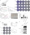
Fig. 4. DTL regulates radiation resistance of breast cancer cells.
A Western blot for DTL and β-actin in MDA-MB-231 DTL stably expressed cells. B Western blot for DTL and β-actin in MDA-MB -231 shDTL stably expressed cells. C, D The stable cell lines underwent a series of radiation treatments at 0, 1, 2, 4, 6, and 8 Gy, followed by an MTT test (n = 3). EH The stable cell lines were subjected to radiation at 0, 1, 2, 4, and 6 Gy, prior to being cloned and evaluated for survival curve (n = 3). I 2 × 10⁵ control or RUVBL1 MDA-MB-231 cells were subcutaneously injected into nude mice (n = 5). Since the 12th day, each group of nude mice were treated with radiation (Every 3 days, 3 Gy each time). Tumor growth curves were shown. Tumors (J) and tumor size (K) of mice were shown. Data presented as mean ± SD, *p < 0.05, statistical differences were assessed using two-tailed unpaired Student’s t-test (C, D, G, H, I, K) (*p < 0.05, **p < 0.01).
Fig. 5
Fig. 5. RUVBL1 regulates radiation resistance of breast cancer cells by DTL.
A Western blot for RUVBL1, DTL and β-actin in MDA-MB-231 control, RUVBL1, RUVBL1-shDTL#1 and RUVBL1-shDTL#2 stably expressed cells. BD MDA-MB-231 control, RUVBL1, RUVBL1-shDTL#1 and RUVBL1-shDTL #2 cells were treated with radiation (0, 1, 2, 4, 6, 8 Gy), followed by MTT assay and clonogenic survival assay (n = 3). E 2 ×10⁵ MDA-MB-231 control, RUVBL1, RUVBL1-shDTL #1 and RUVBL1-shDTL #2 cells were subcutaneously injected into nude mice (n = 5). Since the twelfth day, each group of nude mice were treated with radiation (Every 3 days, 3 Gy each time). Tumor growth curves were shown. Tumors (F) and tumor size (G) of mice were shown. H Western blot assay was used to detect the expression of DTL in MDA-MB-231-RR. I The stable cell lines underwent a series of radiation treatments at 0, 4, and 6 Gy, followed by an MTT test (n = 3). J The MDA-MB-231-RR Control and shDTL cells were subjected to radiation at 0, 4, and 6 Gy, prior to being cloned and evaluated for survival curve (n = 3). K MDA-MB-231-RR cells transfected with shDTL were immunoprecipitated with RUVBL1 antibody and western blot analysis. Data presented as mean ± SD, *p < 0.05, statistical differences were assessed using two-tailed unpaired Student’s t-test (B, D, E, G, I, J). (*p < 0.05, **p < 0.01, ***p < 0.001).
Fig. 6
Fig. 6. DTL-ubiquitinated-RUVBL1 attenuates H4K16 acetylation-mediated HR repair pathway.
A The changes of H4K16ac level after high expression of RUVBL1 and DTL were detected by western blot. B The changes of H4K16Ac level after high expression of RUVBL1 and knockdown of DTL were detected by western blot. C The analysis of the interaction between RUVBL1 and RUVBL2 was carried out in MDA-MB-231-RUVBL1 cells using immunoprecipitation (IP) after 4 h of IR, and it was compared with IgG. DF HEK293T cells were transfected with the indicated plasmids prior to treatment with IR for 4 h before collection. The lysates were incubated with Myc antibody overnight and then subjected to western blot. G MDA-MB-231-RR cells were transfected with the shDTL plasmids prior to treatment with IR for 4 h before collection. The lysates were incubated with RUVBL1 antibody overnight and then subjected to western blot.
Fig. 7
Fig. 7. DTL promotes the binding of RUVBL1 to transcription cofactor β-catenin.
A Volcano plot comparing control and cells high expression of RUVBL1 after IR 4 h. B Heat map from RNA-sequencing analysis. C Enrichment of differentially expressed genes in GO signaling pathways is illustrated in a bar graph. Y-axis represents pathways, and the X-axis represents rich factor. D Enrichment of differentially expressed genes in signaling pathways is illustrated in an advanced bubble chart. Size and color of the bubble are representation of the amount of differentially expressed genes enriched in pathways and their enrichment significance, respectively. E Immunoprecipitation (IP) analysis of the interaction between RUVBL1 and β-catenin, c-Myc, STAT3 in HEK293T cells after IR 4 h. F HEK293T cells transfected with Myc-RUVBL1, DTL, HA-Ub, HA-Ub K63 or HA-Ub K63R were immunoprecipitated with Myc antibody and western blot analysis. G MDA-MB-231-RR cells transfected with Flag-DTL, HA-Ub, HA-Ub K63 or HA-Ub K63R were immunoprecipitated with RUVBL1 antibody and western blot analysis.
Fig. 8
Fig. 8. RUVBL1 affects the expression of NHEJ pathway genes and is regulated by DTL.
A The expressions of NHEJ pathway gene in MDA-MB-231 control and RUVBL1 cell s and corresponding normal cells were determined by western blot, radiate in 5 Gy for 4 h. B The expressions of NHEJ pathway genes in MDA-MB-231 control and DTL cells and corresponding normal cells were determined by western blot, radiate in 5 Gy for 4 h. C DSBs were induced through the I-SceI method, using the NHEJ reporter gene system. Subsequently, flow cytometry was utilized to analyze the impact of RUVBL1 on NHEJ efficiency. D The expressions of NHEJ pathway genes in MDA-MB-231 control, RUVBL1 and RUVBL1-shDTL cells and corresponding normal cells were determined by western blot, radiate in 5 Gy for 4 h. E qRT-PCR detected the mRNA level of the NHEJ pathway genes in MDA-MB-231 control, RUVBL1 and RUVBL1-shDTL cell s, radiate in 5 Gy for 4 h (n = 3). F The expression of DNA-PKcs and 53BP1 in the nude tumor tissues was detected by IHC (×20). Scale bars: 50 μm. Data presented as mean ± SD, statistical differences were assessed using two-tailed unpaired Student’s t-test (C, E). (*p < 0.05, **p < 0.01, ***p < 0.001, ****p < 0.0001).
Fig. 9
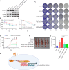
Fig. 9. DTL-RUVBL1-β-catenin regulates radiation resistance of breast cancer cells.
A Western blot for RUVBL1, DTL, β-catenin and β-actin in MDA-MB-231 control, RUVBL1, RUVBL1-DTL, RUVBL1-DTL-shβ-catenin#1 and RUVBL1-DTL-shβ-catenin#2 stably expressed cells (HA-DTL; Myc-RUVBL1). BD The above cell lines were treated with radiation (0, 1, 2, 4, 6, 8 Gy), followed by MTT assay and clonogenic survival assay (n = 3). E 2 × 10⁵ the above cells were subcutaneously injected into nude mice (n = 5). Since the twelfth day, each group of nude mice were treated with radiation (Every 3 days, 3 Gy each time). Tumor growth curves were shown. Tumors (F) and tumor size (G) of mice were shown. H Schematic diagram. RUVBL1 ubiquitination by DTL promotes RUVBL1/2-β-catenin-mediated transcriptional regulation of NHEJ pathway and enhances radiation resistance in breast cancer. Data presented as mean ± SD, statistical differences were assessed using two-tailed unpaired Student’s t-test (B, D, E, G). *p < 0.05, **p < 0.01, ***p < 0.001, ****p < 0.0001, ns non-significant.

Similar articles

Cited by

References

    1. Torre LA, Bray F, Siegel RL, Ferlay J, Lortet-Tieulent J, Jemal A. Global cancer statistics, 2012. CA Cancer J Clin. 2015;65:87–8. doi: 10.3322/caac.21262. - DOI - PubMed
    1. Osuka S, Zhu D, Zhang Z, Li C, Stackhouse CT, Sampetrean O, et al. N-cadherin upregulation mediates adaptive radioresistance in glioblastoma. J Clin Investig. 2021;131:e136098. doi: 10.1172/JCI136098. - DOI - PMC - PubMed
    1. EBCTCG. Effect of radiotherapy after mastectomy and axillary surgery on 10-year recurrence and 20-year breast cancer mortality: meta-analysis of individual patient data for 8135 women in 22 randomised trials. Lancet. 2014;383:2127–35. doi: 10.1016/S0140-6736(14)60488-8. - DOI - PMC - PubMed
    1. Sjostrom M, Lundstedt D, Hartman L, Holmberg E, Killander F, Kovacs A, et al. Response to radiotherapy after breast-conserving surgery in different breast cancer subtypes in the Swedish Breast Cancer Group 91 Radiotherapy Randomized Clinical Trial. J Clin Oncol. 2017;35:3222–9. doi: 10.1200/JCO.2017.72.7263. - DOI - PubMed
    1. Datta K, Jaruga P, Dizdaroglu M, Neumann RD, Winters TA. Molecular analysis of base damage clustering associated with a site-specific radiation-induced DNA double-strand break. Radiat Res. 2006;166:767–81. doi: 10.1667/RR0628.1. - DOI - PMC - PubMed

Publication types