. 2024 Apr;56(4):975-986.
doi: 10.1038/s12276-024-01204-3. Epub 2024 Apr 12.

Central neurocytoma exhibits radial glial cell signatures with FGFR3 hypomethylation and overexpression

Affiliations

Central neurocytoma exhibits radial glial cell signatures with FGFR3 hypomethylation and overexpression

Yeajina Lee et al. Exp Mol Med. 2024 Apr.

Abstract

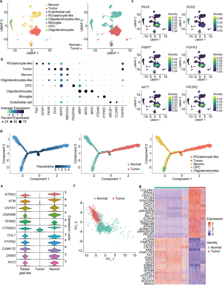
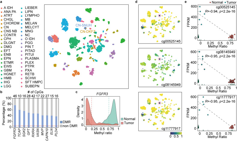
We explored the genomic events underlying central neurocytoma (CN), a rare neoplasm of the central nervous system, via multiomics approaches, including whole-exome sequencing, bulk and single-nuclei RNA sequencing, and methylation sequencing. We identified FGFR3 hypomethylation leading to FGFR3 overexpression as a major event in the ontogeny of CN that affects crucial downstream events, such as aberrant PI3K-AKT activity and neuronal development pathways. Furthermore, we found similarities between CN and radial glial cells based on analyses of gene markers and CN tumor cells and postulate that CN tumorigenesis is due to dysregulation of radial glial cell differentiation into neurons. Our data demonstrate the potential role of FGFR3 as one of the leading drivers of tumorigenesis in CN.


Conflict of interest statement

The authors declare no competing interests.

Figures

Fig. 1
Fig. 1. PI3K-AKT pathway activation in CN.
a Comparison of the tumor mutation burden ratio of CN with other cancers revealed a low score relative to that of other CNS tumors, such as GBM and LGG. b Pathway analysis of significantly differentially expressed genes in CN compared to that in normal brain samples revealed the PI3K-AKT1 pathway as one of the top upregulated pathways in CN. c Enrichment analysis of significantly overexpressed genes in CN compared to that in normal brain samples. d IHC validation of overexpressed PI3K-AKT pathway marker genes along with FGFR3 in CN (scale bar 100 µM). e Functional classification of the PI3K-AKT1 pathway downstream of GSK3-related genes showed that metabolism-, proliferation-, and survival-related genes were upregulated and that neuronal function-related genes were downregulated in CN. (Asterisks represent the following P values: *<0.05; **<0.01; ***<0.001; ****<0.0001).
Fig. 2
Fig. 2. CN shows a similar gene expression pattern to that of radial glial cells in the bulk RNA sequencing data.
a Upper schematic figure showing various stages of neural precursor cells in the normal brain and their progression. The arrow shows the expected paths of the cells during normal neural differentiation. Lower bar charts show the combined gene expression patterns of these different neural developmental cell marker genes in CN and normal brain samples. Significant overexpression of genes related to radial glial and intermediate progenitor cells was detected in CN (asterisks represent the following P values, *<0.05; **<0.01; ***<0.001; ****<0.0001). b Individual marker gene expression patterns showing significant overexpression of all marker genes representing radial glial cells and intermediate progenitor cells in CN compared to the normal brain (asterisks represent the following P values, *<0.05; **<0.01; ***<0.001; ****<0.0001). c IHC validation of radial glial and intermediate progenitor cell marker genes showing that only radial glial marker genes are upregulated in CN, confirming that radial glial cells are the cell type of origin (scale bar 100 µM).
Fig. 3
Fig. 3. Radial glial-like cells deviate from their normal developmental course and give rise to CN.
a snRNA-seq analysis of paired CN tumor and normal brain samples. b Cell-specific gene markers for each identified cell type are shown. FGFR3 can be detected in both tumor and radial glial cells. c Confirmation of overexpression of FGFR3, the PI3K-AKT1 pathway, and radial glial cell markers in tumor cell clusters consistent with the bulk RNA-seq analysis. d Pseudotime analysis of tumor cells, neurons, oligodendrocytes, and radial glial/astrocyte-like cells showing tumor cells positioned between neurons and radial glial/astrocyte-like cells. e Genes expressed in both neurons and radial glial/astrocyte-like cells but not in tumor cells. f PCA plot showing that despite being identified as the same cell type, radial glial/astrocyte-like cells from tumor and normal samples show some differences. g Heatmap of differentially expressed genes in normal and tumor sample radial glial/astrocyte-like cells.
Fig. 4
Fig. 4. FGFR3 hypomethylation in CN.
a t-SNE clustering based on methylation data showing that the CN samples from this study clustered exclusively with the public CNS data from the CN cluster, confirming accurate classification of the samples as CN. CN-SNUH represents the samples from the present study (n = 6), and CN-DFKZ represents the samples from comparison of public methylation data (n = 21). b The FGFR3 gene had the highest percentage among DMR/non-DMR ratios. c FGFR3 was hypomethylated compared to normal brain samples according to methylation expression data. d FGFR3-specific CpG probes (n = 3) showed significant hypomethylation compared to that in other CNS tumors and normal brain samples. The CN samples indicated by red circles contain 6 samples from the present study and 21 samples obtained from the public DFKZ CNS methylation database. e Correlation plot of methylation ratios with FPKM values showing a significant negative correlation for all three FGFR3-specific probes.
Fig. 5
Fig. 5. Schematic representation of the potential mechanism of CN tumorigenesis.
The middle left and upper panels show the normal course of radial glial cell differentiation into mature neurons. The middle right and lower panels show the potential course of CN tumorigenesis, whereby FGFR3 hypomethylation-driven FGFR3-overexpressing radial glial-like cells progress through the ventricle as a result of PIK3-AKT activation and altered neuronal functions such as differentiation and migration.

References

    1. Choudhari KA, et al. Central neurocytoma: a multi-disciplinary review. Br. J. Neurosurg. 2009;23:585–595. doi: 10.3109/02688690903254350. - DOI - PubMed
    1. Yang I, et al. Clinical manifestations of central neurocytoma. Neurosurg. Clin. N. Am. 2015;26:5–10. doi: 10.1016/j.nec.2014.09.011. - DOI - PubMed
    1. Lee SJ, et al. Central neurocytoma: a review of clinical management and histopathologic features. Brain Tumor Res. Treat. 2016;4:49–57. doi: 10.14791/btrt.2016.4.2.49. - DOI - PMC - PubMed
    1. Kim CY, Kim DG, Joo JD, Kim YH. Clinical outcome and quality of life after treatment of patients with central neurocytoma. Neurosurg. Clin. N. Am. 2015;26:83–90. doi: 10.1016/j.nec.2014.09.007. - DOI - PubMed
    1. Hassoun J, et al. Central neurocytoma. An electron-microscopic study of two cases. Acta Neuropathol. 1982;56:151–156. doi: 10.1007/BF00690587. - DOI - PubMed

Publication types

MeSH terms

Substances