

Rebound activation of 5-HT neurons following SSRI discontinuation
- PMID: 38609530
- PMCID: PMC11319583
- DOI: 10.1038/s41386-024-01857-8
Rebound activation of 5-HT neurons following SSRI discontinuation
Abstract
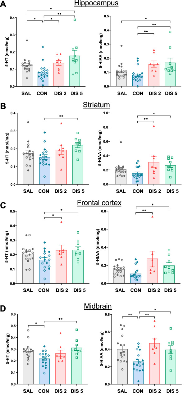
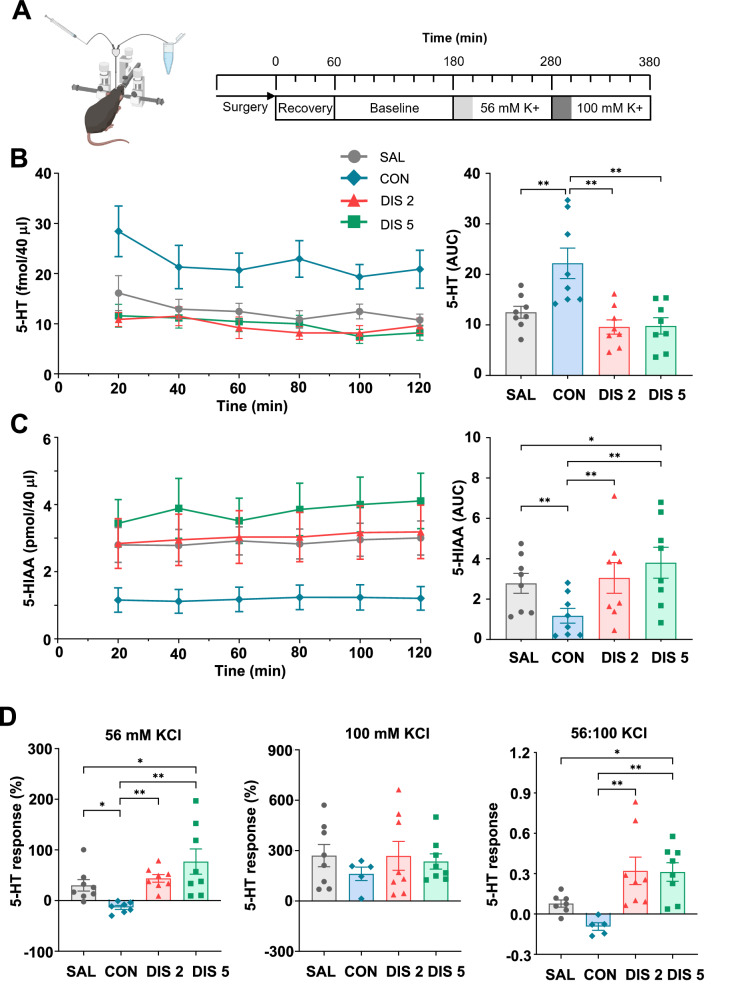
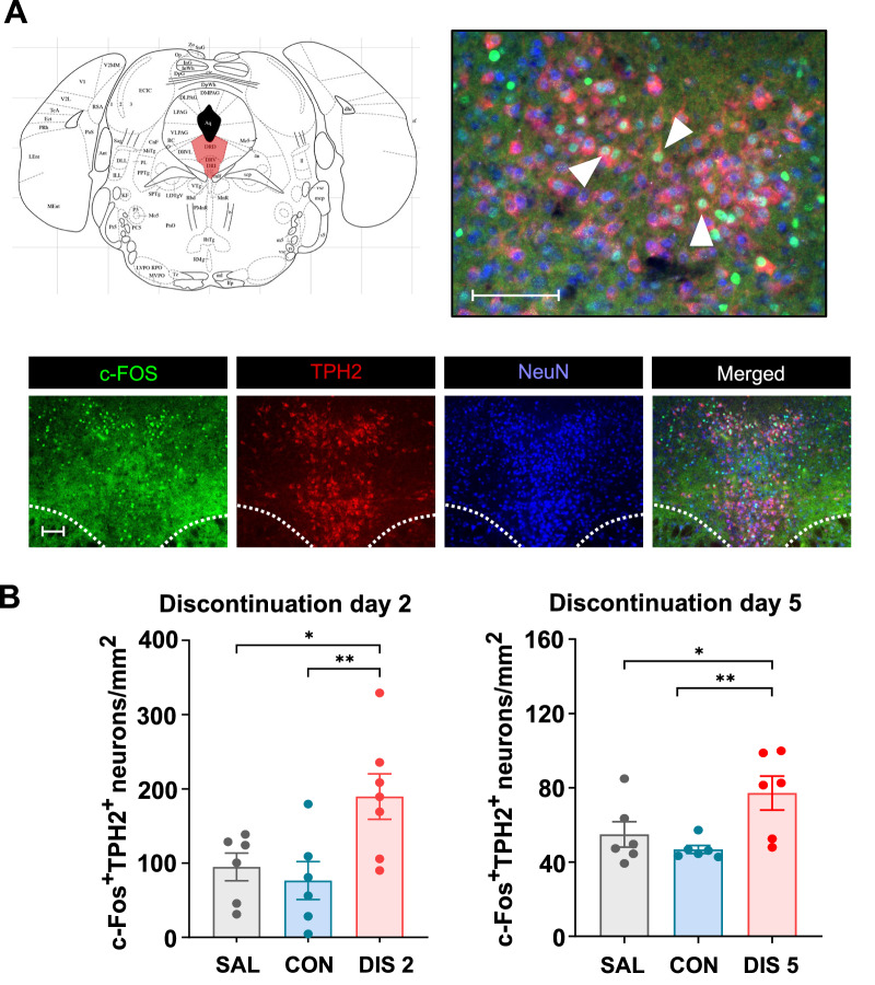
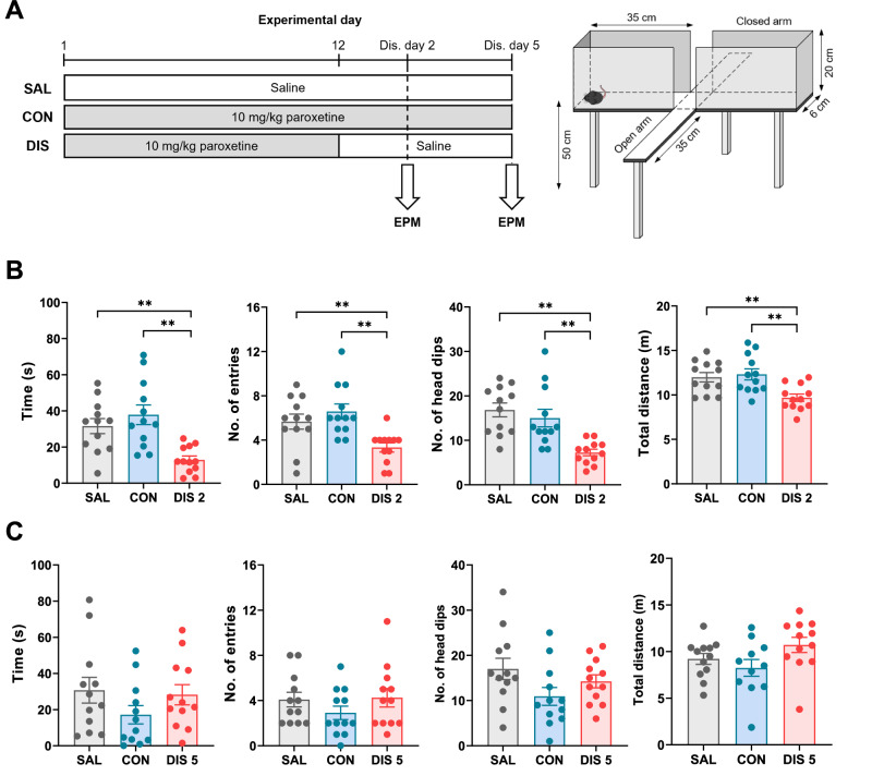
Cessation of therapy with a selective serotonin (5-HT) reuptake inhibitor (SSRI) is often associated with an early onset and disabling discontinuation syndrome, the mechanism of which is surprisingly little investigated. Here we determined the effect on 5-HT neurochemistry of discontinuation from the SSRI paroxetine. Paroxetine was administered repeatedly to mice (once daily, 12 days versus saline controls) and then either continued or discontinued for up to 5 days. Whereas brain tissue levels of 5-HT and/or its metabolite 5-HIAA tended to decrease during continuous paroxetine, levels increased above controls after discontinuation, notably in hippocampus. In microdialysis experiments continuous paroxetine elevated hippocampal extracellular 5-HT and this effect fell to saline control levels on discontinuation. However, depolarisation (high potassium)-evoked 5-HT release was reduced by continuous paroxetine but increased above controls post-discontinuation. Extracellular hippocampal 5-HIAA also decreased during continuous paroxetine and increased above controls post-discontinuation. Next, immunohistochemistry experiments found that paroxetine discontinuation increased c-Fos expression in midbrain 5-HT (TPH2 positive) neurons, adding further evidence for a hyperexcitable 5-HT system. The latter effect was recapitulated by 5-HT1A receptor antagonist administration although gene expression analysis could not confirm altered expression of 5-HT1A autoreceptors following paroxetine discontinuation. Finally, in behavioural experiments paroxetine discontinuation increased anxiety-like behaviour, which partially correlated in time with the measures of increased 5-HT function. In summary, this study reports evidence that, across a range of experiments, SSRI discontinuation triggers a rebound activation of 5-HT neurons. This effect is reminiscent of neural changes associated with various psychotropic drug withdrawal states, suggesting a common unifying mechanism.
© 2024. The Author(s).
Conflict of interest statement
The authors declare no competing interests.
Figures
Similar articles
-
Increased c-Fos immunoreactivity in anxiety-related brain regions following paroxetine discontinuation.Neuropharmacology. 2025 Nov 1;278:110541. doi: 10.1016/j.neuropharm.2025.110541. Epub 2025 Jun 2. Neuropharmacology. 2025. PMID: 40466935
-
Somatodendritic action of pindolol to attenuate the paroxetine-induced decrease in serotonin release from the rat ventral hippocampus: a microdialysis study.Naunyn Schmiedebergs Arch Pharmacol. 2002 May;365(5):378-87. doi: 10.1007/s00210-002-0530-5. Epub 2002 Mar 14. Naunyn Schmiedebergs Arch Pharmacol. 2002. PMID: 12012024
-
Effects of the 5-HT(4) receptor agonist RS67333 and paroxetine on hippocampal extracellular 5-HT levels.Neurosci Lett. 2010 May 31;476(2):58-61. doi: 10.1016/j.neulet.2010.04.002. Epub 2010 Apr 8. Neurosci Lett. 2010. PMID: 20381585
-
Behavioral and serotonergic consequences of decreasing or increasing hippocampus brain-derived neurotrophic factor protein levels in mice.Neuropharmacology. 2008 Nov;55(6):1006-14. doi: 10.1016/j.neuropharm.2008.08.001. Epub 2008 Aug 12. Neuropharmacology. 2008. PMID: 18761360 Review.
-
Mechanisms of SSRI Therapy and Discontinuation.Curr Top Behav Neurosci. 2024;66:21-47. doi: 10.1007/7854_2023_452. Curr Top Behav Neurosci. 2024. PMID: 37955823 Review.
Cited by
-
Selective serotonin reuptake inhibitors induce cardiac toxicity through dysfunction of mitochondria and sarcomeres.Commun Biol. 2025 May 12;8(1):736. doi: 10.1038/s42003-025-08168-8. Commun Biol. 2025. PMID: 40355528 Free PMC article.
-
Deprescribing Antidepressants in Children and Adolescents: A Systematic Review of Discontinuation Approaches, Cross-Titration, and Withdrawal Symptoms.J Child Adolesc Psychopharmacol. 2025 Feb;35(1):3-22. doi: 10.1089/cap.2024.0099. Epub 2024 Oct 29. J Child Adolesc Psychopharmacol. 2025. PMID: 39469761
-
Reduction of Prostate Cancer Risk: Role of Frequent Ejaculation-Associated Mechanisms.Cancers (Basel). 2025 Feb 28;17(5):843. doi: 10.3390/cancers17050843. Cancers (Basel). 2025. PMID: 40075690 Free PMC article. Review.
References
-
- Haddad P. Newer antidepressants and the discontinuation syndrome. J Clin Psychiatry. 1997;58:17–21. - PubMed
-
- Gastaldon C, Schoretsanitis G, Arzenton E, Raschi E, Papola D, Ostuzzi G, et al. Withdrawal Syndrome Following Discontinuation of 28 Antidepressants: Pharmacovigilance Analysis of 31,688 Reports from the WHO Spontaneous Reporting Database. Drug Safety. 2022;45:1539–49. 10.1007/s40264-022-01246-4 - DOI - PMC - PubMed
MeSH terms
Substances
Grants and funding
LinkOut - more resources
Full Text Sources
