. 2024 Apr 12;15(1):107.
doi: 10.1186/s13244-024-01670-3.

O-RADS MRI scoring system: key points for correct application in inexperienced hands

Affiliations

O-RADS MRI scoring system: key points for correct application in inexperienced hands

Lledó Cabedo et al. Insights Imaging. .

Abstract

Objectives: To evaluate the efficacy of the O-RADS MRI criteria in the stratification of risk of malignancy of solid or sonographically indeterminate ovarian masses and assess the interobserver agreement of this classification between experienced and inexperienced radiologists.

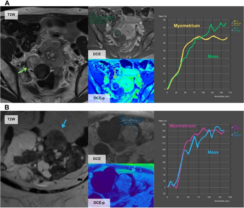
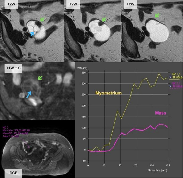
Methods: This single-centre retrospective study included patients from 2019 to 2022 with sonographically indeterminate or solid ovarian masses who underwent MRI with a specific protocol for characterisation according to O-RADS MRI specifications. Each study was evaluated using O-RADS lexicon by two radiologists, one with 17 years of experience in gynaecological radiology and another with 4 years of experience in general radiology. Findings were classified as benign, borderline, or malignant according to histology or stability over time. Diagnostic performance and interobserver agreement were assessed.

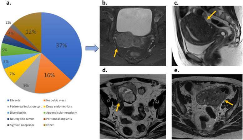
Results: A total of 183 patients with US indeterminate or solid adnexal masses were included. Fifty-seven (31%) did not have ovarian masses, classified as O-RADS 1. The diagnostic performance for scores 2-5 was excellent with a sensitivity, specificity, PPV, and NPV of 97.4%, 100%, 96.2%, and 100%, respectively by the experienced radiologist and 96.1%, 92.0%, 93.9%, and 94.8% by the inexperienced radiologist. Interobserver concordance was very high (Kappa index 0.92). Almost all the misclassified cases were due to misinterpretation of the classification similar to reports in the literature.

Conclusion: The diagnostic performance of O-RADS MRI determined by either experienced or inexperienced radiologists is excellent, facilitating decision-making with high diagnostic accuracy and high reproducibility. Knowledge of this classification and use of assessment tools could avoid frequent errors due to misinterpretation.

Critical relevance statement: Up to 31% of ovarian masses are considered indeterminate by transvaginal US and 32% of solid lesions considered malignant by transvaginal US are benign. The O-RADs MRI accurately classifies these masses, even when used by inexperienced radiologists, thereby avoiding incorrect surgical approaches.

Key points: • O-RADS MRI accurately classifies indeterminate and solid ovarian masses by ultrasound. • There is excellent interobserver agreement between experienced and non-experienced radiologists. • O-RADS MRI is a helpful tool to assess clinical decision-making in ovarian tumours.

Keywords: Cancer; MRI; Ovary.


Conflict of interest statement

The authors declare that they have no competing interests.

Figures

Fig. 1
Fig. 1
Population flowchart. US, ultrasound. Note that in 25 patients two ovarian masses were found; in these cases, only the mass with the greatest O-RAD score was included. In the 57 patients with O-RADS 1, 40 were benign, 8 malignant, and in 9, no mass was found. Borderline and malignant were considered malignant for statistical purposes
Fig. 2
Fig. 2
Distribution of non-adnexal masses and examples. a O-RADS score 1 distribution. b Parametrial fibroid (arrow). c Uterine adenomyosis (arrow). d Appendiceal gastrointestinal stromal tumour (GIST) (arrow) e Sigmoid adenocarcinoma (arrow)
Fig. 3
Fig. 3
Erroneous classifications by the JR and the SR. A A 61-year-old woman presented an incidental right ovarian mass (green arrow) with a high-risk TIC classified as score 5 by both readers but was finally a fibrothecoma. As specified by O-RADS MRI guidelines, unenhanced sequences should be acquired before the contrast bolus injection. However, in this case, there was an error in the acquisition as there were no unenhanced sequences before the injection of the contrast bolus (in both TIC the contrast uptake started at the second 0), and this can distort the TIC results and lead to misinterpretation. In clinical practice, cases like this should be considered O-RADs 0 (incomplete or erroneous MRI technique). B A 56-year-old woman presented an incidental left ovarian mass (blue arrow) with a high-risk TIC that was classified as score 5 by both readers but was finally a luteinised fibrothecoma
Fig. 4
Fig. 4
Errors by the JR due to misinterpretation of the classification. A A 31-year-old woman presented a right ovarian mass with macroscopic fat content (blue arrow) and a high amount of solid-enhancing tissue (T1W FS + C series). The mass does not have a Rokitansky nodule. It was classified as score 2 by the JR but the histological result showed an immature teratoma. Teratomas do not fit the classification well as they can have low, intermediate or high-risk TIC. In this case, the mass had an intermediate TIC. It is difficult to distinguish mature from immature teratoma and thus, it is stipulated that if they present a high amount of solid tissue, they should be classified with a score of 4. B A 79-year-old woman with a left ovarian mass (yellow arrow) with solid hyperenhancing tissue (T1W FS + C series). The TIC was interpreted as low risk by the JR and the mass was misclassified as score 3. Pathological analysis after surgery confirmed that it was a clear cell carcinoma arising from a cystadenoma. In this case, the TIC was an intermediate-risk curve as it had an initial increase lower than the myometrium, followed by a plateau
Fig. 5
Fig. 5
Errors by the JR, paradigmatic examples. A A 33-year-old woman with bilateral adnexal masses with a tree-like morphology (green arrow) and low contrast uptake shown by the TIC. This case was classified as score 3 by the JR as it was considered that it had a low-risk TIC. Pathological analysis after surgery confirmed that it was a borderline serous tumour. B A 67-year-old woman with a left ovarian mass that shows hyperintense T2w content and multiple thin septa (yellow arrow) typical of mucinous tumours. This mass was misclassified as score 3 by the JR as it was interpreted as having a low-risk TIC. The histological results showed metastasis of a mucinous appendix tumour
Fig. 6
Fig. 6
A 74-year-old woman with a right ovarian solid-cystic mass. This case was misclassified as score 4 by the JR considering it as having an intermediate-risk TIC. The postoperative pathological analysis revealed that it was a mucinous cystadenoma mixed with a Brenner tumour. In this case, the JR calculated the TIC out of the ovarian parenchyma surrounding the lesion (green arrow), but the true solid component was the thin septa (blue arrow) that corresponded to a score 3 as the SR perceived. As in this case, false positives may be due to errors performing the TIC

References

    1. Woo YL, Kyrgiou M, Bryant A, Everett T, Dickinson HO. Centralisation of services for gynaecological cancers: a Cochrane systematic review. Gynecol Oncol. 2012;126(2):286–290. doi: 10.1016/j.ygyno.2012.04.012. - DOI - PubMed
    1. Dodge JE, Covens AL, Lacchetti C, et al. (2012) Management of a suspicious adnexal mass: a clinical practice guideline. Curr Oncol. ;19(4). 10.3747/co.19.980 - PMC - PubMed
    1. Siegel R, Naishadham D, Jemal A. (2013) Cancer statistics. CA Cancer J Clin. 2013;63(1):11–30. doi: 10.3322/caac.21166. - DOI - PubMed
    1. Sadowski EA, Paroder V, Patel-Lippmann K, et al. Indeterminate adnexal cysts at US: prevalence and characteristics of ovarian cancer. Radiology. 2018;287(3):1041–1049. doi: 10.1148/radiol.2018172271. - DOI - PubMed
    1. Meys EMJ, Kaijser J, Kruitwagen RFPM, et al. Subjective assessment versus ultrasound models to diagnose ovarian cancer: a systematic review and meta-analysis. Eur J Cancer. 2016;58:17–29. doi: 10.1016/J.EJCA.2016.01.007. - DOI - PubMed

LinkOut - more resources