. 2024 Apr 12;23(1):107.
doi: 10.1186/s12934-024-02369-2.

Deciphering antifungal and antibiofilm mechanisms of isobavachalcone against Cryptococcus neoformans through RNA-seq and functional analyses

Affiliations

Deciphering antifungal and antibiofilm mechanisms of isobavachalcone against Cryptococcus neoformans through RNA-seq and functional analyses

Weidong Qian et al. Microb Cell Fact. .

Abstract

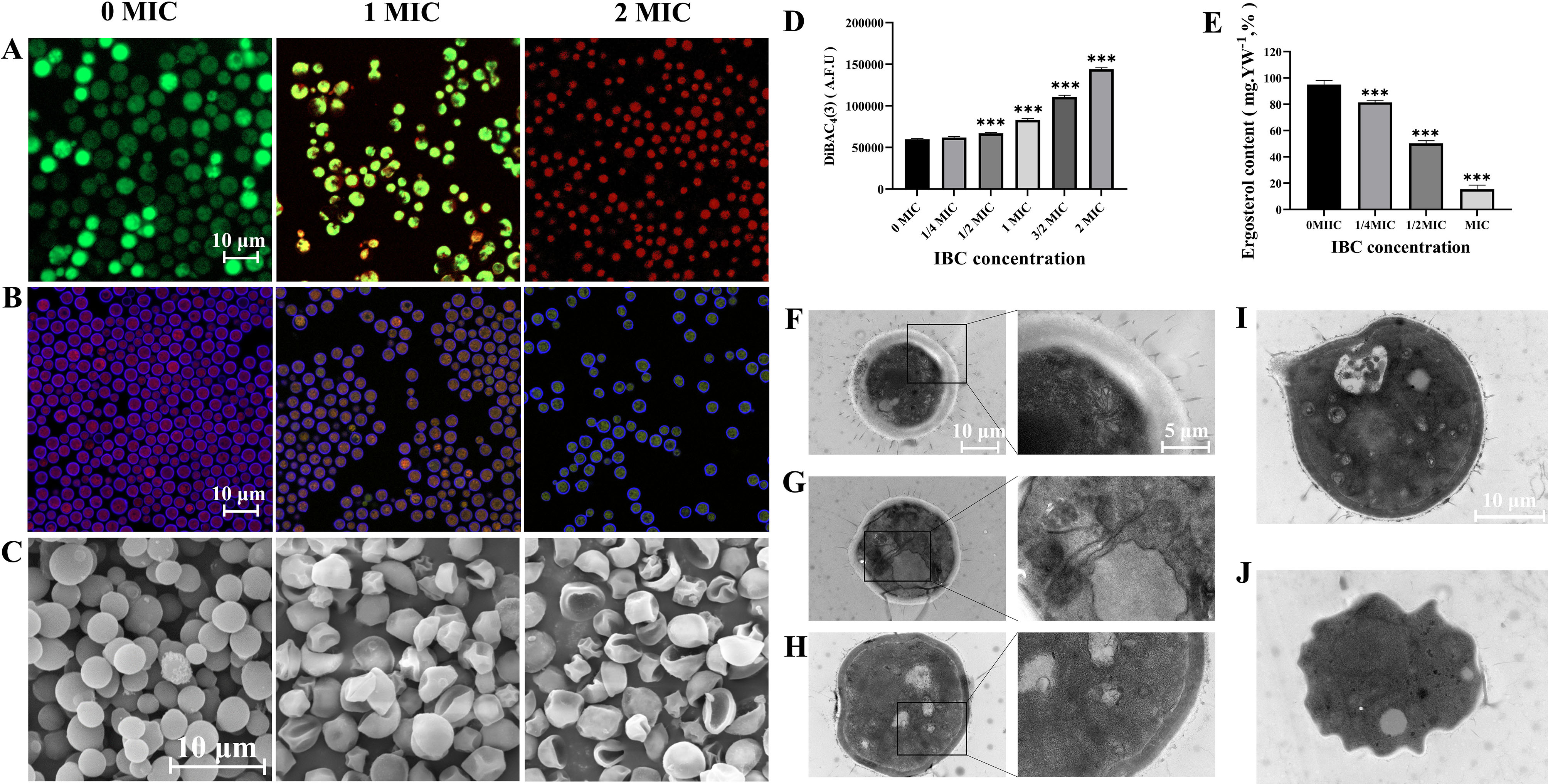
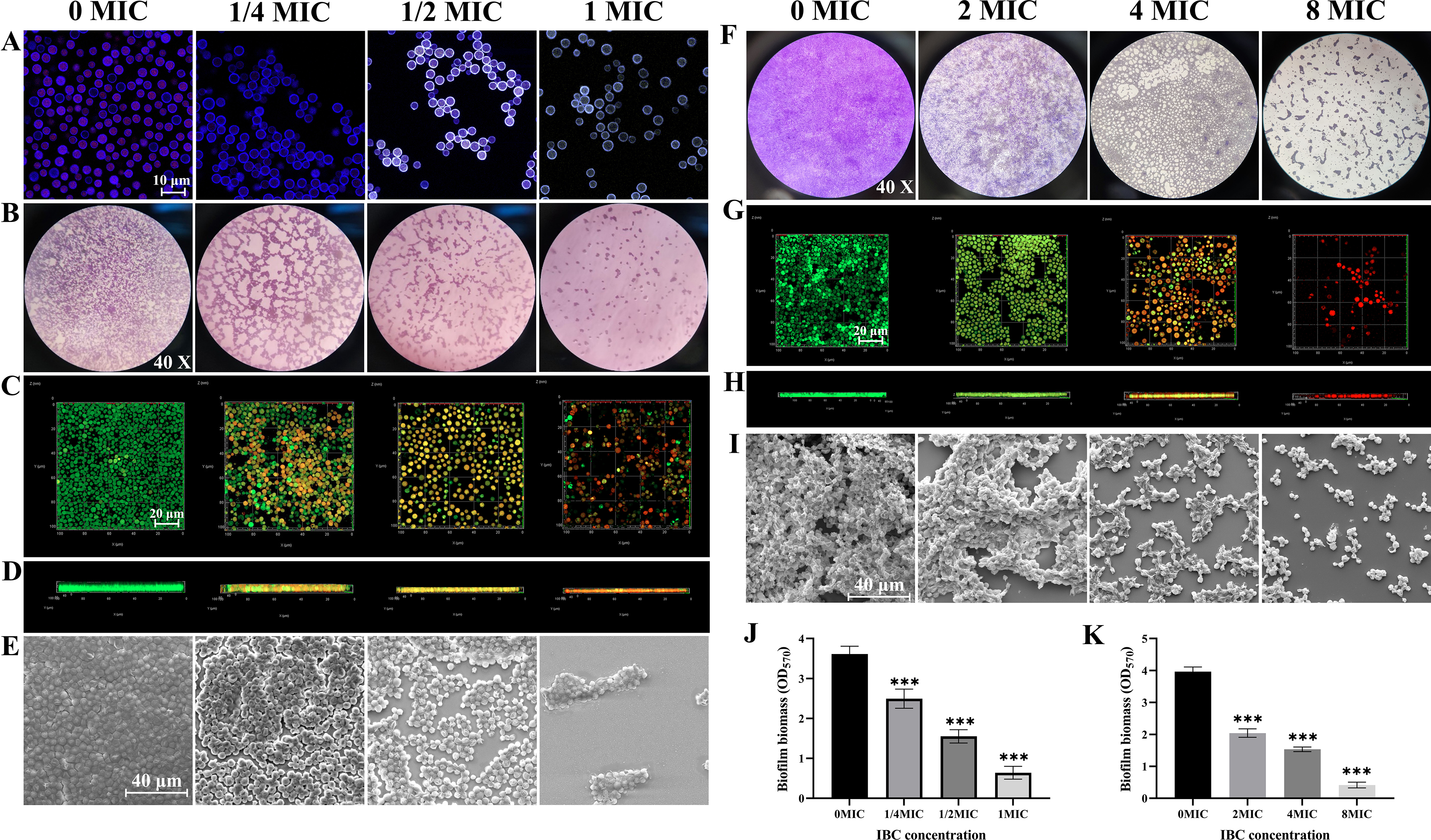
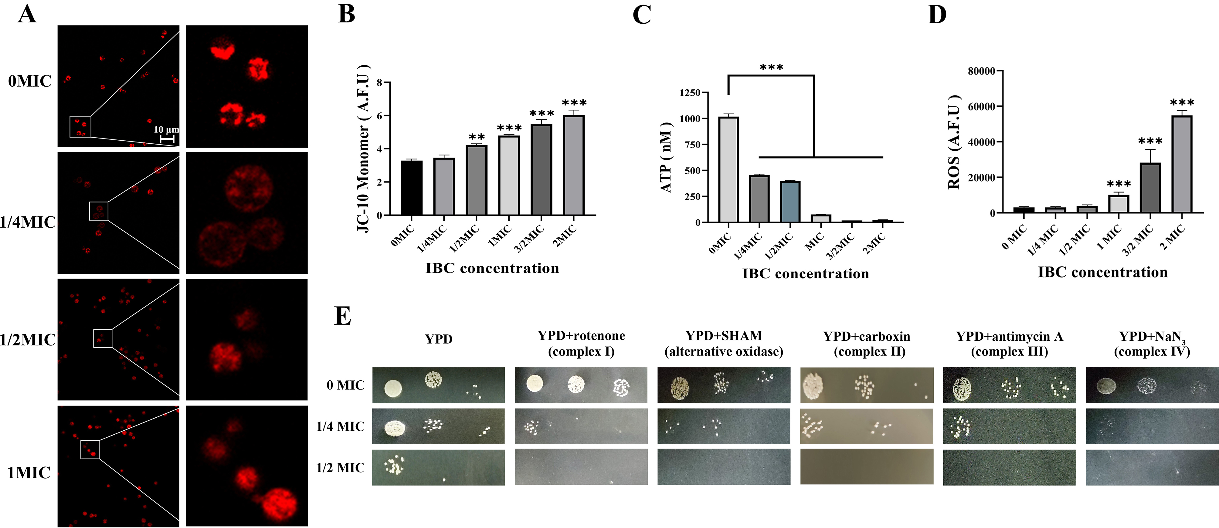
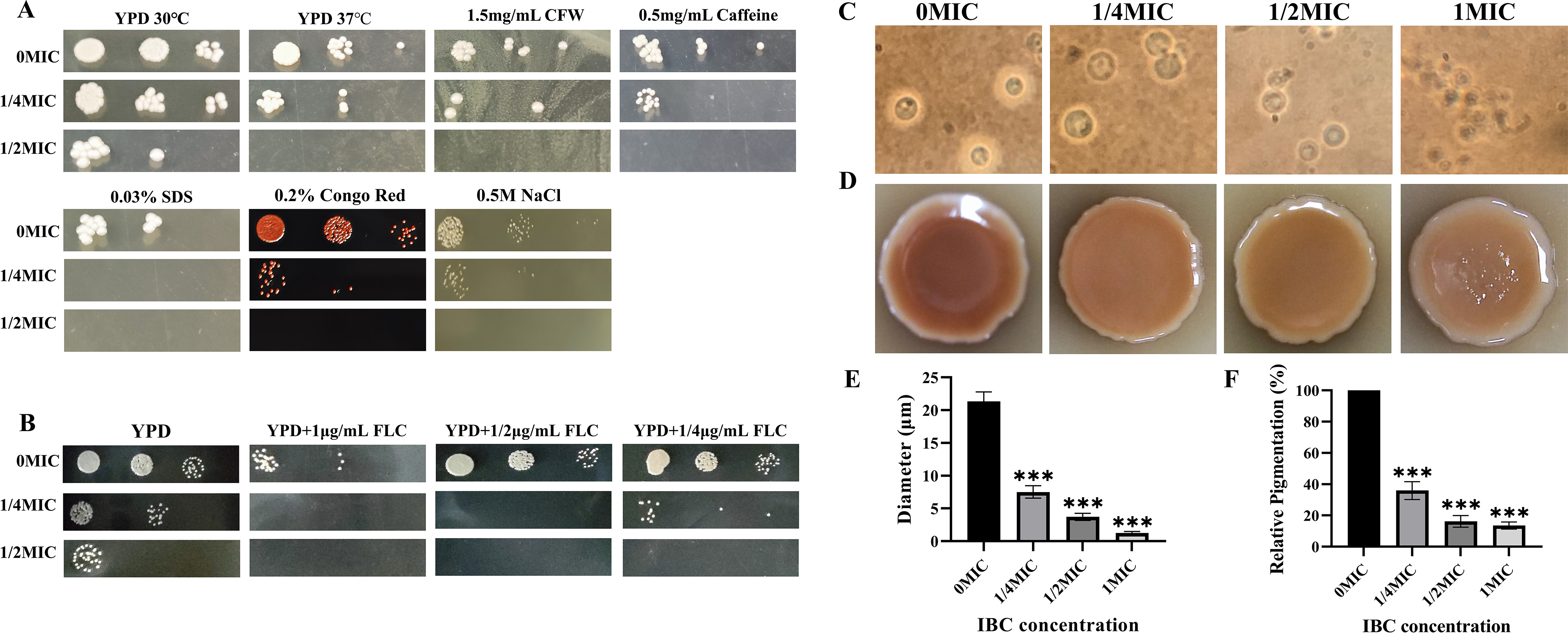
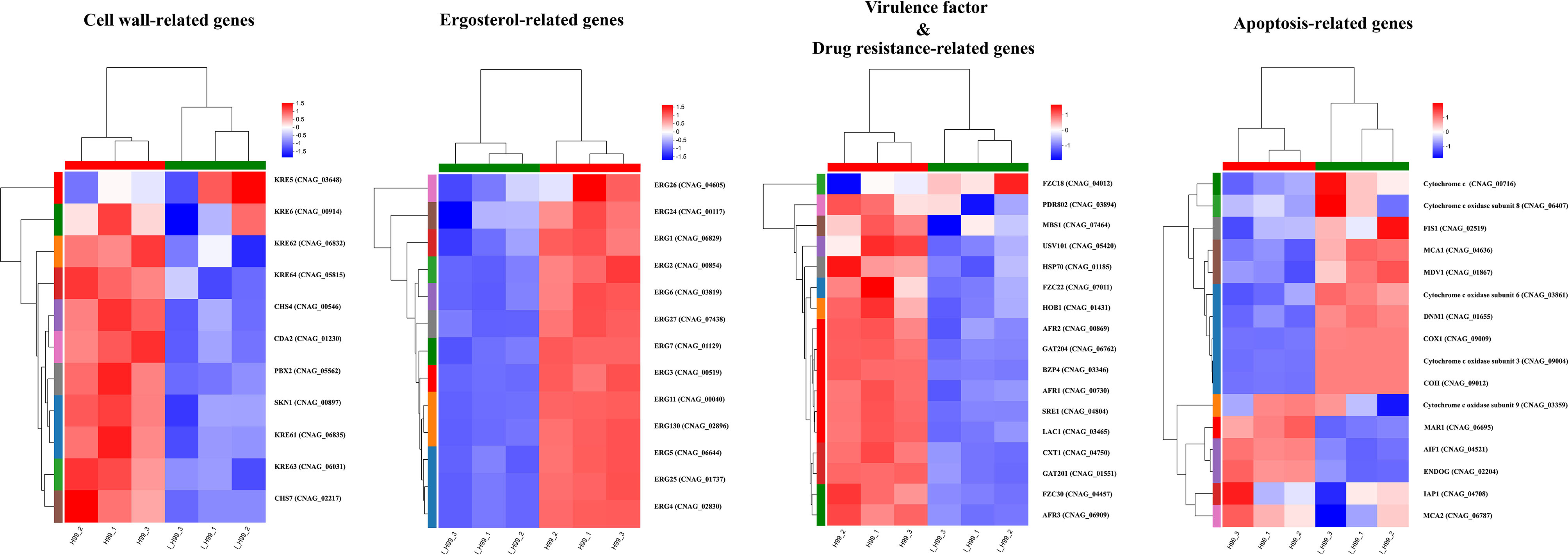
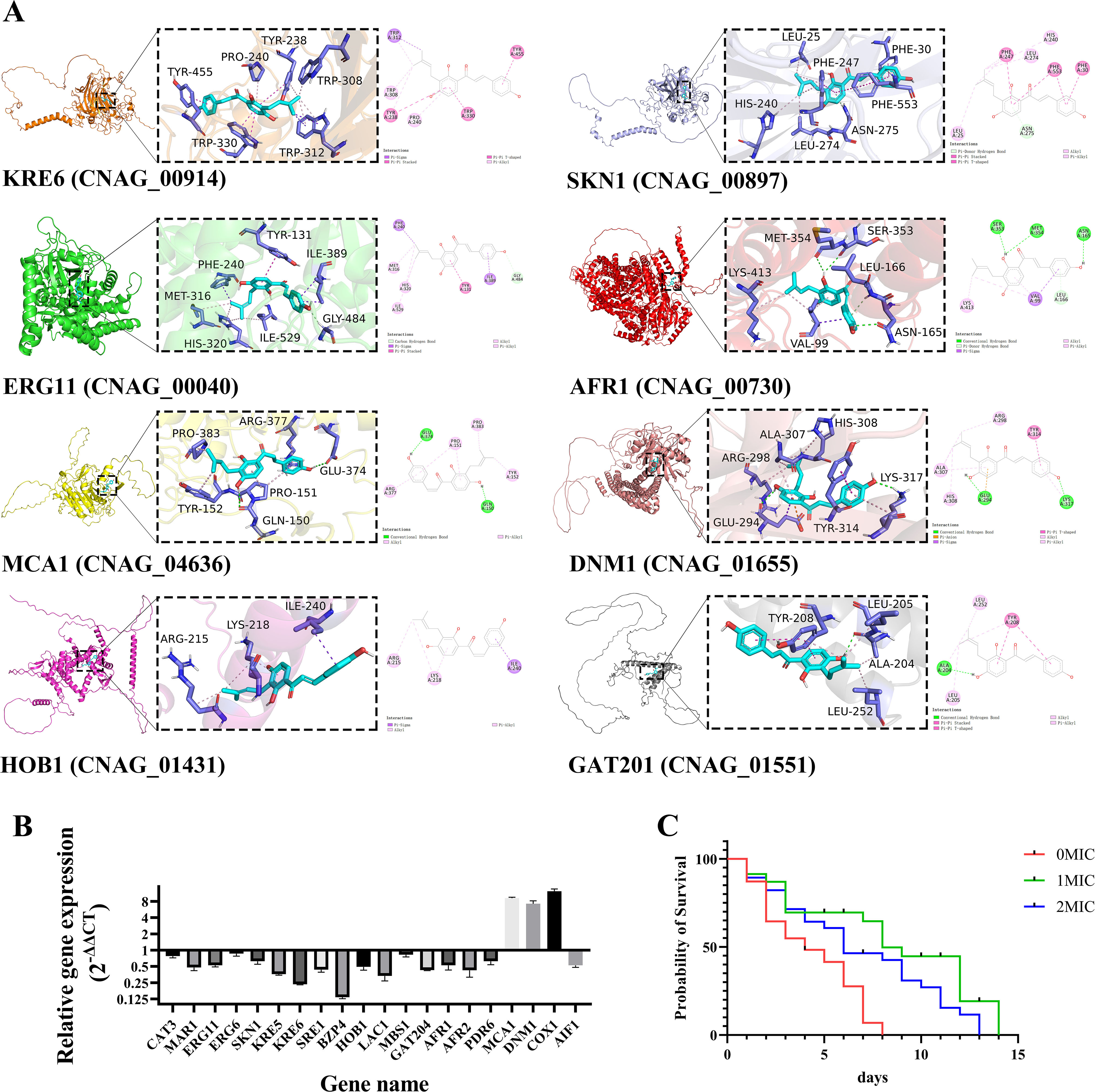
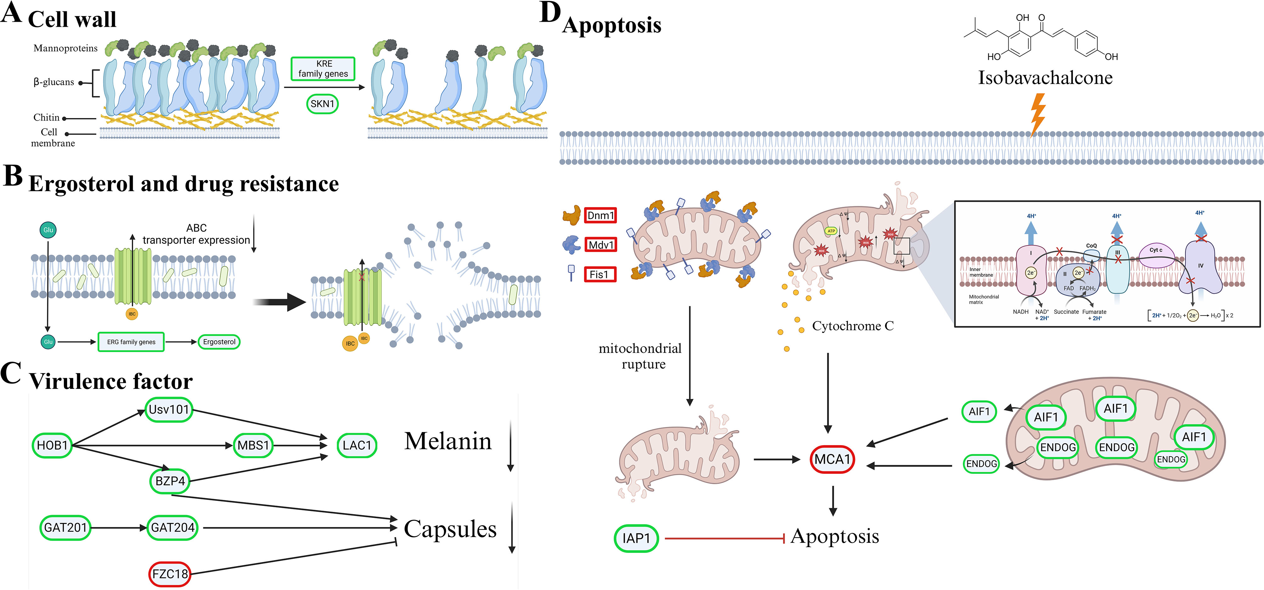
Cryptococcus neoformans has been designated as critical fungal pathogens by the World Health Organization, mainly due to limited treatment options and the prevalence of antifungal resistance. Consequently, the utilization of novel antifungal agents is crucial for the effective treatment of C. neoformans infections. This study exposed that the minimum inhibitory concentration (MIC) of isobavachalcone (IBC) against C. neoformans H99 was 8 µg/mL, and IBC dispersed 48-h mature biofilms by affecting cell viability at 16 µg/mL. The antifungal efficacy of IBC was further validated through microscopic observations using specific dyes and in vitro assays, which confirmed the disruption of cell wall/membrane integrity. RNA-Seq analysis was employed to decipher the effect of IBC on the C. neoformans H99 transcriptomic profiles. Real-time quantitative reverse transcription PCR (RT-qPCR) analysis was performed to validate the transcriptomic data and identify the differentially expressed genes. The results showed that IBC exhibited various mechanisms to impede the growth, biofilm formation, and virulence of C. neoformans H99 by modulating multiple dysregulated pathways related to cell wall/membrane, drug resistance, apoptosis, and mitochondrial homeostasis. The transcriptomic findings were corroborated by the antioxidant analyses, antifungal drug sensitivity, molecular docking, capsule, and melanin assays. In vivo antifungal activity analysis demonstrated that IBC extended the lifespan of C. neoformans-infected Caenorhabditis elegans. Overall, the current study unveiled that IBC targeted multiple pathways simultaneously to inhibit growth significantly, biofilm formation, and virulence, as well as to disperse mature biofilms of C. neoformans H99 and induce cell death.

Keywords: Cryptococcus neoformans; Antifungal activity; Functional profiling; Isobavachalcone; RNA-sequencing.


Conflict of interest statement

The authors declare that the research was performed in the absence of any commercial or financial relationships that can be considered as a potential conflict of interest.

Figures

Fig. 1
Fig. 1
IBC affected the cell membrane integrity (A), metabolic activity (B) and cell morphology (C) of C. neoformans. CLSM images of C. neoformans cells in the presence of various concentrations of IBC (0 MIC, 1 MIC and 2 MIC) were obtained using SYTO9/PI (A), and FUN® 1/CWS (B) double dyes, respectively. Furthermore, C. neoformans treated with various concentrations of IBC at 30 °C for 6 h was investigated using FESEM. The scale bar for CLSM and FESEM images was 10 μm. IBC altered cytoplasmic membrane potential (D), decreased the ergosterol content (E) and disrupted the fungal ultrastructure (F-J) in C. neoformans. Quantitative analysis of cytoplasmic membrane potential and ergosterol contents was performed in IBC-treated C. neoformans cells. In addition, cells were viewed under a TEM microscope, in which fungal cells were incubated in the absence (F, G) and presence (H-J) of IBC at 1 MIC for 24 h. Scale bar was 1 μm for the TEM images, and 500 nm for the extended images. Bars represent the standard deviation (n = 3). *p < 0.05; **p < 0.01; ***p < 0.001
Fig. 2
Fig. 2
IBC showed the inhibitory effect on C. neoformans biofilm formation by inhibiting initial attachment and maturation. (A) The anti-adhesion ability of IBC to the glass surface was examined using FUN® 1/CWS double dyes combined with CLSM. (B-E) The inhibitory effect of IBC on the biofilm formation by C. neoformans was performed using CV staining (B, objective, × 40), CLSM (C, plane images; D, three-dimensional images) and FESEM (E), respectively. Scale bar was 10 μm for CLSM and 40 μm for FESEM, respectively. IBC exhibited the dispersal effect on mature biofilms by C. neoformans. Examination of the ability of high concentrations of IBC to disperse/kill 48-h mature biofilms by C. neoformans was carried out. The dispersal/killing effect of IBC on 48-h mature biofilms was evaluated using CV staining (F, objective, × 40), CLSM (G, plane images; H, three-dimensional images) and FESEM (I), respectively. (J-K) The inhibitory and dispersal effect of IBC on biofilm formation and preformed biofilms were quantitatively assessed using CV staining, respectively. Scale bar was 10 μm for CLSM and 40 μm for FESEM, respectively. Bars represent the standard deviation (n = 3). *p < 0.05; **p < 0.01; ***p < 0.001
Fig. 3
Fig. 3
IBC altered mitochondrial morphologies and induced mitochondrial dysfunction of C. neoformans. (A) Mitochondrial morphologies of C. neoformans were assessed following incubation in YPD medium supplemented with various concentrations of IBC at 30 °C. MitoTracker Red CMXRos was used to stain diffuse, tubular, or fragmented morphologies, and mitochondria were then imaged using CLSM. The right images are the enlarged image of the white square on the left images. (B) MMP of C. neoformans was measured following incubation in YPD medium supplemented with various concentrations of IBC at 30 °C. Mitochondria were stained with JC-10 marker of MMP. The fluorescence microplate reader was used to quantify staining intensity. Experiment was repeated three times to ensure reproducibility. (C) Total intracellular ATP was measured for the C. neoformans following incubation in YPD medium supplemented with various concentrations of IBC at 30 °C. Normalized total cell lysates were determined for total intracellular ATP using a firefly luciferase bioluminescent assay. (D) Total intracellular ROS was measured for C. neoformans following incubation in the absence and presence of various concentrations of IBC. Cells were stained with DCFH-DA as a marker for intracellular ROS. The fluorescence microplate reader was used to quantify staining intensity. (E) IBC affects the electron transport chain function. Growth phenotypes of C. neoformans were assessed in the presence of electron transport chain inhibitors. Cells were normalized by OD600 and subsequently cultured on YPD medium agar plates containing rotenone (0.5 mg/mL), salicylhydroxamic acid (SHAM) (2.5 mM), carboxin (50 µg/mL), antimycin A (3 µg/mL), and sodium azide (NaN3) (0.5 mM) at 30 °C, respectively. Plates were imaged daily. Experiment was repeated two times to ensure reproducibility and representative images are shown
Fig. 4
Fig. 4
IBC improved the susceptibility of C. neoformans to cell stressors and fluconazole and decreased virulence factor production. (A) Growth phenotypes of C. neoformans were assessed in the presence of various cell stressors and different concentrations of IBC. Cells were normalized by OD600 and subsequently diluted. 5 µL aliquots of serial dilutions were subsequently spotted on YPD agar medium supplemented with calcofluor white (1.5 mg/mL), caffeine (0.5 mg/mL), and NaCl (0.5 M), 0.03% SDS and 0.2% Congo red in the presence of various concentrations of IBC at 30 °C, respectively. For temperature stress, the YPD plates were incubated at 37 °C in the presence of various concentrations of IBC. Experiment was repeated three times to ensure reproducibility and representative images are shown. (B) Fluconazole susceptibility was assessed for the IBC-treated C. neoformans cells. Cells were normalized by OD600 and were subsequently grown on YPD medium agar plates in the presence of fluconazole and different concentrations of IBC. Cells were incubated at 30 °C and plates were imaged at the indicated times. (C, E) Representative images of various concentrations of IBC-treated C. neoformans grown in Dulbecco’s Modified Eagle’s Medium (DMEM) for 48 h at 37 °C. Capsules were visualized by India ink staining and examined under a microscope, and the capsule diameter was evaluated. (D, F) C. neoformans cells were grown on L-DOPA agar plates for 72 h at 30 °C with or without the addition of IBC. Reduced amounts of melanin formation in the colonies were observed in a concentration-dependent manner
Fig. 5
Fig. 5
Verification of apoptotic and necrotic cell death using standard methods. CLSM analysis of cells were stained using Annexin V/PI dyes after exposure to different concentrations of IBC.
Fig. 6
Fig. 6
DEGs analysis of untreated and IBC-treated C. neoformans cells using RNA-seq analysis. RNA-seq analysis was performed on total RNA extracted from three replicate samples for each biological group. (A) GO enrichment analysis of DEGs was performed to identify the most significantly enriched pathways at a threshold Padjust of ≤ 0.05. (B) Scatter plot of KEGG enrichment of DEGs. The abscissa Rich factor indicates the number of DEGs located in the KEGG/the total number of genes located in the KEGG metabolic pathway
Fig. 7
Fig. 7
Gene set enrichment analysis plot depicting the enrichment of DEGs in IBC-treated and untreated C. neoformans cells. The expression pattern of candidate DEGs related to cell wall synthesis in IBC-treated C. neoformans cells. The expression pattern of candidate DEGs related to the ergosterol synthesis in IBC-treated C. neoformans cells. The expression pattern of candidate DEGs pertaining to drug efflux in IBC-treated C. neoformans cells. The expression pattern of candidate DEGs involved in the maintenance of mitochondrial homeostasis in IBC-treated C. neoformans cells. The expression pattern of candidate DEGs involved in the melanin and capsule biosynthesis in IBC-treated C. neoformans cells
Fig. 8
Fig. 8
(A) Detailed diagram of molecular docking between IBC and target proteins, including KRE6, SKN1, ERG11, AFR1, MCA1, DNM1, HOB1 and GAT201. The 2D and 3D intermolecular contact between IBC and target proteins. Chemical structures were drawn by ChemDraw Pro 16.0 Suite (PerkinElmer, USA) and analyzed by the Discovery studio visualizer. IBC was represented in light blue, and local magnification of the docking sites and 2D diagram of the interactions were shown. (B) RT-qPCR assay for representative DEGs to validate the result of functional enrichment analysis. Data in the bar graphs are presented as the means and standard deviations of three independent experiments. (C) Effect of IBC on the lifespan of C. neoformans-infected C. elegans nematodes was evaluated, and the survival curve was plotted. Survival of C. elegans was evaluated in different treatment groups. Values represented the mean survival rates of three biological replicates
Fig. 9
Fig. 9
The mechanism diagram of IBC-induced cell death mechanism in C. neoformans cells. (A) IBC exerted antifungal effects through impeding the cell wall biosynthesis by downregulating expression of most of key genes within the KRE family such as Kre5, Kre6, and Skn1 gene. (B) IBC treatment caused damage to cell membrane-mediated C. neoformans cell death by downregulating the expression of most of ergosterol-related (ERG) genes including Erg3, Erg6, and Erg11, and the ABC transporter gene such as Afr1 and Afr2, thereby disrupting the cell membrane integrity and reversing drug resistance. (C) IBC exposure decreased the melanin and capsule synthesis by downregulating Hob1, Usv101, and Lac1 and Gat201. (D) IBC treatment disrupted mitochondrial homeostasis by upregulated the expression of Dnm1, Mdv1 and Fis1 and Mca1 genes, and downregulated the expression of Aif1, which resulted in the mitochondrial-mediated apoptosis of C. neoformans. Genes circled in red and green are significantly upregulated and downregulated, respectively

Similar articles

Cited by

References

    1. Fisher MC, Alastruey-Izquierdo A, Berman J, Bicanic T, Bignell EM, Bowyer P, et al. Tackling the emerging threat of antifungal resistance to human health. Nat Rev Microbiol. 2022;20(9):557–71. doi: 10.1038/s41579-022-00720-1. - DOI - PMC - PubMed
    1. Janbon G, Quintin J, Lanternier F, d’Enfert C. Studying fungal pathogens of humans and fungal infections: fungal diversity and diversity of approaches. Genes Immun. 2019;20(5):403–14. doi: 10.1038/s41435-019-0071-2. - DOI - PubMed
    1. Lee Y, Puumala E, Robbins N, Cowen LE. Antifungal drug resistance: molecular mechanisms in Candida albicans and beyond. Chem Rev. 2021;121(6):3390–411. doi: 10.1021/acs.chemrev.0c00199. - DOI - PMC - PubMed
    1. Cruz R, Wuest WM. Beyond ergosterol: strategies for combatting antifungal resistance in aspergillus fumigatus and Candida Auris. Tetrahedron. 2023;133:133268. doi: 10.1016/j.tet.2023.133268. - DOI - PMC - PubMed
    1. Hu C, Zhou M, Wang W, Sun X, Yarden O, Li S. Abnormal ergosterol biosynthesis activates transcriptional responses to antifungal azoles. Front Microbiol. 2018;9:9. doi: 10.3389/fmicb.2018.00009. - DOI - PMC - PubMed

LinkOut - more resources