. 2024 Apr 12;22(1):154.
doi: 10.1186/s12916-024-03375-2.

PREX2 contributes to radiation resistance by inhibiting radiotherapy-induced tumor immunogenicity via cGAS/STING/IFNs pathway in colorectal cancer

Affiliations

PREX2 contributes to radiation resistance by inhibiting radiotherapy-induced tumor immunogenicity via cGAS/STING/IFNs pathway in colorectal cancer

Mingzhou Li et al. BMC Med. .

Erratum in

Abstract

Background: Colorectal cancer (CRC) lacks established biomarkers or molecular targets for predicting or enhancing radiation response. Phosphatidylinositol-3,4,5-triphosphate-dependent Rac exchange factor 2 (PREX2) exhibits intricate implications in tumorigenesis and progression. Nevertheless, the precise role and underlying mechanisms of PREX2 in CRC radioresistance remain unclear.

Methods: RNA-seq was employed to identify differentially expressed genes between radioresistant CRC cell lines and their parental counterparts. PREX2 expression was scrutinized using Western blotting, real-time PCR, and immunohistochemistry. The radioresistant role of PREX2 was assessed through in vitro colony formation assay, apoptosis assay, comet assay, and in vivo xenograft tumor models. The mechanism of PREX2 was elucidated using RNA-seq and Western blotting. Finally, a PREX2 small-molecule inhibitor, designated PREX-in1, was utilized to enhance the efficacy of ionizing radiation (IR) therapy in CRC mouse models.

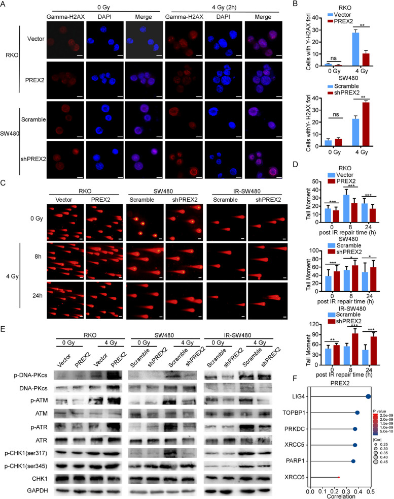
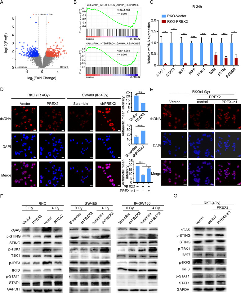
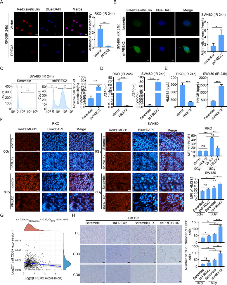
Results: PREX2 emerged as the most significantly upregulated gene in radioresistant CRC cells. It augmented the radioresistant capacity of CRC cells and demonstrated potential as a marker for predicting radioresistance efficacy. Mechanistically, PREX2 facilitated DNA repair by upregulating DNA-PKcs, suppressing radiation-induced immunogenic cell death, and impeding CD8+ T cell infiltration through the cGAS/STING/IFNs pathway. In vivo, the blockade of PREX2 heightened the efficacy of IR therapy.

Conclusions: PREX2 assumes a pivotal role in CRC radiation resistance by inhibiting the cGAS/STING/IFNs pathway, presenting itself as a potential radioresistant biomarker and therapeutic target for effectively overcoming radioresistance in CRC.

Keywords: Colorectal cancer; Immunogenic cell death; PREX2; Radioresistance; cGAS/STING/IFNs.


Conflict of interest statement

The authors declare no competing interests.

Figures

Fig. 1
Fig. 1
PREX2 acts as a radioresistant gene in CRC. A, B heat maps and volcanic maps showed significant differential genes after RNA-seq screening between IR-resistant cell line IR-SW480 and IR-sensitive cell line SW480. C, D qRT-PCR and Western blot analysis were conducted to determine the expression of PREX2 in IR-SW480 and SW480. E The analysis of PREX2 expression in radiotherapy-resistant CRCs compared with radiotherapy-sensitive CRCs in CRC microarray profile (GES145037 responder: 0.674 ± 0.109, non-responder: 0.784 ± 0.096, p = 0.0084; GSE1150082 responder: − 0.261 ± 0.125, non-responder: 0.180 ± 0.143, p = 0.0345). F RSI was calculated using the data from the TCGA database and the expression of PREX2 in the low RSI group and high RSI group was analyzed. G Kaplan–Meier analysis of progression-free interval in all patients with CRC according to PREX2 expression in TCGA dataset (log-rank test p < 0.001). H The expressions of PREX2 protein in specimens, including 54 CRC tissue specimens, were examined by IHC. Representative IHC images (left) and correlation analysis (right) were shown. Scale bar: 50 μm. *p < 0.05, **p < 0.01, ***p < 0.001
Fig. 2
Fig. 2
PREX2 promotes the radiation resistance of CRC cells in vitro. A A basal level of PREX2 was determined using Western blot analysis in several CRC cell lines (including RKO, CaCo2, HCT15, HCT116, SW480, SW620, HT29, and LoVo). B Stable overexpression and interference cell lines were detected by Western blotting. C Survival fraction with multi-target single-hit model. D Flow cytometry was performed to detect apoptosis of stable overexpression and interference cell lines with or without treatment with 4 Gy radiation. E Cell cycle progression was analyzed by flow cytometry. ns not significant, *p < 0.05, **p < 0.01, ***p < 0.001
Fig. 3
Fig. 3
PREX2 inhibits radiosensitization of CRC in vivo. AC Subcutaneous tumor formation in nude mice was established with PREX2-overexpressing or control RKO cells and treated with or without IR (n = 6/group). A The representative image of tumors derived from xenograft mice. B, C The volume and weight of tumors were monitored. Data were shown as means ± SEM. DF Subcutaneous tumor formation in C57/B6J mice was established with PREX2- knockdown or control CMT93 cells and treated with or without IR (n = 6/group). D Representative images of each group were photographed at the end of the experiment. E, F The volume and weight of tumors were monitored. Data were shown as means ± SEM. G Subcutaneous tumors in nude mice were stained with hematoxylin and eosin (H&E, top). The representative IHC images of cleaved-caspase 3 and γH2AX expression in xenograft tumors were shown (below). Scale bar: 100 μm. *p < 0.05, **p < 0.01,***p < 0.001
Fig. 4
Fig. 4
PREX2 modulated radiation-induced DNA damage response by regulating NHEJ repair. A Representative fluorescence images of γH2AX staining. Scale bar: 10 μm. B Quantification of γ-H2AX immunostaining in PREX2 overexpressed RKO cells and PREX2 knocked down SW480 cells with or without 4 Gy radiation. C, D The comet assay of stable overexpression and interference cell lines after IR treatment (scale bar: 50 μm, C) and the statistics result of quantification of tail moment (D). E Western blot detection of the expression levels of DNA-PKc, p-DNA-PKc, p-ATM, ATM, p-ATR, ATR, p-CHK1, and CHK1 in stable overexpression and interference cell lines with or without 4 Gy radiation. F Bioinformatics analysis of the correlation between PREX2 and genes related to NHEJ pathway (XRCC6, XRCC5, LIG4, PARP1, TOPBP1 and PRKDC) in TCGA-CRC. ns, not significant, *p < 0.05, **p < 0.01, ***p < 0.001
Fig. 5
Fig. 5
PREX2 deficiency promotes cytosolic dsDNA accumulation to activate STING signaling. A Volcanic maps showed significant differential genes after RNA-seq screening. B GSEA analyses in RNA-seq data. C qRT-PCR detection of the expression levels of IFN-I response genes in PREX2 overexpressed RKO cells at 24 h after 4 Gy radiation. D Immunofluorescence assay detection of the dsDNA in PREX2 overexpressed RKO cells and PREX2-knockdown SW480 cells after 4 Gy radiation. Scale bar: 20 μm. E Immunofluorescence assay detection of the dsDNA in PREX2 overexpressed RKO cells treated with or without PREX-in1 at 24 h after 4Gy radiation. F Western blot analysis detection of the expression of cGAS, STING, TBK1, STAT1, and IRF3 and the phosphorylation of STING, TBK1, STAT1, and IRF3 in stable overexpression and interference cell lines with or without 4 Gy radiation. G Western blot analysis detection of the expression of cGAS, p-STING, STING, p-TBK1, TBK1, p-STAT1, STAT1, p-IRF3, and IRF3 in PREX2 overexpressed RKO cells treated with or without PREX-in1 at 24 h after 4Gy radiation. *p < 0.05, **p < 0.01, ***p < 0.001
Fig. 6
Fig. 6
PREX2 inhibited radiation-induced immunogenic cell death and CD8+ T cell infiltration. A Immunofluorescence assay detection of the calreticulin (CRT) expression in PREX2 overexpressed RKO cells at 24 h after 4 Gy radiation. Scale bar: 20 μm. B Immunofluorescence assay detection of the CRT expression in PREX2 knocked down SW480 cells at 24 h after 4 Gy radiation. Scale bar: 10 μm. C Flow cytometry analysis confirmed that overexpressing PREX2 reduced CRT protein levels. D Detection of adenosine triphosphate (ATP) secretion by luciferin-based ATP assay kit. E Detection of high mobility group protein B1 (HMGB1) release by ELISA kit. F The representative images of HMGB1 immunofluorescence staining of xenograft tumors were shown (left). Scale bar: 10 μm. Quantification of HMGB1 means fluorescence intensity was analyzed (right). G The correlations between PREX2 expression and the immune score of CD8+ T cells were analyzed by the EPIC algorithm in TCGA-CRC. H Representative HE images and IHC images of CD3 and CD8 staining (left) and statistical analysis (right) of CMT93 tumors. Scale bar: 100 μm. ns, not significant, *p < 0.05, **p < 0.01, ***p < 0.001
Fig. 7
Fig. 7
PREX-in1 sensitizes the efficacy of IR therapy in CRC mouse models. A The overall design of the animal experiments. B The representative image of MC38 and IR-resistant cell line IR-CMT93 tumors treated with IR or IR+PREX-in1. C, D The volume curve and weight of MC38 and IR-CMT93 tumors treated with IR or IR+PREX-in1. E Representative HE images and IHC images of CD3 and CD8 staining (left) and statistical analysis (right) of MC38 and IR-CMT93 tumors. Scale bar: 100 μm. F Representative flow cytometry images (left) and quantitative analysis (right) of tumor-infiltrating lymphocytes in MC38 and IR-CMT93 tumors. ns, not significant, *p < 0.05, **p < 0.01, ***p < 0.001
Fig. 8
Fig. 8
A proposed model for illustrating the function and mechanism of PREX2 in CRC radiosensitization

References

    1. Sung H, Ferlay J, Siegel RL, Laversanne M, Soerjomataram I, Jemal A, et al. Global cancer statistics 2020: GLOBOCAN estimates of incidence and mortality worldwide for 36 cancers in 185 countries. CA Cancer J Clin. 2021;71(3):209–49. - PubMed
    1. Liu S, Jiang T, Xiao L, Yang S, Liu Q, Gao Y, et al. Total neoadjuvant therapy (TNT) versus standard neoadjuvant chemoradiotherapy for locally advanced rectal cancer: a systematic review and meta-analysis. Oncologist. 2021;26(9):e1555–66. - PMC - PubMed
    1. Benson AB, Venook AP, Al-Hawary MM, Cederquist L, Chen YJ, Ciombor KK, et al. Rectal cancer, Version 2.2018, NCCN Clinical Practice Guidelines in Oncology. J Natl Compr Canc Netw. 2018;16(7):874–901. - PMC - PubMed
    1. Janjan NA, Khoo VS, Abbruzzese J, Pazdur R, Dubrow R, Cleary KR, et al. Tumor downstaging and sphincter preservation with preoperative chemoradiation in locally advanced rectal cancer: the M. D. Anderson Cancer Center experience. Int J Radiat Oncol Biol Phys. 1999;44(5):1027–38. - PubMed
    1. J W, J J B, L D, S A R M, Sh F, S M J M. Abscopal effect following radiation therapy in cancer patients: a new look from the immunological point of view. J Biomed Phys Eng. 2020;10(4):537-42. - PMC - PubMed

Publication types

Substances