Review
. 2024 Mar 29;13(7):2011.
doi: 10.3390/jcm13072011.

Endoscopic Management of Post-Sleeve Gastrectomy Complications

Affiliations
Review

Endoscopic Management of Post-Sleeve Gastrectomy Complications

Muaaz Masood et al. J Clin Med. .

Abstract

Obesity is associated with several chronic conditions including diabetes, cardiovascular disease, and metabolic dysfunction-associated steatotic liver disease and malignancy. Bariatric surgery, most commonly Roux-en-Y gastric bypass and sleeve gastrectomy, is an effective treatment modality for obesity and can improve associated comorbidities. Over the last 20 years, there has been an increase in the rate of bariatric surgeries associated with the growing obesity epidemic. Sleeve gastrectomy is the most widely performed bariatric surgery currently, and while it serves as a durable option for some patients, it is important to note that several complications, including sleeve leak, stenosis, chronic fistula, gastrointestinal hemorrhage, and gastroesophageal reflux disease, may occur. Endoscopic methods to manage post-sleeve gastrectomy complications are often considered due to the risks associated with a reoperation, and endoscopy plays a significant role in the diagnosis and management of post-sleeve gastrectomy complications. We perform a detailed review of the current endoscopic management of post-sleeve gastrectomy complications.

Keywords: complications; endoscopic; endoscopy; fistula; gastrectomy; gastric; leak; management; sleeve; stenosis.


Conflict of interest statement

The authors declare no conflict of interest.

Figures

Scheme 1
Scheme 1
Lateral and transverse (top left and top right) views of a computed tomography scan of the abdomen and pelvis reveals air-fluid collections adjacent to the fundus of the stomach (3.3 × 3.0 cm) (arrow, top left), anterior to the stomach (2.2 × 4.6 cm) and in the epigastric region of the abdominal wall (6.2 × 3.0 cm) (arrows, top right), which likely represents abscesses in the setting of staple line dehiscence. Axial views of the computed tomography scan of the abdomen and pelvis (bottom left and bottom right) redemonstrate the somewhat ill-defined collection of gas and (arrows, bottom left and bottom right) fluid likely due to focal dehiscence and an abscess cavity which is the source of a gastrocutaneous fistula.
Scheme 2
Scheme 2
Coronal view of a computed tomography scan of the abdomen and pelvis reveals a small, contained leak (arrow, top left) along the greater curvature of the gastric body in a patient following sleeve gastrectomy. Upper gastrointestinal series demonstrates a small amount of extravasated contrast along the greater curvature of the gastric body with no free spill of extravasated contrast which is consistent with a small, contained perforation (arrow, top right) in a patient post-sleeve gastrectomy. The patient was treated with nothing per os and nasojejunal tube feeds (arrow, bottom left) for two weeks, with the resolution of sleeve leak and the removal of the nasojejunal tube (arrow, bottom right) as demonstrated on a follow-up coronal view of a computed tomography scan of the abdomen and pelvis.
Scheme 3
Scheme 3
Upper endoscopy reveals an inflammatory mucosal reaction (blue arrows; left, middle and right) following the removal of a partially covered, esophageal self-expandable metal stent used for the treatment of a staple line leak in a patient with a sleeve gastrectomy. The patient reported severe regurgitation and chest pain associated with the stent. A through-the-scope clip (purple arrow, left) was used to anchor the removed stent.
Scheme 4
Scheme 4
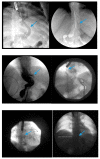
Fluoroscopic image demonstrates the placement of a 23 mm × 15 cm partially-covered esophageal self-expanding metal stent from the distal esophagus to the pylorus (arrow, top left) for the treatment of a leak in a patient following sleeve gastrectomy. Contrast injected endoscopically (arrows, top right and middle left) reveals a small, persistent sleeve leak. The endoscopic removal of the stent was subsequently attempted using a grasper (arrows, middle right and bottom left), but the stent removal was unsuccessful due to tissue ingrowth. A 23 mm × 15 cm, fully-covered, esophageal self-expanding metal stent was placed within the prior stent (arrow, bottom right) to facilitate removal after pressure necrosis of the ingrown tissue.
Scheme 5
Scheme 5
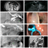
Axial view of a computed tomography scan of the abdomen and pelvis with contrast reveals a uniform, thin-walled, cystic lesion of the pancreas, which measures 90 mm × 52 mm × 48 mm, with peripheral calcifications with a BMI of 38 (arrow, A). Endoscopic ultrasound demonstrates an anechoic and hypoechoic lesion of the pancreatic body with two compartments, no septae or internal debris, and an associated mural nodule which was suggestive of a mucinous cystic neoplasm (arrow, B). The patient subsequently underwent a laparoscopic sleeve gastrectomy and a concurrent, open distal pancreatectomy which was complicated by a persistent pancreatic leak as well as a gastric sleeve leak (blue arrow, C) as demonstrated by contrast injected through a percutaneous drain (silver arrow, C). The percutaneous drain is redemonstrated on upper endoscopy (arrow, D). Note the additional percutaneous drain in the pancreatic bed for the postoperative leak (green arrows, C,G). Endoscopic internal drainage using transgastric double-pigtail stents was performed as demonstrated on fluoroscopic imaging (arrows, E,G) and endoscopy (arrow, F). After a prolonged course, the patient had a complete recovery, and a follow-up CT scan of the abdomen and pelvis confirmed the resolution of the leak and the removal of the double-pigtail stents and percutaneous drains (H).
Figure 1
Figure 1
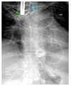
Upper gastrointestinal series reveals a 23 mm × 15 cm fully-covered esophageal self-expanding metal stent from the distal esophagus to the mid portion of the sleeve anchored by two through-the-scope clips (blue arrows) and one over-the-scope clip (green arrow) placed on the proximal edge of the stent.
Scheme 6
Scheme 6
Upper endoscopy reveals the proximal (blue arrows, left) and distal edge (blue arrows, middle) of a fully-covered esophageal self-expanding metal stent anchored by two through-the-scope clips (blue arrows, right) and an over-the-scope clip (green arrows, right). Note copious saliva collected around the non-peristaltic prosthesis.
Figure 2
Figure 2
Upper endoscopy reveals the use of a suturing system (arrow, blue) with a suture in place (arrow, green) at the leak site in a patient following sleeve gastrectomy.
Scheme 7
Scheme 7
Gastric sleeve stenosis in the patient depicted in Scheme 5 was treated with through-the-scope balloon dilation to 20 mm as demonstrated on fluoroscopic image (arrow, left) and upper endoscopy (arrow, middle). Endoscopic contrast injection following dilation revealed improvement in the stenosis and flow of contrast (arrow, right). Note a small residual drain (green arrow, left).
Scheme 8
Scheme 8
Contrast injected endoscopically reveals a sharp angulation and stenosis of the sleeve in a patient following sleeve gastrectomy (blue arrow, left and top right). A pneumatic balloon dilation (green arrow, top right) was performed to 35 mm. Endoscopic contrast injection reveals no post-dilation leak of contrast (bottom right).

References

    1. Koliaki C., Dalamaga M., Liatis S. Update on the Obesity Epidemic: After the Sudden Rise, Is the Upward Trajectory Beginning to Flatten? Curr. Obes. Rep. 2023;12:514–527. doi: 10.1007/s13679-023-00527-y. - DOI - PMC - PubMed
    1. Kloock S., Ziegler C.G., Dischinger U. Obesity and its comorbidities, current treatment options and future perspectives: Challenging bariatric surgery? Pharmacol. Ther. 2023;251:108549. doi: 10.1016/j.pharmthera.2023.108549. - DOI - PubMed
    1. Alalwan A.A., Friedman J., Park H., Segal R., Brumback B.A., Hartzema A.G. US national trends in bariatric surgery: A decade of study. Surgery. 2021;170:13–17. doi: 10.1016/j.surg.2021.02.002. - DOI - PubMed
    1. Buchwald H., Avidor Y., Braunwald E., Jensen M.D., Pories W., Fahrbach K., Schoelles K. Bariatric surgery: A systematic review and meta-analysis. JAMA. 2004;292:1724–1737. doi: 10.1001/jama.292.14.1724. - DOI - PubMed
    1. Clapp B., Ponce J., DeMaria E., Ghanem O., Hutter M., Kothari S., LaMasters T., Kurian M., English W. American Society for Metabolic and Bariatric Surgery 2020 estimate of metabolic and bariatric procedures performed in the United States. Surg Obes. Relat. Dis. 2022;18:1134–1140. doi: 10.1016/j.soard.2022.06.284. - DOI - PubMed