Review
. 2024 Apr 8;13(7):2152.
doi: 10.3390/jcm13072152.

Rationale for Using High-Frequency Ultrasound as a Routine Examination in Skin Cancer Surgery: A Practical Approach

Affiliations
Review

Rationale for Using High-Frequency Ultrasound as a Routine Examination in Skin Cancer Surgery: A Practical Approach

Diana Crisan et al. J Clin Med. .

Abstract

Ultrasound and high-frequency ultrasound assessment of melanoma and non-melanoma skin cancer in the pre-therapeutical setting is becoming increasingly popular in the field of dermatosurgery and dermatooncology, as it can provide clinicians with relevant, "in vivo" parameters regarding tumor lateral and depth extension as well as potential locoregional spread, cancelling the need of more extensive imaging methods and avoiding a delay in diagnosis. Furthermore, preoperative sonography and color Doppler can aid in orienting the clinical diagnosis, being able in numerous situations to differentiate between benign and malignant lesions, which require a different therapeutic approach. This preoperative knowledge is of paramount importance for planning an individualized treatment regimen. Using sonography at the time of diagnosis, important surgical complications, such as neurovascular damage, can be avoided by performing a preoperative neurovascular mapping. Furthermore, sonography can help reduce the number of surgical steps by identifying the lesions' extent prior to surgery, but it can also spare unnecessary surgical interventions in cases of locally advanced lesions, which infiltrate the bone or already present with locoregional metastases, which usually require modern radiooncological therapies in accordance to European guidelines. With this review, we intend to summarize the current indications of sonography in the field of skin cancer surgery, which can help us improve the therapeutic attitude toward our patients and enhance patient counseling. In the era of modern systemic radiooncological therapies, sonography can help better select patients who qualify for surgical procedures or require systemic treatments due to tumoral extension.

Keywords: dermatosurgery; malignant melanoma; non-melanoma skin cancer; skin cancer; ultrasound.


Conflict of interest statement

The authors declare no conflicts of interest.

Figures
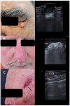
Figure 1
Figure 1
(Clinical aspect—left; HFUS-right): (A) Clinical aspect of a solid erythematous nodule with superficial teleangiectasia, as marked by “↑”; HFUS (17 MHz) and color Doppler show a well-defined, dermal-hypodermal, hypoechoic, homogenous lesion with posterior acoustic enhancement and peripheral vascularization, suspicious of a cystic lesion, which was confirmed by histology—transverse view; (B) Erythematous nodule, marked by “↑”, next to the surgical scar of a previously resected nasal BCC and reconstructed with an advancement flap, marked by “---”, suspicious of local relapse; HFUS and color Doppler identify a localized cartilage protrusion into the dermis, as marked by “c”, due to the performed advancement flap– transverse view; (C) Scar at left temple area after resection of a SCC, as marked by “↑”; HFUS and color Doppler identify next to the scar a hypoechoic dermal-hypodermal, vascularized lesion, suspicious of local satellite metastasis, confirmed by histology—transverse view.
Figure 2
Figure 2
(Clinical aspect—left; HFUS-right): (A) Clinical aspect of an ill-defined, erythematous nodule on the left back side, as marked by “↑”; HFUS and color Doppler show a subcutaneous hypoechoic lesion with a central porus connecting the lesion to the surface and central and peripheral vascularization—transverse view; (B) Ulcerated plaque on the left back side, as marked by “↑”; HFUS shows a hypoechoic, sharply demarcated lesion of the dermis, with multiple hyperechoic spots, as marked by “↑”, suspicious of an ulcerated BCC, and confirmed by histology—transverse view.
Figure 3
Figure 3
(Clinical, dermoscopic, and HFUS aspect—upper image; surgical approach—lower image): Clinical aspect showing an irregular, unsharply demarcated macule at columella level, as marked by “↑”, consistent with a BCC; Dermoscopy shows irregular dot and arborizing vessels; HFUS and color Doppler show a hypoechoic, non-vascularized lesion of the columella (“tu”) with no cartilage infiltration, as marked by “c”; Clinical aspect after complete tumor resection, defect closure with an advancement flap from the columella and clinical aspect at 8 weeks follow-up.
Figure 4
Figure 4
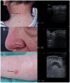
(Clinical aspect—left; HFUS-right): (A) Clinical aspect showing an erythematous plaque of the neck, as marked by “↑”, consistent with a superficial BCC; HFUS showing a superficial, spindle-shaped lesion with a tumor infiltration depth of 0.7 mm—transverse view; (B) Clinical aspect showing an erythematous macule on the left nasal ala, as marked by “↑”, consistent with a BCC; HFUS identifies a hypoechoic lesion with hyperechoic spots within and a tumor infiltration depth of 2.6 mm—longitudinal view; (C) Erythematous patch of the left lower leg, consistent with a BCC, as marked by “↑”,; HFUS and microvascular imaging display a hypoechoic, ill-defined dermal-hypodermal lesion with basal vascularization and a tumor infiltration depth of 2.5 mm; underneath the lesion, section thru the great saphenous vein—transverse view.
Figure 5
Figure 5
(Clinical aspect—left; HFUS, CT, MRI—middle; postoperative aspect—right): (A) Clinical aspect of an exulcerated tumor at the parietal area, as marked by “↑”, consistent with an SCC; HFUS and microvascular imaging showing an irregular hypoechoic, highly vascularized lesion (“tu”) extending up to the bony surface (“b”) with no bone infiltration—transverse view; CT scan showing no bone infiltration by the tumor mass; Clinical aspect after resection and partial defect closure with two rotation flaps; (B) Clinical aspect of an exulcerated SCC of the right parietal area, as marked by “↑”; HFUS and color Doppler display a hypoechoic lesion with suspicion of bone infiltration, as marked by “↑”—transverse view; MRI showing local bone infiltration, as marked by “↑”—coronal view.
Figure 6
Figure 6
(Clinical aspect—left; HFUS—right): (A) Clinical aspect of an erythematous nodule with arborizing vessels, as marked by “↑”, consistent with a BCC; HFUS showing a hypoechoic lesion and numerous hyperechoic spots within, with no contact to the cartilage, as marked by “c”—longitudinal view; (B) Erythematous nodule on the right nasal wall, as marked by “↑”, consistent with a solid BCC; HFUS displaying a hypoechoic, lesion protruding into the dermis with multiple hyperechoic spots and no cartilage infiltration, as marked by “c”- transversal view; (C) Surgical scar at left nasal ala after surgical resection of a BCC, as marked by “↑”; HFUS und microvascular imaging showing a hypoechoic, vascularized lesion beneath the surgical scar, consistent with local relapse, as marked by “tu” – transversal view.
Figure 7
Figure 7
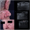
(Clinical aspect—left; HFUS-middle; postoperative aspect—right): (A) Erythematous plaque of the ear helix, as marked by “↑”, consistent with a solid BCC; HFUS and microvascular imaging showing a hypoechoic lesion, as marked by “tu“, with peripheral and basal vascularization with suspicion of cartilage infiltration, marked by “c”—longitudinal view; Clinical aspect 8 weeks after wedge excision; (B) Ulcerated nodule of the left helix, as marked by “↑”, consistent with an ulcerated BCC; HFUS and microvascular imaging show a hypoechoic lesion, as marked by “tu“, without any cartilage infiltration, marked by “c“—longitudinal view; Clinical aspect after performance of a chondrocutaneous flap.
Figure 8
Figure 8
(Clinical aspect—left; HFUS—right): Hyperkeratotic plaque at right temporal area, as marked by “↑”, consistent with an SCC; HFUS and microvascular imaging show a hypoechoic, vascularized lesion, marked by “tu”, protruding above the frontal branch of the superficial temporal artery—longitudinal view.
Figure 9
Figure 9
(Clinical aspect “in vivo” and “ex vivo”—left; HFUS—middle; postoperative aspect—right): Clinical aspect of an ulcerated plaque on the back, consistent with an ulcerated BCC, as seen “in vivo” and “ex vivo”; “ex vivo” HFUS delimitating the tumor margins in relation to the resection margin; Clinical aspect after complete tumor resection.
Figure 10
Figure 10
(Clinical aspect—left; HFUS-right): (A) Erythematous nodule at right frontal area, as marked by “↑”, consistent with an amelanotic MM; HFUS and microvascular imaging show a hypoechoic, intensely vascularized lesion as primary tumor, marked by “tu“ and a secondary dermal-hypodermal, hypoechoic, vascularized lesion situated laterally, suspicious of dermal metastasis, as marked by “met“, and confirmed by histology—transverse view; (B) Ulcerated nodule of the lower leg, as marked by “↑”, consistent with a nodular MM; HFUS and microvascular imaging identify a hypoechoic lesion beneath the primary tumor, marked by “tu“, with central and peripheral vascularization, consistent with a local satellite metastasis, as marked by “met“—transverse view.
Figure 11
Figure 11
(Clinical aspect—left; HFUS—middle, MRI scan- right): Clinical aspect of an ulcerated plaque at the lateral orbital rim, as marked by “↑”, consistent with an ulcerated SCC; HFUS and microvascular imaging of the primary tumor and in-transit area showing no orbital rim infiltration but a suspicious hypoechoic lesion of the parotid gland, as marked by “met”, histologically confirmed as lymph node metastasis—longitudinal view; MRI showing the parotid lesion and the primary tumor with no bony invasion, as marked by “↑”.
Figure 12
Figure 12
(PET-CT—left, US—right): Thoracic PET-CT showing a new lesion in the right axillary area in a MM patient, suspicious of metastatic disease, as marked by “↑”; Intraoperative HFUS showing a subcutaneous hypoechoic lesion, marked by “↑”, guiding the excision margins—transversal view.

Similar articles

Cited by

  • High-Frequency and Ultra-High-Frequency Ultrasound in Dermatologic Diseases and Aesthetic Medicine.
    Argalia G, Reginelli A, Molinelli E, Russo A, Michelucci A, Sechi A, Marzano AV, Desyatnikova S, Fogante M, Patanè V, Granieri G, Tagliati C, Rizzetto G, De Simoni E, Matteucci M, Candelora M, Lanza C, Ventura C, Carboni N, Esposito R, Esposito S, Paolinelli M, Esposto E, Lanni G, Lucidi Pressanti G, Giorgi C, Principi F, Rebonato A, Malinowska SP, Mlosek RK, Giuseppetti GM, Dini V, Romanelli M, Offidani A, Cappabianca S, Wortsman X, Simonetti O. Argalia G, et al. Medicina (Kaunas). 2025 Jan 26;61(2):220. doi: 10.3390/medicina61020220. Medicina (Kaunas). 2025. PMID: 40005337 Free PMC article. Review.

References

    1. Leiter U., Eigentler T., Garbe C. Epidemiology of skin cancer. Adv. Exp. Med. Biol. 2014;810:120–140. doi: 10.1007/978-1-4939-0437-2_7. - DOI - PubMed
    1. Crisan D., Wortsman X., Alfageme F., Catalano O., Badea A., Scharffetter-Kochanek K., Sindrilaru A., Crisan M. Ultrasonography in dermatologic surgery: Revealing the unseen for improved surgical planning. J. Der Dtsch. Dermatol. Ges. 2022;20:913–926. doi: 10.1111/ddg.14781. - DOI - PubMed
    1. Wortsman X. Practical applications of ultrasound in dermatology. Clin. Dermatol. 2021;39:605–623. doi: 10.1016/j.clindermatol.2021.03.007. - DOI - PubMed
    1. Crisan D., Wortsman X., Catalano O., Badea R., Kastler S., Badea A., Manea A., Scharffetter-Kochanek K., Strilciuc S., Crisan M., et al. Pre-operative high-frequency ultrasound: A reliable management tool in auricular and nasal non-melanoma skin cancer. J. Der Dtsch. Dermatol. Ges. 2024;22:357–365. doi: 10.1111/ddg.15308. - DOI - PubMed
    1. Wortsman X., Alfageme F., Roustan G., Arias-Santiago S., Martorell A., Catalano O., Scotto di Santolo M., Zarchi K., Bouer M., Gonzalez C., et al. Guidelines for Performing Dermatologic Ultrasound Examinations by the DERMUS Group. J. Ultrasound Med. 2016;35:577–580. doi: 10.7863/ultra.15.06046. - DOI - PubMed

LinkOut - more resources