Review
. 2024 Mar 27;14(7):706.
doi: 10.3390/diagnostics14070706.

Comments and Illustrations of Ultrasound Findings in Extrapulmonary Tuberculosis Manifestations

Affiliations
Review

Comments and Illustrations of Ultrasound Findings in Extrapulmonary Tuberculosis Manifestations

Kathleen Möller et al. Diagnostics (Basel). .

Abstract

This review describes the appearance of extrapulmonary tuberculosis manifestations in comprehensive and multiparametric ultrasound imaging. The aim is to increase awareness of typical ultrasound findings regarding extrapulmonary tuberculosis, correlate those with pathological features, and facilitate differential diagnosis. Point of care ultrasound protocols can be used as a screening method in high-risk populations, although the negative findings do not exclude tuberculosis. Conversely, the diagnosis of extrapulmonary tuberculosis can never be made using ultrasound alone, as many ultrasound findings in extrapulmonary tuberculosis are non-specific. However, ultrasound-based sampling techniques can significantly facilitate the collection of samples for microbiological or molecular proof of tuberculosis, as well as facilitating the establishment of alternative diagnoses.

Keywords: contrast enhanced ultrasonography; endoscopic ultrasound; extrapulmonary manifestations; tuberculosis; ultrasonography.


Conflict of interest statement

The authors declare no conflict of interest.

Figures
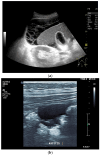
Figure 1
Figure 1
Typical changes in the peritoneum, mesentery, and omentum in patients with tuberculosis. Ascites with pronounced septa (a). Ascites encapsulated between the abdominal wall and colon (b). Omental thickening with non-echoic caseous abscesses and few echogenic contents (c); hypoechoic nodules in hyperechoic thickened peritoneum (d). Significantly thickened hyperechoic peritoneum in B-mode US (right side of image) (marked with arrows). In CEUS, the thickened peritoneum is contrast enhanced in the arterial phase (e). In the parenchymal phase, it shows a decrease in enhancement (f). The thickened hyperechoic peritoneum shows a non-echoic lesion (arrow) (g). In the CEUS, this is not enhanced and instead shows a hyperenhanced rim. This corresponds to caseous necrosis (h).
Figure 1
Figure 1
Typical changes in the peritoneum, mesentery, and omentum in patients with tuberculosis. Ascites with pronounced septa (a). Ascites encapsulated between the abdominal wall and colon (b). Omental thickening with non-echoic caseous abscesses and few echogenic contents (c); hypoechoic nodules in hyperechoic thickened peritoneum (d). Significantly thickened hyperechoic peritoneum in B-mode US (right side of image) (marked with arrows). In CEUS, the thickened peritoneum is contrast enhanced in the arterial phase (e). In the parenchymal phase, it shows a decrease in enhancement (f). The thickened hyperechoic peritoneum shows a non-echoic lesion (arrow) (g). In the CEUS, this is not enhanced and instead shows a hyperenhanced rim. This corresponds to caseous necrosis (h).
Figure 2
Figure 2
A case of extrapulmonary tuberculosis: 33 y/o male from a country at high risk of tuberculosis. Thoracic pain and fever led to a suspicion of pericarditis. There was no pericardial effusion. There were no pulmonary infiltrations or pleural effusions in the chest CT, but there were enlarged mediastinal lymph nodes. Ultrasound and CT also showed subdiaphragmal enlarged lymph nodes and small nodular splenic changes. The diagnosis was confirmed using the transgastric EUS-guided biopsy (19 G Olympus) of the lymph nodes in the hepatic hilum. Enlarged lymph nodes in the hepatic hilum (a); adjacent to the pancreatic head (b); transabdominal ultrasound (TUS) using a linear transducer of 9 MHz. The hypoechoic central parts are conspicuous (a,b). Rounded lymph nodes are observed as being peripancreatic (c) and as being in close proximity to the gallbladder wall (d). The lymph nodes are visualized between the markers. In the EUS, paragastric lymph nodes are enlarged, rounded, very hypoechoic, with hypoechoic central parts, and forming conglomerates. A central vascular hilum cannot be delineated in the CDI (e). The hypoechoic central parts are softer during elastography (f). EUS also shows enlarged hypoechoic lymph nodes in the hepatic hilus, with hypoechoic central parts that lack a central vascular hilum (g). Elastographically, the lymph nodes are indifferent (h). The diagnosis is confirmed using EUS-guided biopsy (i) with evidence of granulomatous inflammation, caseous necrosis, detection of acid-fast rods, and Mycobacterium tuberculosis in the PCR. In the spleen, single hypoechoic lesions < 5 mm are visible during transabdominal ultrasound using a linear transducer of 9 MHz. Otherwise, fine-grained hypoechoic lesions can only be guessed at (j). With magnification using a 9 MHz transducer, multiple hypoechoic lesions < 3 mm can be delineated (k). These do not reveal any vessels in the Power Doppler (l). EUS confirms multiple hypoechoic splenic lesions, in line with splenic tuberculosis (m).
Figure 2
Figure 2
A case of extrapulmonary tuberculosis: 33 y/o male from a country at high risk of tuberculosis. Thoracic pain and fever led to a suspicion of pericarditis. There was no pericardial effusion. There were no pulmonary infiltrations or pleural effusions in the chest CT, but there were enlarged mediastinal lymph nodes. Ultrasound and CT also showed subdiaphragmal enlarged lymph nodes and small nodular splenic changes. The diagnosis was confirmed using the transgastric EUS-guided biopsy (19 G Olympus) of the lymph nodes in the hepatic hilum. Enlarged lymph nodes in the hepatic hilum (a); adjacent to the pancreatic head (b); transabdominal ultrasound (TUS) using a linear transducer of 9 MHz. The hypoechoic central parts are conspicuous (a,b). Rounded lymph nodes are observed as being peripancreatic (c) and as being in close proximity to the gallbladder wall (d). The lymph nodes are visualized between the markers. In the EUS, paragastric lymph nodes are enlarged, rounded, very hypoechoic, with hypoechoic central parts, and forming conglomerates. A central vascular hilum cannot be delineated in the CDI (e). The hypoechoic central parts are softer during elastography (f). EUS also shows enlarged hypoechoic lymph nodes in the hepatic hilus, with hypoechoic central parts that lack a central vascular hilum (g). Elastographically, the lymph nodes are indifferent (h). The diagnosis is confirmed using EUS-guided biopsy (i) with evidence of granulomatous inflammation, caseous necrosis, detection of acid-fast rods, and Mycobacterium tuberculosis in the PCR. In the spleen, single hypoechoic lesions < 5 mm are visible during transabdominal ultrasound using a linear transducer of 9 MHz. Otherwise, fine-grained hypoechoic lesions can only be guessed at (j). With magnification using a 9 MHz transducer, multiple hypoechoic lesions < 3 mm can be delineated (k). These do not reveal any vessels in the Power Doppler (l). EUS confirms multiple hypoechoic splenic lesions, in line with splenic tuberculosis (m).
Figure 3
Figure 3
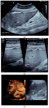
Sonographic and CEUS features of hepatic tuberculosis. Subcapsular liver lesion (a); subcapsular liver lesions (white arrow) and hypoechoic lesion on the capsule (green arrow) using a linear transducer of 12 MHz (b). Caseous necrosis, histologically proven. Hypoechoic lesion with hyperechoic rim using B-mode ultrasound (right side of image). The lesion is hyperenhanced in the arterial phase at the edge of the CEUS; the center is nonenhanced with the exception of some enhanced septa (c). In the portal venous phase, the peripheral areas are hypoenhanced (d). Multiple small, smoothly bordered, non-cystic hypoechoic lesions in the liver, with no evidence of macrovessels when using Color Doppler Imaging (e). CEUS shows mild peripheral enhancement around the lesions. The lesions are without enhancement in the arterial phase (f), portal venous phase (g), and late phase (h).
Figure 3
Figure 3
Sonographic and CEUS features of hepatic tuberculosis. Subcapsular liver lesion (a); subcapsular liver lesions (white arrow) and hypoechoic lesion on the capsule (green arrow) using a linear transducer of 12 MHz (b). Caseous necrosis, histologically proven. Hypoechoic lesion with hyperechoic rim using B-mode ultrasound (right side of image). The lesion is hyperenhanced in the arterial phase at the edge of the CEUS; the center is nonenhanced with the exception of some enhanced septa (c). In the portal venous phase, the peripheral areas are hypoenhanced (d). Multiple small, smoothly bordered, non-cystic hypoechoic lesions in the liver, with no evidence of macrovessels when using Color Doppler Imaging (e). CEUS shows mild peripheral enhancement around the lesions. The lesions are without enhancement in the arterial phase (f), portal venous phase (g), and late phase (h).
Figure 4
Figure 4
Sonographic and CEUS features of splenic tuberculosis. Sonographic and CEUS features of splenic tuberculosis. Subcapsular splenic lesion (arrow) (a), subcapsular splenic lesion using a linear transducer of 12 MHz (arrows) (b). Histologically confirmed tuberculosis from mediastinal lymph nodes. Multiple small splenic lesions (c). Using CEUS with a linear transducer (9 MHz), these are slightly hypoenhanced in the arterial phase (d), and show a progressive washout in the course of the venous phase (arrows) (e). Subcapsular splenic lesion (arrow) with nonenhanced and hypoenhanced parts and hyperenhanced rims indicate a caseous necrosis (f). In another section, an interrupted spleen capsule (arrow) due to a rupture of the caseous necrosis can be assumed (g).
Figure 4
Figure 4
Sonographic and CEUS features of splenic tuberculosis. Sonographic and CEUS features of splenic tuberculosis. Subcapsular splenic lesion (arrow) (a), subcapsular splenic lesion using a linear transducer of 12 MHz (arrows) (b). Histologically confirmed tuberculosis from mediastinal lymph nodes. Multiple small splenic lesions (c). Using CEUS with a linear transducer (9 MHz), these are slightly hypoenhanced in the arterial phase (d), and show a progressive washout in the course of the venous phase (arrows) (e). Subcapsular splenic lesion (arrow) with nonenhanced and hypoenhanced parts and hyperenhanced rims indicate a caseous necrosis (f). In another section, an interrupted spleen capsule (arrow) due to a rupture of the caseous necrosis can be assumed (g).
Figure 5
Figure 5
Pancreatic tuberculosis. Plump large pancreas. There is a lesion with a thick echogenic capsule and hyperechogenic and anechoic contents (between the markers) at the head of the pancreas. This finding was evaluated as a caseous abscess in a patient with tuberculosis.
Figure 6
Figure 6
A case of esophageal tuberculosis. A 52-year-old male non-smoker presented with progressive dysphagia and weight loss (15 kg in 6 months). Upper GI endoscopy revealed two subepithelial esophageal masses covered by normal mucosa (a). A CT scan showed large solid mass lesions in the mediastinum, with small gaseous inclusions (arrowhead) and a thickened esophageal wall (b, arrow). EUS ruled out a subepithelial esophageal tumor, and confirmed a large mediastinal hypoechoic mass infiltrating the esophageal wall, which was up to 12 mm thick with a complete loss of layering (c). Multiple enlarged and confluent hypoechoic lymph nodes were found throughout the mediastinum (d). Contrast-enhanced harmonic EUS showed the hyperenhancement of the mediastinal mass lesion and the thickened esophageal wall with some anechoic necrotic parts (*), and echogenic gaseous reflections (arrow; e). EUS-FNA (22 Gauge) of lymph nodes and of the thickened wall was performed (f; needle is marked with an arrowhead). Turbid fluid was aspirated from one lymph node, and then sent for cytological and microbiological examination and polymerase chain reaction (PCR) for mycobacteria. Positive Ziehl–Neelsen staining and PCR for mycobacterium tuberculosis and cheesy, necrotizing granulomas (g), cytology, Papanicolaou stain: giant cell; (h), histology, hematoxylin-eosin stain: necrotizing granulomas; courtesy Gunnar Schröder, Institute for Pathology Wildau, Germany) established the diagnosis of extrapulmonary tuberculosis involving the mediastinum and esophagus. Bronchoscopy revealed a fistula opening in the left main bronchus marked by the black arrow (i).
Figure 6
Figure 6
A case of esophageal tuberculosis. A 52-year-old male non-smoker presented with progressive dysphagia and weight loss (15 kg in 6 months). Upper GI endoscopy revealed two subepithelial esophageal masses covered by normal mucosa (a). A CT scan showed large solid mass lesions in the mediastinum, with small gaseous inclusions (arrowhead) and a thickened esophageal wall (b, arrow). EUS ruled out a subepithelial esophageal tumor, and confirmed a large mediastinal hypoechoic mass infiltrating the esophageal wall, which was up to 12 mm thick with a complete loss of layering (c). Multiple enlarged and confluent hypoechoic lymph nodes were found throughout the mediastinum (d). Contrast-enhanced harmonic EUS showed the hyperenhancement of the mediastinal mass lesion and the thickened esophageal wall with some anechoic necrotic parts (*), and echogenic gaseous reflections (arrow; e). EUS-FNA (22 Gauge) of lymph nodes and of the thickened wall was performed (f; needle is marked with an arrowhead). Turbid fluid was aspirated from one lymph node, and then sent for cytological and microbiological examination and polymerase chain reaction (PCR) for mycobacteria. Positive Ziehl–Neelsen staining and PCR for mycobacterium tuberculosis and cheesy, necrotizing granulomas (g), cytology, Papanicolaou stain: giant cell; (h), histology, hematoxylin-eosin stain: necrotizing granulomas; courtesy Gunnar Schröder, Institute for Pathology Wildau, Germany) established the diagnosis of extrapulmonary tuberculosis involving the mediastinum and esophagus. Bronchoscopy revealed a fistula opening in the left main bronchus marked by the black arrow (i).
Figure 7
Figure 7
Renal tuberculosis. Adjacent to the renal sinus are cyst-like lesions with smooth borders. Characteristic of caliectasias (a). Small “tubular” structures are noticeable in the kidney (arrow). The parenchyma presents nodular hypoechoic lesions. Perforation of caseous necrosis, and small intrarenal fistulas (b). “Putty kidney”—final stage of renal tuberculosis. Very small (non-functioning) kidney with highly diffused hyperechoic parenchyma (c), surrounded by a perinephritis fluid collection (d).
Figure 7
Figure 7
Renal tuberculosis. Adjacent to the renal sinus are cyst-like lesions with smooth borders. Characteristic of caliectasias (a). Small “tubular” structures are noticeable in the kidney (arrow). The parenchyma presents nodular hypoechoic lesions. Perforation of caseous necrosis, and small intrarenal fistulas (b). “Putty kidney”—final stage of renal tuberculosis. Very small (non-functioning) kidney with highly diffused hyperechoic parenchyma (c), surrounded by a perinephritis fluid collection (d).
Figure 8
Figure 8
Tuberculosis of the uterus and adnexae. Female patients with tuberculosis in India. Pelvis with tubo-ovarian masses: Hypoechoic lesion in right adnexa (arrows) (a) and hypoechoic lesion in left adnexa and a small amount of free fluid (arrows) (b). Free fluid in the Douglas space (arrows) (c). Thickened right fallopian tube (RT FT) (arrows) (d); thickened left fallopian tube (LT FT) (arrows) (e).
Figure 8
Figure 8
Tuberculosis of the uterus and adnexae. Female patients with tuberculosis in India. Pelvis with tubo-ovarian masses: Hypoechoic lesion in right adnexa (arrows) (a) and hypoechoic lesion in left adnexa and a small amount of free fluid (arrows) (b). Free fluid in the Douglas space (arrows) (c). Thickened right fallopian tube (RT FT) (arrows) (d); thickened left fallopian tube (LT FT) (arrows) (e).

References

    1. Houben R.M., Dodd P.J. The Global Burden of Latent Tuberculosis Infection: A Re-estimation Using Mathematical Modelling. PLoS Med. 2016;13:e1002152. doi: 10.1371/journal.pmed.1002152. - DOI - PMC - PubMed
    1. WHO Global Tuberculosis Report 2023. [(accessed on 7 November 2023)]. Available online: www.who.int/tb.
    1. Gupta P., Kumar S., Sharma V., Mandavdhare H., Dhaka N., Sinha S.K., Dutta U., Kochhar R. Common and uncommon imaging features of abdominal tuberculosis. J. Med. Imaging Radiat. Oncol. 2019;63:329–339. doi: 10.1111/1754-9485.12874. - DOI - PubMed
    1. Kulchavenya E. Extrapulmonary tuberculosis: Are statistical reports accurate? Ther. Adv. Infect. Dis. 2014;2:61–70. doi: 10.1177/2049936114528173. - DOI - PMC - PubMed
    1. Patel M.N., Beningfield S., Burch V. Abdominal and pericardial ultrasound in suspected extrapulmonary or disseminated tuberculosis. S. Afr. Med. J. 2011;101:39–42. doi: 10.7196/SAMJ.4201. - DOI - PubMed