Review
. 2024 Apr 5;14(7):772.
doi: 10.3390/diagnostics14070772.

Imaging Considerations before and after Liver-Directed Locoregional Treatments for Metastatic Colorectal Cancer

Affiliations
Review

Imaging Considerations before and after Liver-Directed Locoregional Treatments for Metastatic Colorectal Cancer

David-Dimitris Chlorogiannis et al. Diagnostics (Basel). .

Abstract

Colorectal cancer is a leading cause of cancer-related death. Liver metastases will develop in over one-third of patients with colorectal cancer and are a major cause of morbidity and mortality. Even though surgical resection has been considered the mainstay of treatment, only approximately 20% of the patients are surgical candidates. Liver-directed locoregional therapies such as thermal ablation, Yttrium-90 transarterial radioembolization, and stereotactic body radiation therapy are pivotal in managing colorectal liver metastatic disease. Comprehensive pre- and post-intervention imaging, encompassing both anatomic and metabolic assessments, is invaluable for precise treatment planning, staging, treatment response assessment, and the prompt identification of local or distant tumor progression. This review outlines the value of imaging for colorectal liver metastatic disease and offers insights into imaging follow-up after locoregional liver-directed therapy.

Keywords: Yttrium-90 radioembolization; colorectal cancer; imaging; interventional oncology; liver metastases; locoregional therapy; margin; stereotactic body radiation therapy; thermal ablation.


Conflict of interest statement

The authors declare no conflicts of interest.

Figures
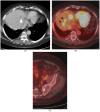
Figure 1
Figure 1
A 75-year-old man with metastatic rectal cancer, prior hepatic arterial infusion pump chemotherapy, and right hepatectomy, referred to interventional radiology clinic for consideration of percutaneous ablation to new 1.7 cm hepatic dome metastasis on CT ((A); white arrowhead). Prior to evaluation in clinic, an FDG PET/CT was obtained showing additional sites of intrahepatic disease ((B); black arrowheads), as well as osseous metastases ((C); left sacrum). The plan for ablation was aborted and systemic chemotherapy was administered.
Figure 2
Figure 2
A 39-year-old woman with metastatic rectal cancer, post-liver resection, and hepatic arterial infusion pump placement, on FOLFIRI chemotherapy with solitary 0.7 cm segment 7 metastasis. The metastasis was subtle on the portal venous phase CT (A) and conspicuous on the hepatobiliary phase of the MRI ((B); white arrowhead). The tumor did not demonstrate increased focal FDG uptake on baseline PET/CT (C). Following a 4-week chemotherapy break, the patient presented to interventional radiology for percutaneous microwave ablation. The target demonstrated FDG-avidity after discontinuation of chemotherapy (D), permitting PET-guided microwave ablation using the split-dose technique. Immediate post-ablation PET/CT confirms a photopenic defect at the tumor site (E).
Figure 3
Figure 3
A 67-year-old man with metastatic rectosigmoid cancer, with imaging consistent with local tumor progression, one year after microwave ablation of segment 7 metastasis. Portal venous phase CT shows 1.3 cm hypoattenuating (relative to liver parenchyma) nodule along the medial aspect of the ablation zone ((A); arrowhead), with corresponding increased focal FDG-uptake on PET/CT (B). The recurrence was treated with repeat microwave ablation.
Figure 4
Figure 4
A 52-year-old woman with liver-dominant metastatic colon cancer, with progressing liver metastases despite hepatic arterial infusion chemotherapy and systemic chemotherapy. Baseline PET/CT demonstrated multiple FDG-avid tumors in the right hepatic lobe (A,B). Following Yttrium-90 radioembolization, follow-up PET/CT after 5 weeks confirmed complete metabolic response (C,D). Based on size criteria alone (RECIST 1.1), this would have been considered stable disease.
Figure 5
Figure 5
A 47-year-old woman with metastatic rectal cancer, status post chemotherapy, primary tumor, and posterior sector liver resection, with 2.9 cm segment 5 liver metastasis, as seen on the hepatobiliary phase of baseline MRI ((A); white arrowhead). This was treated with SBRT (6000 cGy in 5 fractions). PET/CT after 4 months showed persistent uptake in the treatment zone ((B); SUVmax 6.1; liver SUVmean 2.8). After 9 months, there was a continued decrease in FDG uptake in the treatment zone, above that of background liver ((C); SUVmax 3.4; liver SUVmean 2.4). MRI on the same day showed geographic area of arterial enhancement (D), with decreased signal on the hepatobiliary phase (E).
Figure 5
Figure 5
A 47-year-old woman with metastatic rectal cancer, status post chemotherapy, primary tumor, and posterior sector liver resection, with 2.9 cm segment 5 liver metastasis, as seen on the hepatobiliary phase of baseline MRI ((A); white arrowhead). This was treated with SBRT (6000 cGy in 5 fractions). PET/CT after 4 months showed persistent uptake in the treatment zone ((B); SUVmax 6.1; liver SUVmean 2.8). After 9 months, there was a continued decrease in FDG uptake in the treatment zone, above that of background liver ((C); SUVmax 3.4; liver SUVmean 2.4). MRI on the same day showed geographic area of arterial enhancement (D), with decreased signal on the hepatobiliary phase (E).

References

    1. Siegel R.L., Wagle N.S., Cercek A., Smith R.A., Jemal A. Colorectal Cancer Statistics, 2023. CA Cancer J. Clin. 2023;73:233–254. doi: 10.3322/caac.21772. - DOI - PubMed
    1. Bailey C.E., Hu C.-Y., You Y.N., Bednarski B.K., Rodriguez-Bigas M.A., Skibber J.M., Cantor S.B., Chang G.J. Increasing Disparities in the Age-Related Incidences of Colon and Rectal Cancers in the United States, 1975–2010. JAMA Surg. 2015;150:17. doi: 10.1001/jamasurg.2014.1756. - DOI - PMC - PubMed
    1. Hackl C., Neumann P., Gerken M., Loss M., Klinkhammer-Schalke M., Schlitt H.J. Treatment of Colorectal Liver Metastases in Germany: A Ten-Year Population-Based Analysis of 5772 Cases of Primary Colorectal Adenocarcinoma. BMC Cancer. 2014;14:810. doi: 10.1186/1471-2407-14-810. - DOI - PMC - PubMed
    1. Helling T.S., Martin M. Cause of Death from Liver Metastases in Colorectal Cancer. Ann. Surg. Oncol. 2014;21:501–506. doi: 10.1245/s10434-013-3297-7. - DOI - PubMed
    1. Meijerink M.R., Puijk R.S., van Tilborg A.A.J.M., Henningsen K.H., Fernandez L.G., Neyt M., Heymans J., Frankema J.S., de Jong K.P., Richel D.J., et al. Radiofrequency and Microwave Ablation Compared to Systemic Chemotherapy and to Partial Hepatectomy in the Treatment of Colorectal Liver Metastases: A Systematic Review and Meta-Analysis. Cardiovasc. Interv. Radiol. 2018;41:1189–1204. doi: 10.1007/s00270-018-1959-3. - DOI - PMC - PubMed

LinkOut - more resources