. 2024 Mar 24;29(7):1449.
doi: 10.3390/molecules29071449.

Royal Jelly Enhances the Ability of Myoblast C2C12 Cells to Differentiate into Multilineage Cells

Affiliations

Royal Jelly Enhances the Ability of Myoblast C2C12 Cells to Differentiate into Multilineage Cells

Takumi Ito et al. Molecules. .

Abstract

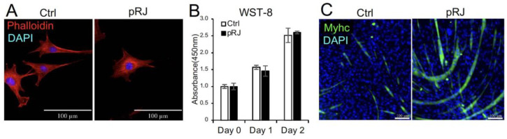
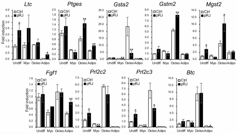
Royal jelly (RJ) is recognized as beneficial to mammalian health. Multilineage differentiation potential is an important property of mesenchymal stem cells (MSCs). C2C12 cells have an innate ability to differentiate into myogenic cells. Like MSCs, C2C12 cells can also differentiate into osteoblast- and adipocyte-lineage cells. We recently reported that RJ enhances the myogenic differentiation of C2C12 cells. However, the effect of RJ on osteoblast or adipocyte differentiation is still unknown. Here in this study, we have examined the effect of RJ on the osteoblast and adipocyte differentiation of C2C12 cells. Protease-treated RJ was used to reduce the adverse effects caused by RJ supplementation. To induce osteoblast or adipocyte differentiation, cells were treated with bone morphogenetic proteins (BMP) or peroxisome proliferator-activated receptor γ (PPARγ) agonist, respectively. RNA-seq was used to analyze the effect of RJ on gene expression. We found that RJ stimulates osteoblast and adipocyte differentiation. RJ regulated 279 genes. RJ treatment upregulated glutathione-related genes. Glutathione, the most abundant antioxidative factor in cells, has been shown to promote osteoblast differentiation in MSC and MSC-like cells. Therefore, RJ may promote osteogenesis, at least in part, through the antioxidant effects of glutathione. RJ enhances the differentiation ability of C2C12 cells into multiple lineages, including myoblasts, osteoblasts, and adipocytes.

Keywords: C2C12; RNA-seq; adipogenesis; myogenesis; osteoblastogenesis; royal jelly; stem cell.


Conflict of interest statement

The authors declare that they have no conflicts of interest. N.O. and H.O. are employees of Yamada Bee Company, Inc.

Figures

Figure 1
Figure 1
Pretreatment with pRJ stimulates myogenic differentiation in C2C12 cells. The cell nuclei and cytoskeleton were visualized by staining with DAPI (Blue) and rhodamine phalloidin (Red), respectively (A). The number of live C2C12 cells pretreated with or without pRJ for 1 week was assessed using the Cell Counting kit-8 on days 0, 1, and 2 (B). The cells were cultured in a myogenic medium for six days. Cells were then stained with an anti-myosin heavy-chain antibody (Green), and cell nuclei were visualized by staining with DAPI (Blue) (C). Data are presented as the mean ± SD (n = 3). Representative images are shown. Similar results were obtained in three independent experiments. Scale = 100 μm (A,C).
Figure 2
Figure 2
pRJ-treated cells have a high potential to differentiate into osteoblasts in response to BMPs. C2C12 cells pretreated with or without pRJ were treated with various concentrations (0, 12.5, 25, or 50 ng/mL) of rhBMP-4. ALP activity was determined on day 3 (A). The cells were treated with 25 ng/mL rhBMP-4. The mRNA levels of Runx2 (B), Osx (C), Alp (D), and Oc (E) were determined by qPCR on days 1 (B,C), 2 (D), and 3 (E). Non-pRJ-pretreated C2C12 cells were treated with 25 ng/mL rhBMP-4 and 0, 0.25, 0.5, or 1.0 mg/mL pRJ solution for 3 days. ALP activity was determined on day 3 (F). Data are presented as mean ± SD (n = 3). ** p < 0.01, * p < 0.05, versus control (Ctrl).
Figure 3
Figure 3
Treatment with pRJ does not affect BMP signaling. C2C12 cells pre-treated with or without pRJ were treated with 100 ng/mL rhBMP-2 or 25 ng/mL rhBMP-4 for 45 min. The protein levels of p-Smad1/5 and GAPDH were determined by Western blotting (A). The cells were treated with 25 ng/mL of rhBMP-4 for 1 h. The mRNA level of Id-1 was determined by qPCR (B). The cells were transfected with IdWT4F-luciferase reporter plasmid along with 100 ng/mL rhBMP-2 or 25 ng/mL rhBMP-4. Luciferase activity was determined on day 1 (C). Representative images are shown. Similar results were obtained in three independent experiments (A). Data are presented as mean ± SD (n = 3).
Figure 4
Figure 4
C2C12 cells pretreated with or without pRJ were treated with adipogenic medium for 5 days, and the mRNA levels of Pparγ2 (A) and Fabp4 (B) were determined by qPCR. Cells were treated with adipogenic medium for 8 days and stained with Oil Red O (C). Data are presented as mean ± SD (n = 3). ** p < 0.01, * p < 0.05, versus control (Ctrl) (A,B). Representative images are shown. Similar results were obtained in three independent experiments Scale = 100 μm (C).
Figure 5
Figure 5
Bar graph (A) and differential expression volcano (B) of genes significantly upregulated or downregulated between the control and pRJ treatment in C2C12 cells. In the differential expression volcano plot, red dots represent genes that were significantly upregulated, and blue dots represent those that were significantly downregulated. X-axis—log2 fold change in gene expression. Y-axis—statistical significance of differential expression in log10 (q-value [fdr, padj]) (B).
Figure 6
Figure 6
C2C12 cells pretreated with or without pRJ were treated with or without myogenic medium (Myo) for 3 days, rhBMP-4 (50 ng/mL) (Osteo) for 2 days, or adipogenic medium (Adipo) for 5 days. Ltc4s, Ptges, Gsta2, Gstm2, Mgst2, Fgf1, Prl2c3, Prl2c2, or Btc were determined by qPCR. Data are presented as mean ± SD (n = 3). ** p < 0.01, * p < 0.05, versus undifferentiation state (Undiff).
Figure 7
Figure 7
The HPLC chromatogram for the extract of pRJ powder.

References

    1. Isidorov V.A., Bakier S., Grzech I. Gas chromatographic-mass spectrometric investigation of volatile and extractable compounds of crude royal jelly. J. Chromatogr. B Analyt. Technol. Biomed. Life Sci. 2012;885–886:109–116. doi: 10.1016/j.jchromb.2011.12.025. - DOI - PubMed
    1. Ahmad S., Campos M.G., Fratini F., Altaye S.Z., Li J. New Insights into the Biological and Pharmaceutical Properties of Royal Jelly. Int. J. Mol. Sci. 2020;21:382. doi: 10.3390/ijms21020382. - DOI - PMC - PubMed
    1. Khazaei M., Ansarian A., Ghanbari E. New Findings on Biological Actions and Clinical Applications of Royal Jelly: A Review. J. Diet. Suppl. 2018;15:757–775. doi: 10.1080/19390211.2017.1363843. - DOI - PubMed
    1. Okumura N., Toda T., Ozawa Y., Watanabe K., Ikuta T., Tatefuji T., Hashimoto K., Shimizu T. Royal Jelly Delays Motor Functional Impairment During Aging in Genetically Heterogeneous Male Mice. Nutrients. 2018;10:1191. doi: 10.3390/nu10091191. - DOI - PMC - PubMed
    1. Okamoto H., Ohkuma A., Kawaguchi M., Shigematsu N., Okumura N. Royal Jelly Increases Hematopoietic Stem Cells in Peripheral Blood: A Double-Blind, Placebo-Controlled, Randomized Trial in Healthy Subjects. Evid. Based Complement. Altern. Med. 2023;2023:7665515. doi: 10.1155/2023/7665515. - DOI - PMC - PubMed