Review
. 2024 Apr 7;17(7):1691.
doi: 10.3390/ma17071691.

Clays and Wound Healing

Affiliations
Review

Clays and Wound Healing

Guangjian Tian et al. Materials (Basel). .

Abstract

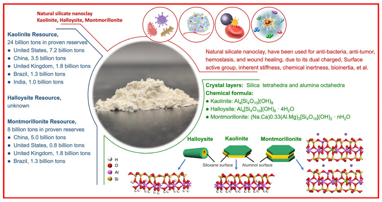
Aluminosilicates, such as montmorillonite, kaolinite, halloysite, and diatomite, have a uniform bidimensional structure, a high surface-to-volume ratio, inherent stiffness, a dual charge distribution, chemical inertness, biocompatibility, abundant active groups on the surface, such as silanol (Si-OH) and/or aluminol (Al-OH) groups. These compounds are on the list of U.S. Food and Drug Administration-approved active compounds and excipients and are used for various medicinal products, such as wound healing agents, antidiarrheals, and cosmetics. This review summarizes the wound healing mechanisms related to the material characteristics and the chemical components. Numerous wound dressings with different active components and multiple forms have been studied. Then, medicinal mineral resources for use in hemostatic materials can be developed.

Keywords: aluminosilicate; clay; hemostasis mechanism; hemostatic material; ionic effect.


Conflict of interest statement

The authors declare no conflict of interest. The funder was not involved in the study design, collection, analysis, interpretation of data, the writing of this article or the decision to submit it for publication.

Figures

Figure 1
Figure 1
An overview diagram for kaolinite, halloysite, and montmorillonite.
Figure 2
Figure 2
An overview of the hemostatic mechanism related to material characteristics. Reprinted with permission from ref. [4]. 2023, Elsevier.
Figure 3
Figure 3
An overview of the hemostatic mechanism related to chemical components. HIF: hypoxia-inducible factor; TNF: tumor necrosis factor; VEGF: vascular endothelial growth factor; ROS: reactive oxygen species. Reprinted with permission from ref. [27]. 2019, Elsevier.
Figure 4
Figure 4
Calcium ions incorporated microporous hydrogel with its (a1,a2) photos and SEM images, (b1,b2) in vitro and in vivo hemostatic evaluation, and (c) wound healing mechanism. 3D-OMS: microporous oxidized maize starch; Ca-OMS: Ca2+ based oxidized maize starch. Reprinted with permission from ref. [36]. 2023, RSC.
Figure 5
Figure 5
Zinc ions incorporated scaffold with its (a) fabrication illustration, (b1b3,c1c3) materials characterization ((b1): SEM; (b2): XRD: (b3): DSL; (c1): SEM; (c2): DSL; (c3): element mappings), (d) in vivo skin burn wound healing evaluation, and (e) wound healing mechanism. PCL: poly(ε-caprolactone). * p < 0.05, ** p < 0.01, *** p < 0.001. Reprinted with permission from ref. [49]. 2023, Wiley.
Figure 6
Figure 6
Iron ion incorporated hydrogel with its (a1a5) fabrication illustration and materials characterization ((a1): fabrication illutration; (a2): photos; (a3): FeS’s TEM; (a4): FeS’s elemental mapping; (a5): FeS/GA hydrogel’s SEM, with red circles indicating FeS), (b1,b2) antibacterial characterization and mechanism, (c) in vivo infected diabetic wound healing evaluation, and (d) wound healing mechanism. GA: glycyrrhizic acid. Scale bars in b1 is 400 nm. Reprinted with permission from ref. [53]. 2023, Elsevier.
Figure 7
Figure 7
Copper ion incorporated hydrogel with its (a) fabrication illustration and materials characterization, (b) schematic illustration with triple stimuli-responsiveness, (c) antibacterial characterization and in vivo infected wound healing evaluation and (d) wound healing mechanism. GSH: glutathione; HA: hyaluronan. Scale bars in a is 300 μm * p < 0.05, ** p < 0.01, *** p < 0.001. Reprinted with permission from ref. [62]. 2022, ACS.
Figure 8
Figure 8
Magnesium ion incorporated hydrogel with its (a1,a2) fabrication illustration and materials characterization, (b) antibacterial characterization (I: QP; II: QP/Mg2+; III: QP/NMN; IV: QP/Mg2+/NMN.), (c) in vivo infected wound healing evaluation, and (d) wound healing mechanism. QP: QCS/PEGSD, quaternized chitosan/poly(glycerol sebacate)–co-poly(ethylene glycol)-g-catechol prepolymer; NMN: nicotinamide mononucleotide. Scale bars in (a2) contains 50 μm (top images) and 200 μm (bottom images). * p < 0.05, ** p < 0.01, *** p < 0.001, **** p < 0.0001. Reprinted with permission from ref. [69]. 2023, Elsevier.
Figure 9
Figure 9
Different forms of clays based wound healing materials, such as (a) kaolinite based hemostatic bandage, (b) halloysite based hemostatic electrospun fibers, and (c) montmorillonite based wound healing hydrogel. Reprinted with permission from refs. [4,73]. 2023, Elsevier.
Figure 10
Figure 10
Commercial bandage with its (a,b) photos and SEM images, and (c) in vitro hemostatic evaluation. Reprinted with permission from ref. [74]. 2023, Elsevier.
Figure 11
Figure 11
Montmorillonite-based hydrogel with its (a) hemostatic mechanism, (b1b3) photo and SEM images, (c) hemolysis assays, and (d1d7) hemostatic experiment in rabbits ((d1): separated femoral artery and transected artery; (d2): the wound caused hemorrhage. (d3): the GMCS was compressed on the wound; (d4): hemostasis was achieved; (d5): the wound was cleaned, and a clot formed (white arrow); (d6): the wound healing image, the white arrows denote the residue GMCS). MMT: montmorillonite; CGS: cross-linked GO sponge; GMCS: GO-MMT composite sponge. Reprinted with permission from ref. [14]. 2016, ACS.
Figure 12
Figure 12
Halloysite-based electrospun fibers with its (a) SEM images, (b) in vitro hemostatic evaluation, (c) hemostatic mechanism, and (d) the blood components-electrospun fibers interaction. PLA: polyactic acid; HNMs: halloysite nanotube microspheres. Reprinted with permission from ref. [74]. 2023, Elsevier.

References

    1. Wang L., You X., Dai C., Tong T., Wu J. Hemostatic Nanotechnologies for External and Internal Hemorrhage Management. Biomater. Sci. 2020;8:4396–4412. doi: 10.1039/D0BM00781A. - DOI - PubMed
    1. Liu L., Hu E., Yu K., Xie R., Lu F., Lu B., Bao R., Li Q., Dai F., Lan G. Recent Advances in Materials for Hemostatic Management. Biomater. Sci. 2021;9:7343–7378. doi: 10.1039/D1BM01293B. - DOI - PubMed
    1. Wang X.X., Liu Q., Sui J.X., Ramakrishna S., Yu M., Zhou Y., Jiang X.Y., Long Y.Z. Recent Advances in Hemostasis at the Nanoscale. Adv. Healthc. Mater. 2019;8:1900823. doi: 10.1002/adhm.201900823. - DOI - PubMed
    1. Tian G., Zhang Y., Jiang Y., Hu P., Wang H., Zhang Y. Nanoscale Zerovalent Iron-Incorporated Kaolinite for Hemostatic and Antibacterial Applications. Appl. Surf. Sci. 2023;636:157879. doi: 10.1016/j.apsusc.2023.157879. - DOI
    1. Teng L., Shao Z., Bai Q., Zhang X., He Y.S., Lu J., Zou D., Feng C., Dong C.M. Biomimetic Glycopolypeptide Hydrogels with Tunable Adhesion and Microporous Structure for Fast Hemostasis and Highly Efficient Wound Healing. Adv. Funct. Mater. 2021;31:2105628. doi: 10.1002/adfm.202105628. - DOI

LinkOut - more resources