. 2024 Mar 22;25(7):3593.
doi: 10.3390/ijms25073593.

Serum Induces the Subunit-Specific Activation of NF-κB in Proliferating Human Cardiac Stem Cells

Affiliations

Serum Induces the Subunit-Specific Activation of NF-κB in Proliferating Human Cardiac Stem Cells

Kazuko E Schmidt et al. Int J Mol Sci. .

Abstract

Cardiovascular diseases (CVDs) are often linked to ageing and are the major cause of death worldwide. The declined proliferation of adult stem cells in the heart often impedes its regenerative potential. Thus, an investigation of the proliferative potential of adult human cardiac stem cells (hCSCs) might be of great interest for improving cell-based treatments of cardiovascular diseases. The application of human blood serum was already shown to enhance hCSC proliferation and reduce senescence. Here, the underlying signalling pathways of serum-mediated hCSC proliferation were studied. We are the first to demonstrate the involvement of the transcription factor NF-κB in the serum-mediated proliferative response of hCSCs by utilizing the NF-κB inhibitor pyrrolidine dithiocarbamate (PDTC). RNA-Sequencing (RNA-Seq) revealed ATF6B, COX5B, and TNFRSF14 as potential targets of NF-κB that are involved in serum-induced hCSC proliferation.

Keywords: NF-κB; PDTC; RNA-Seq; human blood serum; human cardiac stem cells; proliferation.


Conflict of interest statement

The authors declare no conflicts of interest.

Figures

Figure 1
Figure 1
PDTC strongly reduces the proliferation of serum-treated hCSCs. (A) After starvation, hCSCs were treated with human blood serum and the NF-κB inhibitor PDTC. (B) Increasing amounts of PDTC strongly reduced the proliferation of hCSCs after serum treatment with a maximum effect at 500 nM. (C) The IC50 value of PDTC is 11.34 nM. (D) The application of 500 nM PDTC significantly reduced the proliferative effect of human blood serum on hCSCs below the cell count of untreated cells, therefore indicating cell death. (hCSC donor n = 2, human blood serum donors n = 6; technical replicates n = 3, Mann–Whitney U, * p < 0.05, *** p < 0.001).
Figure 2
Figure 2
Human blood serum induces NF-κB-activation by nuclear translocation of the subunits RelA, c-Rel, and RelB. (A) Subsequent to confocal laser scanning microscopy, the nuclear fluorescence intensity was quantified and normalized to untreated cells to determine the subunit translocation from cytosol to the nucleus. Exemplary pictures of c-Rel are shown. (BD) All three NF-κB subunits containing a transactivation domain showed the highest degree of nuclear translocation after 10 to 20 min of treatment in hCSCs, while the maximum effect of serum was visible for c-Rel translocation to the nucleus. (C,D) In addition, RelB and c-Rel show a higher activation in response to serum than to TNFα. (hCSC n = 1, serum from male donors n = 6 (pooled), Kruskal-Wallis test, * p < 0.05; *** p < 0.0005; **** p < 0.0001, ns = not significant).
Figure 3
Figure 3
PDTC significantly reduces the nuclear translocation of NF-κB subunits upon serum treatment. Nuclear fluorescence intensity was quantified and normalized to untreated cells to determine the subunit translocation from cytosol to the nucleus. The induction of the subunit activation of RelA, RelB, and c-Rel was significantly reduced via PDTC after treatment with human blood serum. (hCSC n = 1, serum from male donors n = 6 (pooled), serum from female donors n = 6 (pooled), Kruskal-Wallis test, *** p < 0.0005; **** p < 0.0001).
Figure 4
Figure 4
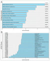
Pathway analysis of hCSCs after serum and PDTC treatment. (A) GO-Term analysis reveals the upregulation of cellular responses to metal ions in serum and PDTC-treated hCSCs. (B) KEGG pathway analysis shows downregulation of cell cycle in serum and PDTC-treated hCSCs.
Figure 5
Figure 5
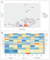
Differential gene expression analysis of hCSCs after treatment with the NF-κB inhibitor PDTC and human blood serum. (A) Vulcano plot reveals significant upregulation of 212 genes and significant downregulation of 66 genes. (B) Heatmap shows differential expression of MT genes after serum and PDTC treatment, highlighted by red box.
Figure 6
Figure 6
ATF6B, TNFRSF14, and COX5B are involved in the NF-κB-regulated response to serum in hCSCs. Immunocytochemical staining of hCSCs shows induction of ATF6B, COX5B, and TNFRSF14 protein expression upon serum treatment compared to untreated cells. This effect is reduced in serum and NF-κB inhibitor PDTC-treated cells compared to serum-treated cells, thereby indicating potential gene expression regulation of ATF6B, COX5B, and TNFRSF14 via NF-κB.

References

    1. López-Otín C., Blasco M.A., Partridge L., Serrano M., Kroemer G. The Hallmarks of Aging. Cell. 2013;153:1194–1217. doi: 10.1016/j.cell.2013.05.039. - DOI - PMC - PubMed
    1. Laflamme M.A., Murry C.E. Heart Regeneration. Nature. 2011;473:326–335. doi: 10.1038/nature10147. - DOI - PMC - PubMed
    1. Aguilar-Sanchez C., Michael M., Pennings S. Cardiac Stem Cells in the Postnatal Heart: Lessons from Development. Stem Cells Int. 2018;2018:1247857. doi: 10.1155/2018/1247857. - DOI - PMC - PubMed
    1. Beltrami A.P., Barlucchi L., Torella D., Baker M., Limana F., Chimenti S., Kasahara H., Rota M., Musso E., Urbanek K., et al. Adult Cardiac Stem Cells Are Multipotent and Support Myocardial Regeneration. Cell. 2003;114:763–776. doi: 10.1016/S0092-8674(03)00687-1. - DOI - PubMed
    1. Bearzi C., Rota M., Hosoda T., Tillmanns J., Nascimbene A., De Angelis A., Yasuzawa-Amano S., Trofimova I., Siggins R.W., LeCapitaine N., et al. Human Cardiac Stem Cells. Proc. Natl. Acad. Sci. USA. 2007;104:14068–14073. doi: 10.1073/pnas.0706760104. - DOI - PMC - PubMed