. 2024 Mar 23;25(7):3611.
doi: 10.3390/ijms25073611.

Co-Culture of P. gingivalis and F. nucleatum Synergistically Elevates IL-6 Expression via TLR4 Signaling in Oral Keratinocytes

Affiliations

Co-Culture of P. gingivalis and F. nucleatum Synergistically Elevates IL-6 Expression via TLR4 Signaling in Oral Keratinocytes

Lucas Yáñez et al. Int J Mol Sci. .

Abstract

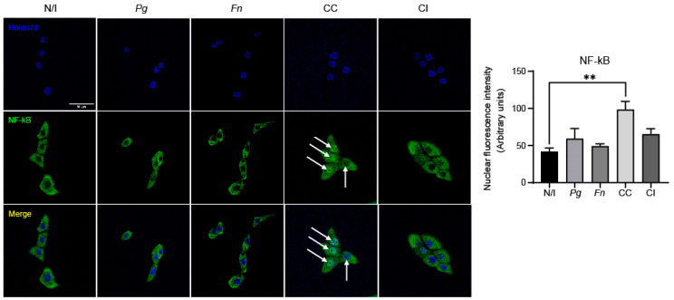
Periodontitis, characterized by persistent inflammation in the periodontium, is intricately connected to systemic diseases, including oral cancer. Bacteria, such as Porphyromonas gingivalis and Fusobacterium nucleatum, play a pivotal role in periodontitis development because they contribute to dysbiosis and tissue destruction. Thus, comprehending the interplay between these bacteria and their impacts on inflammation holds significant relevance in clinical understanding and treatment advancement. In the present work, we explored, for the first time, their impacts on the expressions of pro-inflammatory mediators after infecting oral keratinocytes (OKs) with a co-culture of pre-incubated P. gingivalis and F. nucleatum. Our results show that the co-culture increases IL-1β, IL-8, and TNF-α expressions, synergistically augments IL-6, and translocates NF-kB to the cell nucleus. These changes in pro-inflammatory mediators-associated with chronic inflammation and cancer-correlate with an increase in cell migration following infection with the co-cultured bacteria or P. gingivalis alone. This effect depends on TLR4 because TLR4 knockdown notably impacts IL-6 expression and cell migration. Our study unveils, for the first time, crucial insights into the outcomes of their co-culture on virulence, unraveling the role of bacterial interactions in polymicrobial diseases and potential links to oral cancer.

Keywords: Fusobacterium nucleatum; Porphyromonas gingivalis; co-culture; cytokines; inflammation; oral cancer; periodontitis; toll-like receptor 4.


Conflict of interest statement

The authors declare no conflicts of interest.

Figures

Figure 1
Figure 1
P. gingivalis W50 and F. nucleatum ATCC 10953 are present in a similar ratio after being co-cultured for 24 h. (A) Numbers of colony-forming units per milliliter determined for P. gingivalis W50 and F. nucleatum ATCC 10953 following the co-culture of both bacteria is shown (n = 3). Differences were not significant according to t-tests. (B) Quantification of copy numbers of 16S rRNA for P. gingivalis strain W50 (Pg W50) and of nusG gene for F. nucleatum strain ATCC 10953 (Fn 10953) by qPCR following co-culturing for 24 h. No differences were detected; unpaired t-test; n = 3. (CF) Scanning electron microscopy images of aliquots from monocultures of P. gingivalis W50 (C) and F. nucleatum ATCC 10953 (D) and the co-culture of both bacteria (E,F). White arrows suggest physical interactions between P. gingivalis and F. nucleatum. Scale bar, 1 µm (C,D,F) and 10 µm (E).
Figure 2
Figure 2
The co-culture of P. gingivalis and F. nucleatum synergistically increases the expressions of pro-inflammatory cytokines. The fold increases in the relative expressions (ΔΔCTs) of pro-inflammatory cytokines TNF-α (A), IL-1β (B), IL-8 (C), and IL-6 (D) in OKF6/TERT2 cells at 24 h post infection. Multiple comparisons were made with the non-infected control. One-way ANOVA; Dunnett post-test. * indicates significant difference of p < 0.05; n ≥ 4. N/I = non-infected control; Pg = P. gingivalis; Fn = F. nucleatum; CC = co-culture; CI = co-infection with both bacteria previously grown separately.
Figure 3
Figure 3
The co-culture of P. gingivalis and F. nucleatum promotes NF-kB but not STAT3 phosphorylation. Representative images of (A) phospho-NF-kB(S536) and total NF-kB and (B) phospho-STAT3 (Y705) and total STAT3 for western blot analysis of OKF6/TERT2 cells at 2 h post infection (representative blots to the left), and quantifications by scanning densitometry are shown (graphs to the right). Data were analyzed using one-way ANOVA followed by a Dunnett post-test. * indicates significant difference (p < 0.05; n = 3). N/I = non-infected control; Pg = P. gingivalis; Fn = F. nucleatum; CC = co-culture; CI = co-infection with both bacteria previously grown separately.
Figure 4
Figure 4
The co-culture of P. gingivalis and F. nucleatum promotes NF-kB translocation to the cell nucleus at 2 h post infection. Representative images are shown for NF-kB fluorescence (green channel), nuclear staining (blue channel), and the merger of these obtained in OKF6/TERT2 cells at 2 h post infection. Nuclear fluorescence of NF-kB was quantified for all the experimental conditions using imageJ software(version 2.9.0). White arrows indicate nuclear NF-kB. Data were analyzed using one-way ANOVA followed by a Dunnett post-test. ** indicates significant difference (p < 0.005; n = 3). N/I = non-infected control; Pg = P. gingivalis; Fn = F. nucleatum; CC = co-culture; CI = co-infection with both bacteria previously grown separately. Images taken at ×63 magnification. Scale bar, 50 µm.
Figure 5
Figure 5
The co-culture of P. gingivalis and F. nucleatum does not trigger STAT3 nuclear translocation at 2 h post infection in OKs. Representative images of STAT3 fluorescence (green channel), nuclear staining (blue channel), and the merger of these observed in OKF6/TERT2 cells at 2 h post infection are shown. Nuclear fluorescence of STAT3 was quantified for all the experimental conditions using imageJ software(version 2.9.0). Data were analyzed using one-way ANOVA followed by a Dunnett post-test (n = 3). N/I = non-infected control; Pg = P. gingivalis; Fn = F. nucleatum; CC = co-culture; CI = co-infection with both bacteria previously grown separately. Images taken at ×63 magnification. Scale bar, 50 µm.
Figure 6
Figure 6
The co-culture of P. gingivalis and F. nucleatum is not cytotoxic and increases the cell migration of oral keratinocytes. Cell viability of OKF6/TERT2 cells at 24 h post infection was evaluated using MTS (A) and trypan blue assays (B). Data were analyzed using one-way ANOVA followed by a Dunnett post-test (n = 3). (C) OKF6/TERT2 cells were infected for 1.5 h, then migration after 2 h was measured. Data correspond to the average number of migrated cells observed in 7 fields. Data were analyzed using one-way ANOVA followed by a Dunnett post-test (*, **, and *** indicate significant differences of p < 0.05, p < 0.005, and p < 0.0005, respectively; n = 3). N/I = non-infected control; Pg = P. gingivalis; Fn = F. nucleatum; CC = co-culture; CI = co-infection with both bacteria previously grown separately.
Figure 7
Figure 7
The co-culture of P. gingivalis and F. nucleatum does not affect TLR4 levels. Representative image of TLR4 levels revealed by western blot analysis of OKF6/TERT2 cells at 24 h post infection (left panel), and quantification by scanning densitometry (right panel). Data were analyzed using one-way ANOVA followed by a Dunnett post-test (n = 5). N/I = non-infected control; Pg = P. gingivalis; Fn = F. nucleatum; CC = co-culture; CI = co-infection with both bacteria previously grown separately.
Figure 8
Figure 8
The co-culture of P. gingivalis and F. nucleatum regulates the expression of IL-6 and cell migration through TLR4 signaling. (A) The fold increases in the relative expressions (ΔΔCTs) of pro-inflammatory cytokines TNF-α (A), IL-1β (B), IL-8 (C), and IL-6 (D) observed in shTLR4 cells at 24 h post infection are shown. Multiple comparisons were made against the shTLR4 non-infected control. Data were analyzed using one-way ANOVA followed by a Dunnett post-test (* and ** indicate significant differences of p < 0.05 and p < 0.005, respectively; n = 3). (E) OKF6/TERT2 cells, shTLR4 cells, and shScrambled cells were infected for 1.5 h, then migration after 2 h was measured. Data correspond to the average numbers of migrated cells observed in 7 fields. Multiple comparisons were carried out using one-way ANOVA followed by Tukey’s post-test (** and *** indicate significant differences of p < 0.005 and p < 0.0001, respectively; n = 3). WT = wild-type control; N/I = non-infected control; Pg = P. gingivalis; Fn = F. nucleatum; CC = co-culture; CI = co-infection with both bacteria previously grown separately.
Figure 9
Figure 9
Proposed model of this work. The following model is proposed. For the first time, we demonstrate that the co-culture of P. gingivalis and F. nucleatum activates the expression of IL-6 and cell migration. We suggest that the pathway involved includes TLR4 because the knockdown of TLR4 blocked this expression. We also observed a correlation between NF-kB phosphorylation and nuclear translocation upon infecting OKs with the bacterial co-culture, suggesting the participation of NF-kB in this process. Moreover, both the co-culture and the monoculture of P. gingivalis regulate cell migration through TLR4 signaling (potential connections, as indicated, are illustrated with dashed arrows).

References

    1. Larsson L., Kavanagh N.M., Nguyen T.V.N., Castilho R.M., Berglundh T., Giannobile W.V. Influence of epigenetics on periodontitis and peri-implantitis pathogenesis. Periodontol. 2000. 2022;90:125–137. doi: 10.1111/prd.12453. - DOI - PubMed
    1. Papapanou P.N., Susin C. Periodontitis epidemiology: Is periodontitis under-recognized, over-diagnosed, or both? Periodontol. 2000. 2017;75:45–51. doi: 10.1111/prd.12200. - DOI - PubMed
    1. Abusleme L., Dupuy A.K., Dutzan N., Silva N., Burleson J.A., Strausbaugh L.D., Gamonal J., Diaz P.I. The subgingival microbiome in health and periodontitis and its relationship with community biomass and inflammation. ISME J. 2013;7:1016–1025. doi: 10.1038/ismej.2012.174. - DOI - PMC - PubMed
    1. Ferlay J., Colombet M., Soerjomataram I., Mathers C., Parkin D.M., Pineros M., Znaor A., Bray F. Estimating the global cancer incidence and mortality in 2018: GLOBOCAN sources and methods. Int. J. Cancer. 2019;144:1941–1953. doi: 10.1002/ijc.31937. - DOI - PubMed
    1. Ye L., Jiang Y., Liu W., Tao H. Correlation between periodontal disease and oral cancer risk: A meta-analysis. J. Cancer Res. Ther. 2016;12:C237–C240. doi: 10.4103/0973-1482.200746. - DOI - PubMed

LinkOut - more resources