Review
. 2024 Mar 23;25(7):3618.
doi: 10.3390/ijms25073618.

Multidisciplinary Approach to the Diagnosis of Idiopathic Interstitial Pneumonias: Focus on the Pathologist's Key Role

Affiliations
Review

Multidisciplinary Approach to the Diagnosis of Idiopathic Interstitial Pneumonias: Focus on the Pathologist's Key Role

Stefano Lucà et al. Int J Mol Sci. .

Abstract

Idiopathic Interstitial Pneumonias (IIPs) are a heterogeneous group of the broader category of Interstitial Lung Diseases (ILDs), pathologically characterized by the distortion of lung parenchyma by interstitial inflammation and/or fibrosis. The American Thoracic Society (ATS)/European Respiratory Society (ERS) international multidisciplinary consensus classification of the IIPs was published in 2002 and then updated in 2013, with the authors emphasizing the need for a multidisciplinary approach to the diagnosis of IIPs. The histological evaluation of IIPs is challenging, and different types of IIPs are classically associated with specific histopathological patterns. However, morphological overlaps can be observed, and the same histopathological features can be seen in totally different clinical settings. Therefore, the pathologist's aim is to recognize the pathologic-morphologic pattern of disease in this clinical setting, and only after multi-disciplinary evaluation, if there is concordance between clinical and radiological findings, a definitive diagnosis of specific IIP can be established, allowing the optimal clinical-therapeutic management of the patient.

Keywords: desquamative interstitial pneumonia; diffuse alveolar damage; idiopathic interstitial pneumonias; idiopathic pulmonary fibrosis; interstitial lung disease; multidisciplinary discussion; nonspecific interstitial pneumonia; organizing pneumonia; pleuroparenchymal fibroelastosis; usual interstitial pneumonia.


Conflict of interest statement

The authors declare no conflicts of interest.

Figures
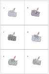
Figure 2
Figure 2
Schematic representation of normal SPL and key histopathological changes of IIPs. Normal secondary pulmonary lobule consisting of several primary lobules, supplied by the bronchiolovascular bundle (preterminal bronchiole and a branch of the pulmonary artery) and surrounded incompletely by fibrous interlobular septa (A). Heterogeneous appearance of UIP pattern with characteristic “patchwork” due to the patchy scarred fibrosis associated with a normal lung parenchyma (B). Temporal and geographical uniformity of NSIP pattern with diffuse and uniform interstitial fibrosing process characterizing the fibrotic type of disease (C). Patchy airspace filled by Masson bodies is the histopathological hallmark of the OP pattern (D). The basic element of the pathological DIP pattern is alveolar filling by pigmented macrophages, termed as “smokers’ macrophages” (E). Diffuse and dense cellular chronic interstitial infiltrate with a primarily alveolar septal distribution, with extensive infiltration and thickening of the alveolar septa, is the histological hallmark of the LIP pattern (F). (SPL, secondary pulmonary lobule; IIPs, Idiopathic Interstitial Pneumonias; UIP, Usual Interstitial Pneumonia; NSIP, Nonspecific Interstitial Pneumonia; OP, Organizing Pneumonia; DIP, Desquamative Interstitial Pneumonia; LIP, Lymphoid Interstitial Pneumonia).
Figure 3
Figure 3
UIP pattern. Temporal and spatial heterogeneity diagnostic of UIP pattern ((A,B); hematoxylin and eosin, original magnifications ×500 and ×200). Temporal heterogeneity is defined by fibroblast foci localized at the interface between dense fibrosis and normal lung parenchyma ((B); hematoxylin and eosin, original magnifications ×200). Architectural distortion with normal alveolar structure replaced by scar-like fibrosis and/or honeycomb changes (C,D). Honeycomb change consists of reorganized, enlarged, and irregularly dilated cystic airspaces at least partially lined by ciliated (respiratory tract) epithelium ((C); hematoxylin and eosin, original magnifications ×1000). Normal lung parenchyma replaced by scar-like fibrosis define the end-stage lung disease ((D); hematoxylin and eosin, original magnifications ×500).
Figure 1
Figure 1
Current classification of Interstitial Lung Diseases (ILDs) [9]. Idiopathic Interstitial Pneumonias (IIPs), Idiopathic Pulmonary Fibrosis (IPF), Idiopathic Nonspecific Interstitial Pneumonia (iNSIP), Cryptogenic Organizing Pneumonia (COP), Acute Interstitial Pneumonia (AIP), Idiopathic Lymphoid Interstitial Pneumonia (iLIP), Idiopathic Desquamative Interstitial Pneumonia (iDIP), Idiopathic Pleuroparenchymal Fibroelastosis (iPPFE), Respiratory Bronchiolitis–Interstitial Lung Disease (RB-ILD), Acute Fibrinous and Organizing Pneumonia (AFOP), Rheumatoid Arthritis (RA), Systemic Sclerosis (SSc), Mixed Connective Tissue Disease (MCTD), Systemic Lupus Erythematosus (SLE), Hypersensitivity Pneumonitis (HP), Eosinophilic Pneumonia (EP), Langerhans Cell Histiocytosis (LCH), Pulmonary Alveolar Proteinosis (PAP), Lymphangioleiomyomatosis (LAM). * Occupational: asbestosis, silicosis, coal miner, berylliosis, others.
Figure 4
Figure 4
NSIP pattern. Temporal and spatial uniformity of NSIP pattern ((A,B); hematoxylin and eosin, original magnifications ×1000 and ×500). Diffuse and uniform interstitial chronic inflammatory infiltrate of cNSIP ((C); hematoxylin and eosin, original magnifications ×200). Diffuse and uniform interstitial fibrosis with associated lympho-plasmacytic infiltrate of variable degrees of fNSIP ((D); hematoxylin and eosin, original magnifications ×500). (cNSIP, cellular Nonspecific Interstitial Pneumonia; fNSIP, fibrotic Nonspecific Interstitial Pneumonia).
Figure 5
Figure 5
OP pattern. Patchy airspace filled with Masson bodies. Masson bodies are clustered in airspaces within and around small bronchioles and alveolar ducts, and they vary in shape according to the airspace’s size—round to elongated when localized in bronchioles, serpiginous and branching when localized in alveolar ducts and small and round when localized in alveolar spaces ((A,B); hematoxylin and eosin, original magnifications ×500 and ×200). Reactive alveolar pneumocyte hyperplasia in OP ((C); hematoxylin and eosin, original magnifications ×50).
Figure 6
Figure 6
DAD pattern. Hyaline membranes are the histologic hallmark of the DAD exudative phase; they are associated with airspace epithelial denudation ((A,B); hematoxylin and eosin, original magnifications ×100 and ×100).
Figure 7
Figure 7
PPFE pattern. The basic elements of the PPFE pattern are temporally uniform elastofibrosis involving the pleura and subpleural lung parenchyma ((A,B); hematoxylin and eosin, original magnifications ×500 and ×200).
Figure 8
Figure 8
Thoracic computed tomography of the main Idiopathic Interstitial Pneumonias (IIPs). Respiratory Bronchiolitis–Interstitial Lung Disease (RB-ILD) is characterized by poorly formed centrolobular nodules (arrows); Idiopathic Nonspecific Interstitial Pneumonia (iNSIP) is characterized by patchy ground-glass opacities (GGOs) possibly with traction bronchiectasis (circle) and reticulations (arrow); Desquamative Interstitial Pneumonia (DIP) is characterized by homogeneous fibrotic GGOs with traction bronchiectasis (circle) possibly associated with cysts; Idiopathic Pulmonary Fibrosis (IPF) is associated with patchy areas of honeycombing with traction bronchiectasis (circle) with reticulation (arrow); Pleuroparenchymal Fibroelastosis (PPFE) is characterized by bilateral irregular apex thickening (circles); Cryptogenetic Organizing Pneumonia (COP) is associated with peribronchial or peripheral consolidative areas (arrows) possibly associated with perilobular pattern or reverse halo sign (circle).

References

    1. Guler S.A., Corte T.J. Interstitial Lung Disease in 2020: A History of Progress. Clin. Chest Med. 2021;42:229–239. doi: 10.1016/j.ccm.2021.03.001. - DOI - PubMed
    1. Antoniou K.M., Margaritopoulos G.A., Tomassetti S., Bonella F., Costabel U., Poletti V. Interstitial lung disease. Eur. Respir. Rev. 2014;23:40–54. doi: 10.1183/09059180.00009113. - DOI - PMC - PubMed
    1. Carvajalino S., Reigada C., Johnson M.J., Dzingina M., Bajwah S. Symptom prevalence of patients with fibrotic interstitial lung disease: A systematic literature review. BMC Pulm. Med. 2018;18:78. doi: 10.1186/s12890-018-0651-3. - DOI - PMC - PubMed
    1. Lucà S., Zannini G., Morgillo F., Della Corte C.M., Fiorelli A., Zito Marino F., Campione S., Vicidomini G., Guggino G., Ronchi A., et al. The prognostic value of histopathology in invasive lung adenocarcinoma: A comparative review of the main proposed grading systems. Expert. Rev. Anticancer Ther. 2023;23:265–277. doi: 10.1080/14737140.2023.2179990. - DOI - PubMed
    1. Lucà S., Franco R., Napolitano A., Soria V., Ronchi A., Zito Marino F., Della Corte C.M., Morgillo F., Fiorelli A., Luciano A., et al. PATZ1 in Non-Small Cell Lung Cancer: A New Biomarker That Negatively Correlates with PD-L1 Expression and Suppresses the Malignant Phenotype. Cancers. 2023;15:2190. doi: 10.3390/cancers15072190. - DOI - PMC - PubMed

LinkOut - more resources