. 2024 Mar 26;25(7):3693.
doi: 10.3390/ijms25073693.

FL118 Is a Potent Therapeutic Agent against Chronic Myeloid Leukemia Resistant to BCR-ABL Inhibitors through Targeting RNA Helicase DDX5

Affiliations

FL118 Is a Potent Therapeutic Agent against Chronic Myeloid Leukemia Resistant to BCR-ABL Inhibitors through Targeting RNA Helicase DDX5

Kengo Takeda et al. Int J Mol Sci. .

Abstract

Chronic myeloid leukemia (CML) is induced by the expression of the fused tyrosine kinase BCR-ABL, which is caused by a chromosomal translocation. BCR-ABL inhibitors have been used to treat CML; however, the acquisition of resistance by CML cells during treatment is a serious issue. We herein demonstrated that BCR-ABL induced the expression of the RNA helicase DDX5 in K562 cells derived from CML patients in a manner that was dependent on its kinase activity, which resulted in cell proliferation and survival. The knockout of DDX5 decreased the expression of BIRC5 (survivin) and activated caspase 3, leading to apoptosis in K562 cells. Similar results were obtained in cells treated with FL118, an inhibitor of DDX5 and a derivative compound of camptothecin (CPT). Furthermore, FL118 potently induced apoptosis not only in Ba/F3 cells expressing BCR-ABL, but also in those expressing the BCR-ABL T315I mutant, which is resistant to BCR-ABL inhibitors. Collectively, these results revealed that DDX5 is a critical therapeutic target in CML and that FL118 is an effective candidate compound for the treatment of BCR-ABL inhibitor-resistant CML.

Keywords: BCR-ABL; DDX5; FL118; apoptosis; chronic myeloid leukemia (CML).


Conflict of interest statement

The authors declare no conflicts of interest.

Figures
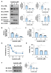
Figure 1
Figure 1
The treatment with imatinib reduces the expression of DDX5 in K562 cells. (A,B) K562 cells were treated with imatinib (1 and 2 μM) for 24 h. (A) The phosphorylation and/or expression levels of BCR-ABL, STAT5, BIRC5, and cleaved caspase-3 were examined by immunoblotting. The relative phosphorylation or expression of each protein is shown in the graphs. *, **, and *** indicate p < 0.05, p < 0.01, and p < 0.001, respectively. (B,C) The mRNA expression of DDX5 and BIRC5 was analyzed by RT-PCR. The mRNA expression level of β2-microglobulin was used as an internal control. *, **, and *** indicate p < 0.05, p < 0.01 and p < 0.001, respectively. (D) K562 cells (2 × 10⁴ cells/100 μL) were treated with imatinib (0.125, 0.25, 0.5, 1, and 2 μM) for 48 h. Cell proliferation was assessed by the WST assay. *** indicates p < 0.001. (E) K562 cells (4 × 10⁶ cells/4 mL) were treated with imatinib (1 and 2 μM) for 48 h. Cell viability was evaluated by the Trypan blue staining method. *** indicates p < 0.001. (F) The expression level of DDX5 was examined by immunoblotting and the relative expression of DDX5 is shown in the graph. * indicates p < 0.05.
Figure 2
Figure 2
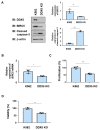
The knockout of DDX5 induces apoptosis in K562 cells. DDX5-KO cells were established by knocking out DDX5 in K562 cells by genome editing. (A,B) K562 cells and DDX5-KO cells were cultured in RPMI medium containing 1% FBS for 24 h. (A) Whole-cell lysates were prepared and the expression of BIRC5, cleaved caspase-3, or β-actin was examined by immunoblotting. The relative expression levels of BIRC5 and cleaved caspase-3 are shown in the graphs. ** and *** indicate p < 0.01 and p < 0.001, respectively. (B) The mRNA expression of BIRC5 was analyzed by RT-PCR. The mRNA expression level of β2-microglobulin was used as an internal control. * indicates p < 0.01. (C) Transduced K562 cells (1 × 10⁴ cells/100 μL) were cultured in RPMI containing 1% FBS for 24 h. Cell proliferation was assessed by the WST assay. *** indicates p < 0.001. (D) Transduced K562 cells (1 × 10⁵ cells/1 mL) were cultured in RPMI containing 1%FBS for 24 h. Cell viability was evaluated by the Trypan blue staining method. ** indicates p < 0.01.
Figure 3
Figure 3
FL118 inhibits the activity of Topo I and suppresses the expression of DDX5. (A) The chemical structures of camptothecin (CPT) and FL118 are shown. (B) The effects of CPT and FL118 on Topo I activity were measured using recombinant Topo I and supercoiled DNA. After the reaction at 37 °C for 30 min, electrophoresis was performed using a 1% agarose gel. As a control, relaxed DNA was electrophoresed. (C) K562 cells were treated with CPT (125, 250, and 500 nM) or FL118 (125, 250, and 500 nM) for 24 h. Whole-cell lysates were prepared, the expression of DDX5 or β-actin was examined by immunoblotting, and the relative expression level of DDX5 is shown in the graph. * and ** indicate p < 0.05 and p < 0.01, respectively. (D) K562 cells were treated with FL118 (500 nM and DMSO (0.1%) or MG-132 (5 μM) for 12 h. Whole-cell lysates were prepared, the expression of DDX5 or β-actin was examined by immunoblotting, and the relative expression level of DDX5 is shown in the graph. *, **, and *** indicate p < 0.05, p < 0.01, and p < 0.001, respectively.
Figure 4
Figure 4
FL118 reduces the expression of BIRC5 and activates caspase-3 in K562 cells. (A,B) K562 cells were treated with CPT or FL118 at the indicated concentrations for 24 h. (A) Whole-cell lysates were prepared and the expression of BIRC5, cleaved caspase-3, or β-actin was examined by immunoblotting. The relative expression level of BIRC5 and cleaved caspase-3 is shown in the graphs. ** and *** indicate p < 0.01 and p < 0.001, respectively. ## and ### indicate p < 0.01 and p < 0.001, respectively. (B) The mRNA expression of BIRC5 was analyzed by RT-PCR. The mRNA expression level of β2-microglobulin was used as an internal control. *, **, and *** indicate p < 0.05, p < 0.01, and p < 0.001, respectively. # and ### indicate p < 0.05 and p < 0.001, respectively.
Figure 5
Figure 5
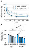
FL118 reduced cell proliferation and induced cell death more strongly than CPT in K562 cells. (A) K562 cells (2 × 10⁴ cells/100 μL) were treated with CPT or FL118 at various concentrations for 48 h. Cell proliferation was assessed by WST assay. *** and ### indicate p < 0.001. (B) Transduced K562 cells (4 × 10⁶ cells/4 mL) were treated with CPT (125, 250, and 500 nM) or FL118 (125, 250, and 500 nM) for 48 h. Cell viability was evaluated by the Trypan blue staining method. *, **, and *** indicate p < 0.05, p < 0.01, and p < 0.001, respectively. ## and ### indicate p < 0.01 and p < 0.001, respectively.
Figure 6
Figure 6
Exogenous expression of BCR-ABL and its T315I mutant induces the expression of DDX5 in Ba/F3 cells. (A) The structures of BCR-ABL and its T315I mutant are shown. (BF) Ba/F3 cells expressed BCR-ABL and its T315I mutant by retrovirus infection. Ba/F3 cells expressing BCR-ABL or the T315I mutant were incubated with RPMI containing 10% FBS for 24 h. (B) Whole-cell lysates were prepared and immunoblotting was performed. The relative expression level of DDX5 is shown in the graph. *** indicates p < 0.001. (C) Whole-cell lysates were prepared and immunoblotting was performed. The relative phosphorylation level of BCR-ABL or STAT5 and the relative expression of BIRC5, cleaved caspase-3, or DDX5 are shown in the graphs. ** and *** indicate p < 0.01 and p < 0.001, respectively. (D) The mRNA expression of Birc5 was analyzed by RT-PCR. The mRNA expression level of β2-microglobulin was used as an internal control. *** indicates p < 0.001. (E) Ba/F3 cells expressing BCR-ABL or the T315I mutant (1 × 10⁴ cells/100 μL) were treated with imatinib at various concentrations for 24 h. Cell proliferation was assessed by the WST assay. (F) Ba/F3 cells expressing BCR-ABL or the T315I mutant (5 × 10⁵ cells/1 mL) were treated with imatinib (2 μM) for 18 h. Cell viability was evaluated by the Trypan blue staining method. *** indicates p < 0.001.
Figure 7
Figure 7
The knockout of DDX5 induces cell death in Ba/F3 cells expressing BCR-ABL or the BCR-ABL T315I mutant. DDX5 was knocked out in Ba/F3 cells expressing BCR-ABL and Ba/F3 cells expressing the BCR-ABL T315I mutant by genome editing. These cells were cultured in RPMI medium containing 10% FBS for 24 h. (A) Whole-cell lysates were prepared and the expression of DDX5, BIRC5, or β-actin was examined by immunoblotting. The relative expression levels of BIRC5 are shown in the graph. ** indicates p < 0.01. (B) The mRNA expression of Birc5 was analyzed by RT-PCR. The mRNA expression level of β2-microglobulin was used as an internal control. * and ** indicate p < 0.05 and p < 0.01, respectively. (C) Transduced Ba/F3 cells (1 × 10⁴ cells/100 μL) were cultured in RPMI containing 1% FBS for 24 h. Cell proliferation was assessed by the WST assay. *** indicates p < 0.001. (D) Transduced Ba/F3 cells (1 × 10⁵ cells/1 mL) were cultured in RPMI containing 1% FBS for 24 h. Cell viability was evaluated by the Trypan blue staining method. ** and *** indicate p < 0.01 and p < 0.001, respectively.
Figure 8
Figure 8
FL118 reduces the expression of BIRC5 and activates caspase-3 in Ba/F3 cells expressing BCR-ABL or the T315I mutant. (A,B) Ba/F3 cells expressing BCR-ABL or the T315I mutant were treated with CPT (50 and 100 nM) or FL118 (50 and 100 nM) for 24 h. (A) Whole-cell lysates were prepared and immunoblotting was performed. The relative expression levels of DDX5, BIRC5, and cleaved caspase-3 are shown in the graphs. *, **, and *** indicate p < 0.05, p < 0.01, and p < 0.001, respectively. ## and ### indicate p < 0.01 and p < 0.001, respectively. (B) The mRNA expression of Birc5 was analyzed by RT-PCR. The mRNA expression level of β2-microglobulin was used as an internal control. *, **, and *** indicate p < 0.05, p < 0.01, and p < 0.001, respectively. # and ### indicate p < 0.05 and p < 0.001, respectively.
Figure 9
Figure 9
FL118 induces apoptosis in Ba/F3 cells expressing BCR-ABL or the T315I mutant. (A) Ba/F3 cells expressing BCR-ABL or the T315I mutant (1 × 10⁴ cells/100 μL) were treated at various concentrations for 24 h. Cell proliferation was assessed by the WST assay. *, **, and *** indicate p < 0.05, p < 0.01, and p < 0.001, respectively. ### indicates p < 0.001. (B) Ba/F3 cells expressing BCR-ABL or the T315I mutant (5 × 10⁵ cells/1 mL) were treated with CPT (50 and 100 nM) or FL118 (50 and 100 nM) for 18 h. Cell viability was evaluated by the Trypan blue staining method. **, and *** indicate p < 0.01, and p < 0.001, respectively. # and ## indicate p < 0.05, and p < 0.001, respectively.
Figure 10
Figure 10
FL118, a DDX5 inhibitor, may be a novel therapeutic agent for CML that is resistant to BCR-ABL inhibitors. BCR-ABL promotes the proliferation and survival of CML cells by inducing the expression of DDX5. Due to the appearance of a point mutation (T315) in BCR-ABL during treatment with the BCR-ABL inhibitor imatinib, CML cells acquire resistance to BCR-ABL inhibitors. Similar to camptothecin (CPT), FL118 inhibits Topo I, but also suppresses the expression of DDX5. Therefore, FL118 has potential as a new treatment for CML that overcomes the drug resistance of CML cells expressing the BCR-ABL T315I mutation.

Similar articles

References

    1. Rowley J.D. Letter: A new consistent chromosomal abnormality in chronic myelogenous leukaemia identified by quinacrine fluorescence and Giemsa staining. Nature. 1973;243:290–293. doi: 10.1038/243290a0. - DOI - PubMed
    1. de Klein A., van Kessel A.G., Grosveld G., Bartram C.R., Hagemeijer A., Bootsma D., Spurr N.K., Heisterkamp N., Groffen J., Stephenson J.R. A cellular oncogene is translocated to the Philadelphia chromosome in chronic myelocytic leukaemia. Nature. 1982;300:765–767. doi: 10.1038/300765a0. - DOI - PubMed
    1. Pane F., Intrieri M., Quintarelli C., Izzo B., Muccioli G.C., Salvatore F. BCR/ABL genes and leukemic phenotype: From molecular mechanisms to clinical correlations. Oncogene. 2002;21:8652–8667. doi: 10.1038/sj.onc.1206094. - DOI - PubMed
    1. Zhao X., Ghaffari S., Lodish H., Malashkevich V.N., Kim P.S. Structure of the Bcr-Abl oncoprotein oligomerization domain. Nat. Struct. Biol. 2002;9:117–120. - PubMed
    1. Sonoyama J., Matsumura I., Ezoe S., Satoh Y., Zhang X., Kataoka Y., Takai E., Mizuki M., Machii T., Wakao H., et al. Functional cooperation among Ras, STAT5, and phosphatidylinositol 3-kinase is required for full oncogenic activities of BCR/ABL in K562 cells. J. Biol. Chem. 2002;277:8076–8082. doi: 10.1074/jbc.M111501200. - DOI - PubMed

MeSH terms

Substances