Review
. 2024 Apr 3;25(7):3995.
doi: 10.3390/ijms25073995.

Neuroinflammation in Neurodegenerative Disorders: Current Knowledge and Therapeutic Implications

Affiliations
Review

Neuroinflammation in Neurodegenerative Disorders: Current Knowledge and Therapeutic Implications

Paras Mani Giri et al. Int J Mol Sci. .

Abstract

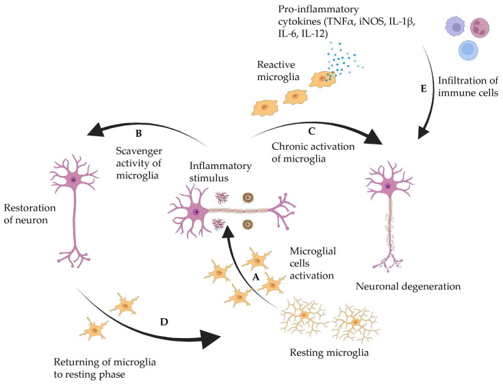
Neurodegenerative disorders (NDs) have become increasingly common during the past three decades. Approximately 15% of the total population of the world is affected by some form of NDs, resulting in physical and cognitive disability. The most common NDs include Alzheimer's disease, Parkinson's disease, amyotrophic lateral sclerosis, and Huntington's disease. Although NDs are caused by a complex interaction of genetic, environmental, and lifestyle variables, neuroinflammation is known to be associated with all NDs, often leading to permanent damage to neurons of the central nervous system. Furthermore, numerous emerging pieces of evidence have demonstrated that inflammation not only supports the progression of NDs but can also serve as an initiator. Hence, various medicines capable of preventing or reducing neuroinflammation have been investigated as ND treatments. While anti-inflammatory medicine has shown promising benefits in several preclinical models, clinical outcomes are often questionable. In this review, we discuss various NDs with their current treatment strategies, the role of neuroinflammation in the pathophysiology of NDs, and the use of anti-inflammatory agents as a potential therapeutic option.

Keywords: Alzheimer’s disease; Huntington’s disease; Parkinson’s disease; amyotrophic lateral sclerosis; anti-inflammatory drugs; neurodegenerative disorders.


Conflict of interest statement

The authors declare no conflicts of interest.

Figures

Figure 1
Figure 1
Role of microglia in AD progression. (A) Inactive microglia become activated by stimuli like amyloid β or tau protein deposition. (B) Complete clearance of amyloid β leads to restoration of neurons, and (D) microglia returns to the resting phase. In contrast, (C) failure in amyloid β clearance leads to chronic microglia activation that releases proinflammatory cytokines and neurotoxicity. (E) Entry of the peripheral immune system leads to neurodegeneration. This figure was created using Biorender.com.
Figure 2
Figure 2
(A) Sections of hippocampus and subiculum immunostained for ionized calcium binding adaptor molecule 1 (Iba1) in 5xFAD mice untreated, treated with 1 mg/kg/day, and treated with 5 mg/kg/day of fingolimod. Untreated 3 month old 5xFAD mice showed significant increase in the number of activated microglial cells. (B) Quantitation of total, active, and resting variants of microglia in the hippocampus of 5xFAD untreated and fingolimod-treated groups. The number of activated Iba1-positive microglia significantly decreased in the hippocampus of 1 mg/kg/day, and 5 mg/kg/day fingolimod-treated groups compared with untreated group (* p < 0.05), (n = 8–10 mice/group) This figure is reprinted under the terms of the Creative Commons Attribution 4.0 International License from reference [99].
Figure 3
Figure 3
Role of inflammation in PD pathogenesis. (A1 and A2): Pathological conditions like genetic mutation, protein aggregation, aging, and cytokine released from infiltrated T cells activate microglia. (B) The release of inflammatory mediators from microglia activates astrocytes. (C) Release of IL-B, TNFα, and ROS from activated astrocytes causes dopaminergic neuron degeneration. (D) Degeneration of dopaminergic neurons due to chemokine release from astrocytes. (E) The chemicals released from degenerating neurons activate glial cells and aggravate inflammatory responses. This figure is adapted under the terms of the Creative Commons Attribution 4.0 International Li-cense from reference [111].
Figure 4
Figure 4
Effect of Hidrox® on behavioral impairments and histological parameters induced by rotenone intoxication. (A,A1) Motor function was assessed using a Pole test. At 28 days, mice exhibited a significant motor dysfunction as indicated by an increase in “Time to turn” and “Total time” spent to descend to the floor following injection of rotenone compared with the Sham group. Hidrox® administration notably reduced “Total time” and “Time to turn”. (A) *** p < 0.001 vs. Sham; ** p < 0.01 vs. rotenone; (A1) *** p < 0.001 vs. Sham; ** p < 0.01 vs. rotenone. (B) At 28 days, using a Rotarod apparatus, mice exhibited a significant motor dysfunction as indicated by a decrease in time spent on the Rotarod. Hidrox® treatment blunted the motor dysfunction in mice. (B) ** p < 0.01 vs. Sham; * p < 0.05 vs. rotenone. (C) Catalepsy was evaluated according to the standard bar hanging procedure; this motor test showed that the Hidrox® treatment reduced behavioral impairment induced by rotenone. (C) *** p < 0.001 vs. Sham; *** p < 0.001 vs. rotenone. Values are the mean ± SEM of 10 mice for each group. Sham+Hidrox® and Sham+vehicle groups showed no evidence of degenerating cells in the SN (D,E), whereas degeneration of neuromelanin-pigmented cells was evident in the SN of the rotenone-treated animals (F). Hidrox® treatment restored the architecture compared to the control mice (G). The data are representative of at least three independent experiments and are expressed as the mean ± SEM of 5 mice for each group. (H) * p < 0.05 vs. Sham; * p < 0.05 vs. rotenone. Scale bar: 50 μm. This figure is reprinted under the terms of the Creative Commons Attribution 4.0 International License from reference [122].
Figure 5
Figure 5
Treatment with RD2RD2 rescued significant numbers of neurons in the motor cortex of SOD1*G93A mice. Analysis of neurons in the brain stem (A) and motor cortex (B) revealed a significant loss in placebo-treated SOD1*G93A mice, while the count of neurons in RD2RD2-treated mice was similar to the count in non-transgenic mice. Data are presented as mean ± SEM. Statistical calculations were conducted by one-way ANOVA with Fisher’s LSD post hoc analysis, ntg n = 11, RD2RD2 n = 11, placebo n = 8 (brain stem) and ntg n = 10, RD2RD2 n = 10, placebo n = 10 (motor cortex). Lozenges and asterisks (*) indicate a significance between treatment groups (ntg vs. RD2RD2 or ntg vs. placebo: ## p = 0.01 and RD2RD2 vs. placebo: * p = 0.05). IR: immunoreactivity. Circles: placebo-treated ntg; triangles: RD2RD2-treated SOD1*G93A mice and squares: placebo-treated SOD1*G93A mice. This figure is reprinted under the terms of the Creative Commons Attribution 4.0 International License from reference [129].

References

    1. Kormas P., Moutzouri A. Current Psychological Approaches in Neurodegenerative Diseases. In: Vlamos P., Kotsireas I.S., Tarnanas I., editors. Handbook of Computational Neurodegeneration. Springer International Publishing; Cham, Switzerland: 2020. pp. 1–29.
    1. Morén C., deSouza R.M., Giraldo D.M., Uff C. Antioxidant Therapeutic Strategies in Neurodegenerative Diseases. Int. J. Mol. Sci. 2022;23:9328. doi: 10.3390/ijms23169328. - DOI - PMC - PubMed
    1. Bezprozvanny I.B. Calcium signaling and neurodegeneration. Acta Naturae. 2010;2:72–82. doi: 10.32607/20758251-2010-2-1-72-80. - DOI - PMC - PubMed
    1. Feigin V.L., Vos T., Nichols E., Owolabi M.O., Carroll W.M., Dichgans M., Deuschl G., Parmar P., Brainin M., Murray C. The global burden of neurological disorders: Translating evidence into policy. Lancet Neurol. 2020;19:255–265. doi: 10.1016/S1474-4422(19)30411-9. - DOI - PMC - PubMed
    1. 2023 Alzheimer’s disease facts and figures. Alzheimers Dement. 2023;19:1598–1695. doi: 10.1002/alz.13016. - DOI - PubMed