. 2024 Apr 7;25(7):4095.
doi: 10.3390/ijms25074095.

Characterization of the First Secreted Sorting Nexin Identified in the Leishmania Protists

Affiliations

Characterization of the First Secreted Sorting Nexin Identified in the Leishmania Protists

Olympia Tziouvara et al. Int J Mol Sci. .

Abstract

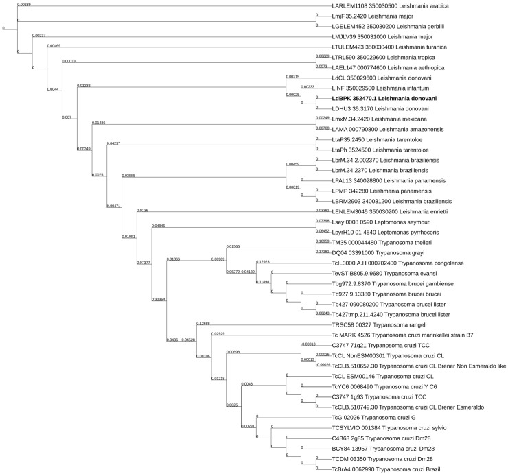
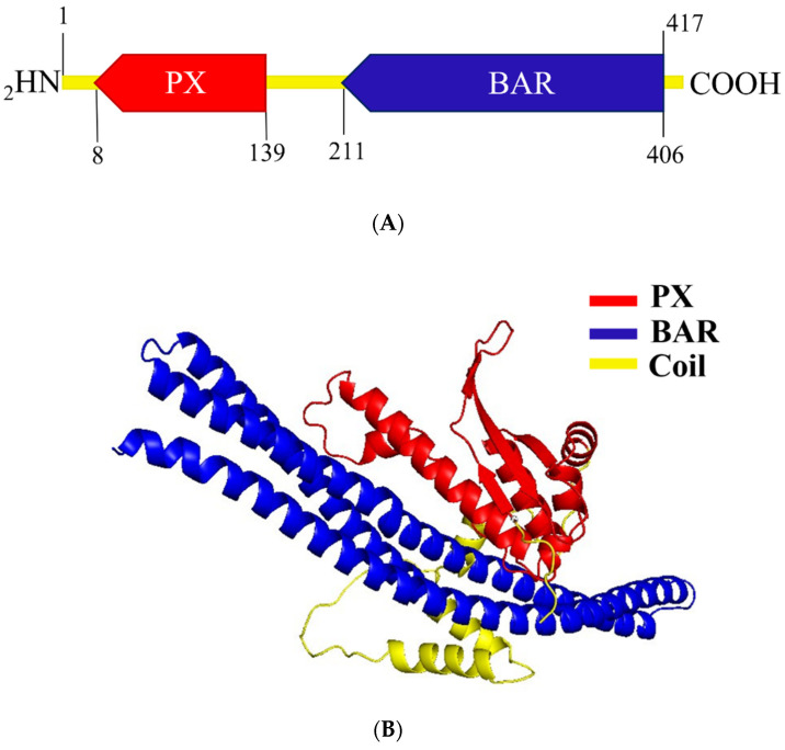
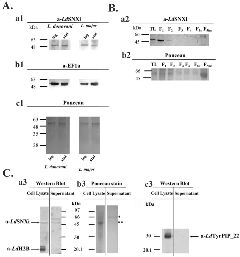
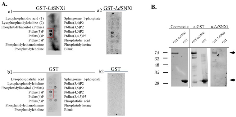
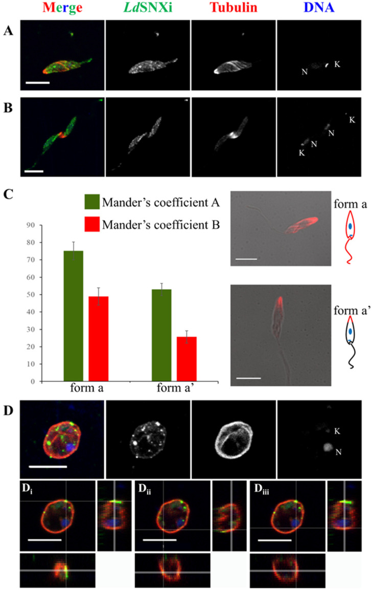
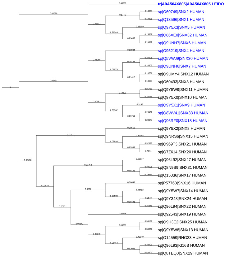
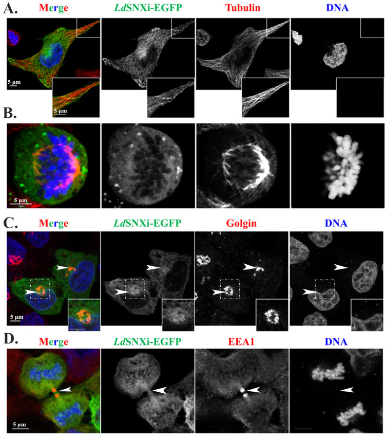
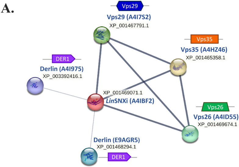
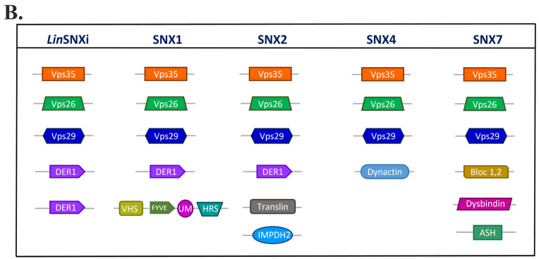
Proteins of the sorting nexin (SNX) family present a modular structural architecture with a phox homology (PX) phosphoinositide (PI)-binding domain and additional PX structural domains, conferring to them a wide variety of vital eukaryotic cell's functions, from signal transduction to membrane deformation and cargo binding. Although SNXs are well studied in human and yeasts, they are poorly investigated in protists. Herein, is presented the characterization of the first SNX identified in Leishmania protozoan parasites encoded by the LdBPK_352470 gene. In silico secondary and tertiary structure prediction revealed a PX domain on the N-terminal half and a Bin/amphiphysin/Rvs (BAR) domain on the C-terminal half of this protein, with these features classifying it in the SNX-BAR subfamily of SNXs. We named the LdBPK_352470.1 gene product LdSNXi, as it is the first SNX identified in Leishmania (L.) donovani. Its expression was confirmed in L. donovani promastigotes under different cell cycle phases, and it was shown to be secreted in the extracellular medium. Using an in vitro lipid binding assay, it was demonstrated that recombinant (r) LdSNXi (rGST-LdSNXi) tagged with glutathione-S-transferase (GST) binds to the PtdIns3P and PtdIns4P PIs. Using a specific a-LdSNXi antibody and immunofluorescence confocal microscopy, the intracellular localization of endogenous LdSNXi was analyzed in L. donovani promastigotes and axenic amastigotes. Additionally, rLdSNXi tagged with enhanced green fluorescent protein (rLdSNXi-EGFP) was heterologously expressed in transfected HeLa cells and its localization was examined. All observed localizations suggest functions compatible with the postulated SNX identity of LdSNXi. Sequence, structure, and evolutionary analysis revealed high homology between LdSNXi and the human SNX2, while the investigation of protein-protein interactions based on STRING (v.11.5) predicted putative molecular partners of LdSNXi in Leishmania.

Keywords: BAR domain; Leishmania donovani; PX domain; SNX-BAR; SNX2; phosphoinositide binding protein; sorting nexin.


Conflict of interest statement

The authors declare no conflict of interest.

Figures

Figure 1
Figure 1
Evolutionary relationships of the LdBPK 352470.1 gene orthologs in Trypanosomatids. Comprehensive neighbor-joining phylogenetic tree, generated using iTOL, based on multiple sequence alignment (ClustalW) of the LdBPK 352470.1 gene product and its orthologs in Leishmania and Trypanosoma spp.; iTOL link: https://itol.embl.de/tree/1939213722931771674054468, accessed on 10 June 2023. Default scaling factors were used except for the vertical and horizontal scaling, which were adjusted to 1.2. Branch lengths corresponding to evolutionary distances are displayed on each corresponding branch of the phylogram. Statistically significant % identities of the sequences are shown in Table 1. LdSNXi is depicted in bold.
Figure 2
Figure 2
Domain organization and tertiary structure prediction of A0A504X805. (A) Schematic representation of the A0A504X805 domain architecture. The N′ and C′ terminal aa residues are indicated above the schematic diagram while the beginning and end of the PX and BAR domains are indicated below. (B) Tertiary structure prediction of LdSNXi with PyMol tool based on data retrieved from Alpha Fold. Red depicts the PX domain, blue depicts the BAR domain, and yellow shows the coil region.
Figure 3
Figure 3
Biochemical detection of LdSNXi in cultured Leishmania cells and in the extracellular medium. (A) Detection of the endogenous SNXi in wild type L. donovani (LG13) and L. major (Friedlin) promastigotes. Total lysates (40 μg) (Section 4) were analyzed by SDS-PAGE and Western blot probed with the a-LdSNXi pAb (0.5 μg/mL) (a1). The same membrane, after stripping, was re-blotted with the a-EF1a mAb (1:10,000) (b1). LdEF1a was used as a loading indicator. The membrane stained with Ponceau-S is shown (c1) as a second loading indicator. (B) Subcellular distribution of LdSNXi in wt L. donovani (LG13) fractions generated by stepwise solubilization of stationary phase promastigote pellets with digitonin and Triton X-100 (Section 4). Western blot with the rabbit a-LdSNXi pAb (a2). Ponceau-S of respective membrane regions is shown in (b2) as loading indicator. (C) Biochemical detection of the endogenous LdSNXi in wt L. donovani (LG13) promastigotes and in the extracellular material of the promastigote culture (secreted fraction). (a3) Western blot with the purified rabbit a-LdSNXi pAb (0.5 μg/mL) and rabbit a-LdHistone2B (H2B) serum (1:1000) used together. (c3) Western blot with the a-LdTyrPIP_22 mouse pAb (1:2500) after stripping of the Abs used in (a3). The membrane stained with Ponceau-S is shown in (b3) as loading indicators. The black arrows point to the bands identified by the a-LdSNXi and a-LdTyrPIP_22 specific pAbs at the expected migration position, while the gray arrow points to the band identified by the a-LdH2B specific rabbit pAb. Molecular weights are indicated in kDa in the middle of the two panels. The asterisk indicates the migration position where the major secreted protein GP63 is expected, while the two asterisks indicate the major cytoskeleton protein tubulin. Molecular weights are indicated in kDa on the left of the panels. Different molecular weight markers were used in A–C (Section 4). TL: total lysate. F1–F5ins: the detergent fractionation fractions.
Figure 4
Figure 4
PI binding specificity of GST-LdSNXi. (A) Recombinant GST-LdSNXi (a1,a2) and GST (b1,b2) were incubated with nitrocellulose membranes (PIP strips) spotted with phosphoinositides and other lipids, as described in Section 4. In (a1,b1), equal ng/mL protein were used (i.e., 50 ng/mL). In (a2,b2) were used equimolar protein concentrations (i.e., 50 ng/mL for GST-LdSNXi and 17 ng/mL for GST respectively). The signal for PtdIns(3)P and PtdIns(4)P is framed by a red rectangle in (a1,b1). (B) Purified GST-LdSNXi and GST from lysates of the corresponding E. coli BL21 recombinant clones used in the PI binding assay were analyzed by SDS-PAGE (Coomassie) and Western blot (a-GST and a-LdSNXi). The arrows on the right indicate the protein species with the expected size for GST-LdSNXi (upper arrow) and GST (lower arrow), respectively.
Figure 5
Figure 5
Localization of endogenous LdSNXi in L. donovani promastigotes and axenic amastigotes. Localization of LdSNXi in different cell cycle-dependent morphological forms of cultured L. donovani promastigotes; co-staining for LdTubulin. Maximum intensity projection images from each case are shown. Single FL images are shown in black and white (BW) for better contrast while images of the merged FL signals are shown in color. The molecule highlighted in the BW images with single-color FL is indicated at the top in the same color as the respective FL signal. (A) Procyclic-like promastigote. (Β) Dividing promastigote with the intercellular bridge highlighted by the tubulin staining. (C) Mander’s coefficient graphs depicting the percentage of pixels in the co-localization channel, as calculated by IMARIS (Section 4). Two different forms of promastigotes depending on the observed microtubules’ staining pattern were grouped together. Merged images of phase contrast and FL images of microtubule staining (Red FL) in the two representative parasite forms are shown on the right, together with schematic representations of the microtubule staining distribution. (D) Amastigote-like parasite. (i,ii) are orthogonal images of the same parasite showing the position of different LdSNXi stained vesicles in relation to the parasite’s surface membrane. (iii) is an orthogonal image of the same parasite showing the co-localization of LdSNXi epitopes and subpellicular microtubules. Red FL: Tubulin. Green FL: LdSNXi. Κ: kinetoplast DNA. N: nuclear DNA. Scale bar size: 5 μm.
Figure 6
Figure 6
Evolutionary relationship between the LdBPK 352470.1 gene product (A0A504X805) and the human sorting nexins. Comprehensive neighbor-joining phylogenetic trees generated using iTOL based on multiple sequence alignment (ClustalW) of the LdBPK 352470.1 gene product and all reviewed human sorting nexins (retrieved from UniProt) https://itol.embl.de/tree/14123716952488761694711835 accessed on 10 June 2023. The members of the human SNX-BAR subfamily (according to UniProt) are shown in blue. Branch lengths are displayed on each corresponding branch of the phylogram. Statistically significant % identities of the sequences are shown in Table 2. LdSNXi is depicted in bold.
Figure 7
Figure 7
Localization of rLdSNXi-EGFP heterologously expressed in human HeLa cells. HeLa cells were transfected with the pEGFP-N3-ldsnxi plasmid for 24 h, followed by pre-extraction and immunostaining, as described in Section 4. (A). An Interphase cell. (B). A metaphase cell. (C). Interphase cells stained for Golgi. (D). A cell in late cytokinesis stained for the early endosome protein EEA1. Insets indicate the area framed in the main figure in 2× zoom. Arrows point to the Golgi apparatus and the intercellular bridge. Single FL images are shown in BW for better contrast, while images of the merged FL signals are shown in color. The molecule highlighted in the BW images with single color FL is indicated at the top in the same color as the respective FL signal. Scale bar size: 5 μm.
Figure 8
Figure 8
Molecular partners of LdSNXi and human SNXs according to STRING database analysis. (A) Protein–protein interactions network of LinSNXi (ortholog protein of the LdSNXi in Leishmania infantum) and its partners according to STRING analysis based on experimental evidence. The characteristic domain of each protein (according to Pfam) is depicted next to each node of the network. (B) Comparative illustration for the characteristic domains of the molecular partners of LinSNXi and human SNX1, SNX2, SNX4, and SNX7.
Figure 8
Figure 8
Molecular partners of LdSNXi and human SNXs according to STRING database analysis. (A) Protein–protein interactions network of LinSNXi (ortholog protein of the LdSNXi in Leishmania infantum) and its partners according to STRING analysis based on experimental evidence. The characteristic domain of each protein (according to Pfam) is depicted next to each node of the network. (B) Comparative illustration for the characteristic domains of the molecular partners of LinSNXi and human SNX1, SNX2, SNX4, and SNX7.

Similar articles

Cited by

References

    1. Teasdale R.D., Collins B.M. Insights into the PX (phox-homology) domain and SNX (sorting nexin) protein families: Structures, functions and roles in disease. Biochem. J. 2012;441:39–59. doi: 10.1042/BJ20111226. - DOI - PubMed
    1. Hanley S.E., Cooper K.F. Sorting Nexins in Protein Homeostasis. Cells. 2020;10:17. doi: 10.3390/cells10010017. - DOI - PMC - PubMed
    1. Haft C.R., Sierra M.d.l.L., Barr V.A., Haft D.H., Taylor S.I. Identification of a family of sorting nexin molecules and characterization of their association with receptors. Mol. Cell. Biol. 1998;18:7278–7287. doi: 10.1128/MCB.18.12.7278. - DOI - PMC - PubMed
    1. Peter B.J., Kent H.M., Mills I.G., Vallis Y., Butler P.J.G., Evans P.R., McMahon H.T. BAR domains as sensors of membrane curvature: The amphiphysin BAR structure. Science. 2004;303:495–499. doi: 10.1126/science.1092586. - DOI - PubMed
    1. Chishti A.H., Kim A.C., Marfatia S.M., Lutchman M., Hanspal M., Jindal H., Liu S.-C., Low P.S., Rouleau G.A., Mohandas N., et al. The FERM domain: A unique module involved in the linkage of cytoplasmic proteins to the membrane. Trends Biochem. Sci. 1998;23:281–282. doi: 10.1016/S0968-0004(98)01237-7. - DOI - PubMed