Review
. 2024 Apr 8;25(7):4110.
doi: 10.3390/ijms25074110.

X-Linked Epilepsies: A Narrative Review

Affiliations
Review

X-Linked Epilepsies: A Narrative Review

Pia Bernardo et al. Int J Mol Sci. .

Abstract

X-linked epilepsies are a heterogeneous group of epileptic conditions, which often overlap with X-linked intellectual disability. To date, various X-linked genes responsible for epilepsy syndromes and/or developmental and epileptic encephalopathies have been recognized. The electro-clinical phenotype is well described for some genes in which epilepsy represents the core symptom, while less phenotypic details have been reported for other recently identified genes. In this review, we comprehensively describe the main features of both X-linked epileptic syndromes thoroughly characterized to date (PCDH19-related DEE, CDKL5-related DEE, MECP2-related disorders), forms of epilepsy related to X-linked neuronal migration disorders (e.g., ARX, DCX, FLNA) and DEEs associated with recently recognized genes (e.g., SLC9A6, SLC35A2, SYN1, ARHGEF9, ATP6AP2, IQSEC2, NEXMIF, PIGA, ALG13, FGF13, GRIA3, SMC1A). It is often difficult to suspect an X-linked mode of transmission in an epilepsy syndrome. Indeed, different models of X-linked inheritance and modifying factors, including epigenetic regulation and X-chromosome inactivation in females, may further complicate genotype-phenotype correlations. The purpose of this work is to provide an extensive and updated narrative review of X-linked epilepsies. This review could support clinicians in the genetic diagnosis and treatment of patients with epilepsy featuring X-linked inheritance.

Keywords: X-linked; developmental and epileptic encephalopathies (DEEs); epilepsy; genetics.


Conflict of interest statement

None of the authors has conflicts of interest to declare in relation to the present review.

Figures

Figure 1
Figure 1
X-linked modes of inheritance, influenced by X-chromosome inactivation in females, epigenetic regulation, mosaic distribution of pathogenic variants.
Figure 2
Figure 2
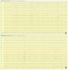
Poly-graphic recording, including EEG and electromyography (EMG) channels of a focal seizure recorded at 7 year and 3 months of age in a girl with PCDH19-related DEE. Interictal sleep EEG (A) showing slow background activity with poor representation of physiological sleeping figures. Seizure onset (B) with a bilateral ictal theta activity over both parieto-temporal regions, followed by bilateral high amplitude slow waves (C) and a rhythmic fast ictal discharge persisting over the bilateral temporal regions. Twelve seconds later (D), a sudden termination of the ictal discharge with electro-decrementation is visible. Seizure semiology: (1) eyes opening and staring; (2) behavior arrest; (3) right eye deviation, bilateral blinking and minor arm and hand movements, followed by desaturation and post ictal sleep. Poly-graphic recording, including EEG (bipolar montage: blue lines are the right electrodes, black lines are the left electrodes; red lines are the vertex electrodes) and electromyography (EMG1: left deltoid; EMG2: right deltoid) of a focal seizure recorded.
Figure 3
Figure 3
Poly-graphic recording, including EEG and electromyography (EMG) channels, in a 10 year and 2 months years old girl with PCDH19-gene related DEE. Interictal awake (A,B) and sleep (C,D) EEG study showing slow background activity and bilateral frontal paroxysmal activity, which increases during sleep, spreading over the temporal regions. Poly-graphic recording, including EEG (bipolar montage: blue lines are the right electrodes, black lines are the left electrodes; red lines are the vertex electrodes) and electromyography (EMG1: left deltoid; EMG2: right deltoid) in a 10 year and 2 months.
Figure 4
Figure 4
EEG recording (bipolar montage) of a 9 year and 2 months old girl with CDKL5-gene related DEE. (A) Interictal EEG during sleep showing continuous spikes and slow wave activity, prominent over the frontal regions. Sleep figures are poorly represented. (B) Ictal recording showing a tonic seizure corresponding to an electro-decremental change (B) followed by low voltage diffuse fast activity and high-voltage sharp waves (C,D), lasting 40 s.
Figure 5
Figure 5
Video-EEG recording of a 14 year and 6 months old girl with typical Rett Syndrome MECP2-related. Interictal EEG during wakefulness showing a background activity characterized by monomorphic theta activity (6 Hz), topographically undifferentiated (A). During sleep, asymmetrical and disorganized slow activity was present, with poor physiological sleep figures representation (B). Video-EEG recording (bipolar montage: blue lines are the right electrodes, black lines are the left electrodes; red lines are the vertex electrodes).
Figure 6
Figure 6
Electroencephalographic (EEG) recording (performed at 25 years old) of a seizure in a female patient with MECP2 duplication syndrome (MDS) and clinically featuring a Lennox Gastaut Syndrome (A). Ictal EEG starts with a generalized wave with an electro-decremental component followed by desynchronization of the background activity; after 6 s, slow delta activity with interspersed spikes appears. After about 20 s, irregular 2–2.5 Hz, high voltage spike-wave or poly-spike–wave complexes appear, with diffuse wide spreading prevalent over the bilateral frontal regions. The event terminates with post ictal delta activity (1.5 Hz) (A). Clinically, the episode is characterized by: (a) sudden jerk involving the proximal limbs with impairment of awareness; (b) neck and face myoclonia; (c) right eyes and neck deviation (d) prolonged facial and right distal upper limb myoclonia which subsequently (e) involves asynchronously the left distal upper limb. (B) Polygraphic recording including EEG and EMG (Right Trapezius) recording of a male patient affected with MDS and showing slow background activity, diffuse spike and wave epileptiform activity and an ictal spasm of the upper limbs time-locked with the electrodecremental high amplitude wave.

References

    1. Deng H., Zheng W., Song Z. Genetics, molecular biology, and phenotypes of X-linked epilepsy. Mol. Neurobiol. 2014;49:1166–1180. doi: 10.1007/s12035-013-8589-1. - DOI - PubMed
    1. Stevenson R.E., Holden K.R., Rogers R.C., Schwartz C.E. Seizures and X-linked intellectual disability. Eur. J. Med. Genet. 2012;55:307–312. doi: 10.1016/j.ejmg.2012.01.017. - DOI - PMC - PubMed
    1. den Veyver I.B.V. Skewed X inactivation in X-linked disorders. Semin. Reprod. Med. 2001;19:183–191. doi: 10.1055/s-2001-15398. - DOI - PubMed
    1. Germain D.P. General aspects of X-linked diseases. In: Mehta A., Beck M., Sunder-Plassmann G., editors. Fabry Disease: Perspectives from 5 Years of FOS. Oxford PharmaGenesis; Oxford, UK: 2006. Chapter 7. - PubMed
    1. Lyon M.F. Gene action in the X-chromosome of the mouse (Mus musculus L.) Nature. 1961;190:372–373. doi: 10.1038/190372a0. - DOI - PubMed