. 2024 Apr 8;25(7):4115.
doi: 10.3390/ijms25074115.

The Oligomeric State of Vasorin in the Plasma Membrane Measured Non-Invasively by Quantitative Fluorescence Fluctuation Spectroscopy

Affiliations

The Oligomeric State of Vasorin in the Plasma Membrane Measured Non-Invasively by Quantitative Fluorescence Fluctuation Spectroscopy

Junyi Liang et al. Int J Mol Sci. .

Abstract

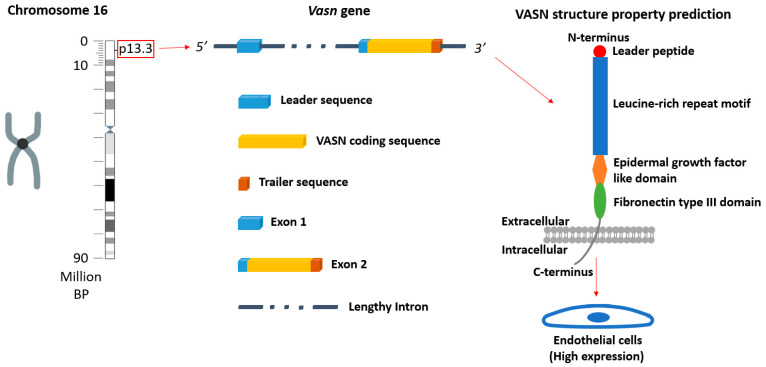
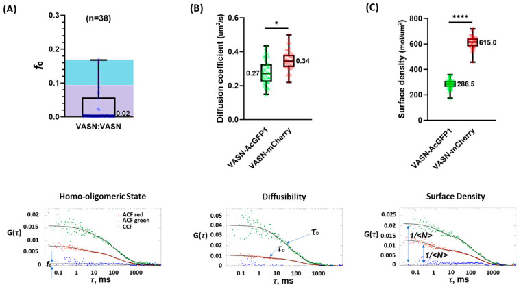
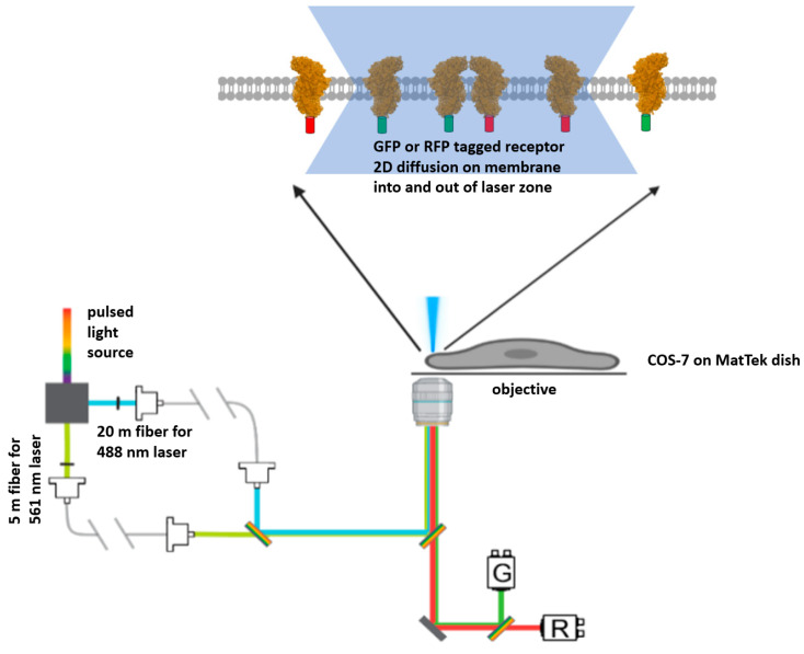
Vasorin (VASN), a transmembrane protein heavily expressed in endothelial cells, has garnered recent interest due to its key role in vascular development and pathology. The oligomeric state of VASN is a crucial piece of knowledge given that receptor clustering is a frequent regulatory mechanism in downstream signaling activation and amplification. However, documentation of VASN oligomerization is currently absent. In this brief report, we describe the measurement of VASN oligomerization in its native membranous environment, leveraging a class of fluorescence fluctuation spectroscopy. Our investigation revealed that the majority of VASN resides in a monomeric state, while a minority of VASN forms homodimers in the cellular membrane. This result raises the intriguing possibility that ligand-independent clustering of VASN may play a role in transforming growth factor signaling.

Keywords: fluorescence fluctuation spectroscopy; fluorescent proteins; membrane protein multimerization; vasorin (VASN).


Conflict of interest statement

The authors declare no conflict of interest.

Figures

Figure 1
Figure 1
The location of the vasn gene in a human chromosome (chromosome 16), the structural properties of the gene, and a prediction of the VASN protein structure (predicted by RaptorX http://raptorx.uchicago.edu (accessed on 2 April 2024)) [15,16].
Figure 2
Figure 2
The biophysical characterization of VASN in the cellular membrane including its oligomeric state (* and **** are markers denoting slight and notable statistical significance). (A) The lower panel is a representative set of single-cell PIE-FCCS data. The fraction correlated (fc) can be obtained from the amplitude of the CCF as the CCF is dependent on the distribution of monomers, dimers, trimers, and multimers. The upper panel is VASN’s fc as measured from 38 cells, which clearly established that the vast majority of VASN resided in the membrane in a monomeric state. (B) The mobility of VASN-AcGFP1 and VASN-mCherry, as denoted as the diffusion coefficient in the (upper panel), was computed with the information of the diffusers’ average dwell time available in the measurement curve (lower panel). (C) ACF’s amplitude is an indicator of the average number of diffusers as these two are in an inverse proportional relationship.
Figure 3
Figure 3
The instrumental set up: Simplified schematic of the PIE-FCCS optical path. A pulsed light source is used to generate two laser beams for excitation. The beams pass through optical fibers with a length difference of 15 m to hard write a 50 ns difference in excitation time. (Note that 50 ns is roughly an order of magnitude longer than AcGFP1 (GFP) and that mCherry (RFP) needs to decay back to the ground state from the excited state.) Fluorescent tagged membrane proteins (a general receptor shown here in this figure) would diffuse into and out of the confocal area under laser excitation, resulting in fluctuations in fluorescence intensity captured by photon counting.
Figure 4
Figure 4
Design strategy for the single-plasmid-based, dual-expression construct. The two regular expression plasmids are the starting material. For one plasmid, nearly the entirety was amplified out with primer 1 (p1) and primer 2 (p2), including the gene encoding VASN-AcGFP1 and the regulatory units (enhancer and promoter (E&P) with transcription terminator (TT)) and survival units (origin of replication in prokaryote (Ori_P) and in eukaryote (Ori_E) and antibiotic resistance gene (ARG)). For the other plasmid, the gene encoding VASN-mCherry and the regulatory units were amplified out with primer 3 (p3) and primer 4 (p4). The fitting sticky ends were supplied onto the end of the linearized DNA in the PCR. The linear PCR products were purified and digested. The digestive products were then purified and ligated into a single circular DNA. This circularized DNA is the single plasmid for the dual expression construct.

References

    1. Ikeda Y., Imai Y., Kumagai H., Nosaka T., Morikawa Y., Hisaoka T., Manabe I., Maemura K., Nakaoka T., Imamura T., et al. Vasorin, a transforming growth factor β-binding protein expressed in vascular smooth muscle cells, modulates the arterial response to injury in vivo. Proc. Natl. Acad. Sci. USA. 2004;101:10732–10737. doi: 10.1073/pnas.0404117101. - DOI - PMC - PubMed
    1. Louvet L., Lenglet G., Krautzberger A.M., Mentaverri R., Hague F., Kowalewski C., Mahtal N., Lesieur J., Bonnet A., Andrique C., et al. Vasorin plays a critical role in vascular smooth muscle cells and arterial functions. J. Cell. Physiol. 2022;237:3845–3859. doi: 10.1002/jcp.30838. - DOI - PMC - PubMed
    1. Bonnet A.-L., Chaussain C., Broutin I., Rochefort G.Y., Schrewe H., Gaucher C. From Vascular Smooth Muscle Cells to Folliculogenesis: What About Vasorin? Front. Med. 2018;5:335. doi: 10.3389/fmed.2018.00335. - DOI - PMC - PubMed
    1. Luong T.T.D., Estepa M., Boehme B., Pieske B., Lang F., Eckardt K.-U., Voelkl J., Alesutan I. Inhibition of vascular smooth muscle cell calcification by vasorin through interference with TGFβ1 signaling. Cell. Signal. 2019;64:109414. doi: 10.1016/j.cellsig.2019.109414. - DOI - PubMed
    1. Korte L., Widmer-Teske R., Donde K., Dutzmann J., Daniel J.M., Bauersachs J., Sedding D.G. 13Vasorin controls smooth muscle cell proliferation by regulating EGFR activation. Cardiovasc. Res. 2018;114((Suppl. S1)):S3. doi: 10.1093/cvr/cvy060.003. - DOI

Substances

LinkOut - more resources