. 2024 Aug;245(2):271-288.
doi: 10.1111/joa.14045. Epub 2024 Apr 13.

Microanatomy of the human tunnel of Corti structures and cochlear partition-tonotopic variations and transcellular signaling

Affiliations

Microanatomy of the human tunnel of Corti structures and cochlear partition-tonotopic variations and transcellular signaling

Dina Giese et al. J Anat. 2024 Aug.

Abstract

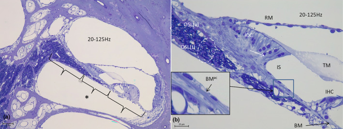
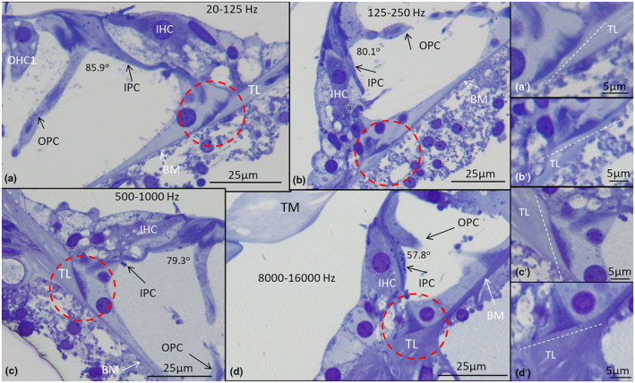
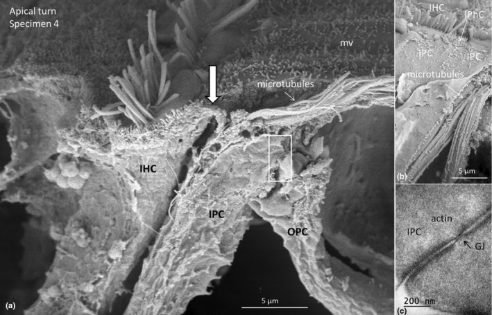
Auditory sensitivity and frequency resolution depend on the optimal transfer of sound-induced vibrations from the basilar membrane (BM) to the inner hair cells (IHCs), the principal auditory receptors. There remains a paucity of information on how this is accomplished along the frequency range in the human cochlea. Most of the current knowledge is derived either from animal experiments or human tissue processed after death, offering limited structural preservation and optical resolution. In our study, we analyzed the cytoarchitecture of the human cochlear partition at different frequency locations using high-resolution microscopy of uniquely preserved normal human tissue. The results may have clinical implications and increase our understanding of how frequency-dependent acoustic vibrations are carried to human IHCs. A 1-micron-thick plastic-embedded section (mid-modiolar) from a normal human cochlea uniquely preserved at lateral skull base surgery was analyzed using light and transmission electron microscopy (LM, TEM). Frequency locations were estimated using synchrotron radiation phase-contrast imaging (SR-PCI). Archival human tissue prepared for scanning electron microscopy (SEM) and super-resolution structured illumination microscopy (SR-SIM) were also used and compared in this study. Microscopy demonstrated great variations in the dimension and architecture of the human cochlear partition along the frequency range. Pillar cell geometry was closely regulated and depended on the reticular lamina slope and tympanic lip angle. A type II collagen-expressing lamina extended medially from the tympanic lip under the inner sulcus, here named "accessory basilar membrane." It was linked to the tympanic lip and inner pillar foot, and it may contribute to the overall compliance of the cochlear partition. Based on the findings, we speculate on the remarkable microanatomic inflections and geometric relationships which relay different sound-induced vibrations to the IHCs, including their relevance for the evolution of human speech reception and electric stimulation with auditory implants. The inner pillar transcellular microtubule/actin system's role of directly converting vibration energy to the IHC cuticular plate and ciliary bundle is highlighted.

Keywords: cochlea; human; microanatomy; microtubules; synchrotron radiation phase‐contrast imaging.


Conflict of interest statement

The authors declare that the research was conducted in the absence of any commercial or financial relationships that could be construed as a potential conflict of interest. Disclosures: The Western University Foundation has received a donation from Med‐El GmbH. However, this work was carried out independent of industry involvement or oversight.

Figures

FIGURE 1
FIGURE 1
(a) Mid‐modiolar cross‐section of an archival human cochlea (~1 μm semi‐thin section, osmium/toluidine blue staining, specimen1). The OCs are magnified in Figure 2. (b) SR‐PCI and 3D reconstruction of a matched cochlea with delineated octave bands constructed from Greenwood's formula (Greenwood, 1961). Insets show the appraised frequency map.
FIGURE 2
FIGURE 2
Light microscopy of the OC (a–f) (as shown in Figure 1, specimen 1) at different frequency locations shown at same magnification. Scale bar is 25 μm. Cell structure is well preserved. The different OC have been mirrored to better compare dimensions and architectures. The most apical region A (lagena) contains no sensory epithelium. The tectorial membrane (TM) has partly separated from the RL. Beneath is a frequency map of the basilar membrane (BM) developed according to Greenwood's equation. Estimated longitudinal distances and rotational angles for the different OC are shown at frequency bands along the BM (vertical arrows).
FIGURE 3
FIGURE 3
Dimensions of the OC and pillar cell lengths at different frequencies. IPC length is similar at all frequencies. A best‐fit trend‐line is shown for each variable (specimen 1).
FIGURE 4
FIGURE 4
Dimensions in the inter‐scalar septum (and trend‐lines) at different frequency locations (specimen 1).
FIGURE 5
FIGURE 5
(a) LM of the most basal part of the human cochlea shows the width of the basilar membrane and osseous spiral lamina (OSL) (staples) (specimen 1). (b) The OSL reaches the habenular opening and the tympanic lip on which the IPC foot is anchored and the BM attaches. Some spiral lamina fibers attach to the OSL bone and continue medially under the inner sulcus (IS) (arrows and in inset). Some of these fibers attach to the bone of the vestibular plate of the OSL (OSLv). The inset shows the framed area at higher magnification. Ax, axons; B, bone; IHC, inner hair cell; OSL(t), tympanic plate of the OSL; RM, Reissner's membrane; SL, spiral lamina; TM, tectorial membrane. Osmium/toluidine staining.
FIGURE 6
FIGURE 6
(a) At 500–1000 Hz, a small soft bridge (74 μm in length) of the spiral lamina lacks bone (asterisk) (specimen 1). (b) Higher magnification shows the axons exiting the opening of the OSL (circle). A fibrous lamina continues medially from the tympanic lip and the BM under the floor of the IS to the spiral limbus, here named the BMac. It runs fairly independently of the OSL with a few fibers merging with the vestibular plate of the OSLv. Ax, axons; IHC, inner hair cell; OSL(t), tympanic plate of the OSL; RM, Reissner's membrane; TCL, Tympanic covering layer; TM, tectorial membrane.
FIGURE 7
FIGURE 7
(a) At the 250–500 Hz region, the OSL bridge is 193 μm (middle staple, marked with an asterisk) (specimen 1, mirrored). (b) An “accessory basilar membrane” (BMac) continues medially under the floor of the IS into the spiral lamina. A few fibers merge with the vestibular plate of the OSL (arrow). BM, basilar membrane; IDC, interdental cells; IHC, inner hair cell; OSL(t), tympanic plate of the OSL; OSL(v), vestibular plate of the OSL; OSL, osseous spiral lamina; RM, Reissner's membrane; TM, tectorial membrane. Osmium/toluidine staining.
FIGURE 8
FIGURE 8
(a) At 125–250 Hz, the width of the bridge is 160 μm (middle staple, marked with an asterisk) (specimen 1). (b) BMac runs medially under the floor of the inner sulcus into the spiral limbus. Framed area is magnified in the inset and shows a BMac fiber connecting to the vestibular plate of the OSL (OSLv). Osmium/toluidine staining. (c) Positive collagen type II expression in the BM (arrow) and BMac beneath the IS and the tectorial membrane (TM) (from Hayashi et al., 2015). There is no OSL collagen staining at the bridge (staple). (d) and (e) TEM of the BMac (d) and BM (e) show identical radially arranged fibers with a diameter around 15 nm (Specimen 2, scale bars 200 nm). Ax, axon; BM, basilar membrane; BMac, accessory basilar membrane; IDC, interdental cell; IHC, inner hair cell; IPC, inner pillar cell; OPC, outer pillar cells; OSL(t), tympanic plate of the osseous spiral lamina; RM, Reissner's membrane; TCL, tympanic covering layer; TL, tympanic lip.
FIGURE 9
FIGURE 9
(a) At 20–125 Hz, the width of the bridge is 228 μm (middle staple, marked with an asterisk) (specimen 1, mirrored). Scale bar is 75 μm. (b) BMac thickness is around 3.2 μm and continues from the tympanic lip beneath the IPC foot as a collagen sheet (framed area is magnified in the inset), the fibrils of which spread out in the spiral limbus. A few fibers even reach the interdental cells (IDCs, arrow), while some are connected to the vestibular plate of the OSL (OSLv). Scale bar is 25 μm. BMac, accessory basilar membrane; IHC, inner hair cell; IPC, inner pillar cell; IS, inner sulcus; OSL(t), tympanic plate of the OSL; OSL, osseous spiral lamina; RM, Reissner's membrane; TM, tectorial membrane; BM, basilar membrane. Osmium/toluidine staining.
FIGURE 10
FIGURE 10
SR‐PCI of the same cochlea as shown in Figure 1b (Top left, scale bar is 1 mm) using a bone algorithm to show the different degree of ossification in the spiral lamina at different frequency locations (a–d). The length of the non‐ossified spiral lamina (soft bridge) increases against the apical low‐frequency region and is marked with a staple. IAC, internal auditory canal; ICV, inferior cochlear vein; IHC, inner hair cell; IPC, inner pillar cell; OSL, osseous spiral lamina; RM, Reissner's membrane; SL, spiral ligament; SP, spiral prominence; SV, stria vascularis.
FIGURE 11
FIGURE 11
(a‐d) The inner pillar cell (IPC) cytoskeletal rod and its foot sole (encircled) form a near straight angle with the slope of the tympanic lip (TL) at different frequency regions in the human cochlea (specimen 1). The IPC rod parallels mostly the long axis of the IHC. The height of the IPCs and OPCs, but not the IHCs, increases against the low frequencies, as does the angle between the IPCs and OPCs. (a′–d′) Demonstrate the encircled IPC foot angles at different frequency locations. BM, basilar membrane; OHC1, outer hair cell in the first row; OPC, outer pillar cell; TM, tectorial membrane.
FIGURE 12
FIGURE 12
Illustration showing different architecture of the human cochlear partition at low and high frequencies in specimen 1 (125–250 Hz/8000–16,000 Hz). The geometry of the IPC rod cytoskeleton (encircled) is closely regulated by the angle of the tympanic lip and is related to the RL slope. At low frequencies, a fibrous lamina (BMac) extends medially from the tympanic lip under the inner sulcus. A non‐bony SL or bridge contributes to increasing the overall compliance of the CP. Graph shows variations in the tympanic lip/IPC foot and RL angles relative to the BM at different frequency locations. The angles are inversely related, with the largest difference at 125–250 Hz corresponding to the human fundamentals. Best‐fit trend‐lines are shown. The different angles of the tympanic lip maintain a straight‐angle relationship between the IPC microtubule rod and run parallel with the IHC. This may create optimal conditions to relay mechanical vibrations from the IPC base to the IHC along the CP at different frequencies.
FIGURE 13
FIGURE 13
(a) SEM of the human inner hair cell (IHC) at the low‐frequency region (<500 Hz) in a human cochlea (specimen 4). The pillar heads of the inner pillar cell (IPC) and outer pillar cell (OPC) extend up to the surface, where they bend and form part of the reticular lamina. There is a close relationship between the IPC and IHC (white arrow), with no interphalangeal cell (IPhC). (b) Surface SEM view of the IPC and reticular lamina with a ruptured membrane surface exposing an armamentarium of microtubules extending to the first row of OHCs. (c) A corresponding TEM section of the framed area shown in A with several intercellular membrane specializations, including gap junctions (GJ) (specimen 2).
FIGURE 14
FIGURE 14
(a) TEM ultrathin section of a human IPC/IHC junction at low‐frequency region (specimen 1). The IPC surfoskelosome (yellow broken line) is intimately associated with the IHC CP (red broken line). (b) The junction contains sub‐membranous electron‐dense material. The junctional region forms a potential route for acoustic vibrations to reach the CP actin meshwork and hair bundle rootlets. CP, cuticular plate; IHC, inner hair cell; IPC, inner pillar cell; OPC, outer pillar cell; ZA, zonula adherens; ZO, zonula occludens.
FIGURE 15
FIGURE 15
(a) Tangential TEM section of two IHCs in the human apical turn (specimen 2). Framed areas are magnified in b and c. A “star‐shaped” conglomerate of electron‐dense material at the cytoplasmic cell surfaces are seen. The IPC interacts with two IHC cuticular plates (CP). The CPs are structurally linked to the IPC junctions (broken arrows in right cell). There are several tight junctions between the IPCs and IHCs (arrow in c). (d) IHC/IPC interaction in the lower turn of the human cochlea. (specimen 2). The CP of the IHC is closely related to the electron‐dense surfoskelosome of the IPC (broken white line). C, cilia, CP, cuticular plate; IHC, inner hair cell; IPC, inner pillar cell; IPhC, inner phalangeal cell; mt, microtubules; OPC, outer pillar cell.
FIGURE 16
FIGURE 16
(a) Super‐resolution immunohistochemistry (SR‐SIM) of actin/LMO7 co‐expression in the low‐frequency region of a human cochlea (specimen 2). The cuticular plate (CP) of the inner hair cell (IHC) and outer hair cell of the first row (OHC1) express LMO7. (b) The actin/cytoskeletal network of the inner pillar cell (IPC, long arrow) is closely associated with the apical junctional area and CP of the IHC (arrows). Positive LMO7 staining extends to the lateral cell membrane reaching the IPC head. (c) Antibody labeling shows positive staining for actin in the CP of the IHC and IPC/OPC heads and bases. BM, basilar membrane; RL, reticular lamina; TL, tympanic lip.
FIGURE 17
FIGURE 17
(a) Actin immunocytochemistry of the IPC foot. (b) TEM of microtubules (mt) of the inner pillar cell (IPC) in the middle turn. The microtubule diameter is around 25 nm. (c) Tubulin‐β1 expression in the IPC and LMO7 in the cuticular plate (CP) of the IHC in the middle turn organ. (d) Tubulin‐β1 and LMO7 expression in the organ of Corti. (e and f) TEM section through a basal surfoskelosome of the IPC at the tympanic lip at 4–8,000 Hz. Actin material is linked with the transcellular array of mt and the electron‐dense layer of the basal plasma membrane of the pillar foot. (g) Graphic illustration of principal force vectors focusing toward the IPC/CP junction (Illustration by Karin Lodin). BL, basal lamina; BM, basilar membrane; IHC, inner hair cell; OPC, outer pillar cell; TCF, tunnel crossing fiber; TL, tympanic lip.

Similar articles

Cited by

References

    1. Angelborg, C. & Engström, H. (1972) Supporting elements in the organ of Corti I. Fibrillar structures in the supporting cells of the organ of Corti of mammals. Acta Oto‐Laryngologica, 73, 49–60. - PubMed
    1. Antonellis, P.J. , Pollock, L.M. , Chou, S.W. , Hassan, A. , Geng, R. , Chen, X. et al. (2014) ACF7 Is a hair‐bundle antecedent, positioned to integrate cuticular plate Actin and somatic tubulin. The Journal of Neuroscience, 34, 305–312. - PMC - PubMed
    1. Arnold, W. (1987) Myelination of the human spiral ganglion. Acta Oto‐Laryngologica, 104, 76–84. - PubMed
    1. Békésy, G. (1960) Experiments in hearing. New York, NY: McGraw‐Hill.
    1. Bieniussa, L. , Jain, I. , Bosch Grau, M. , Juergens, L. , Hagen, R. , Janke, C. et al. (2023) Microtubule and auditory function – an underestimated connection. Seminars in Cell & Developmental Biology, 137, 74–86. - PubMed

Publication types

MeSH terms