. 2024 Apr 25;187(9):2224-2235.e16.
doi: 10.1016/j.cell.2024.03.008. Epub 2024 Apr 12.

NINJ1 mediates plasma membrane rupture by cutting and releasing membrane disks

Affiliations

NINJ1 mediates plasma membrane rupture by cutting and releasing membrane disks

Liron David et al. Cell. .

Abstract

The membrane protein NINJ1 mediates plasma membrane rupture in pyroptosis and other lytic cell death pathways. Here, we report the cryo-EM structure of a NINJ1 oligomer segmented from NINJ1 rings. Each NINJ1 subunit comprises amphipathic (⍺1, ⍺2) and transmembrane (TM) helices (⍺3, ⍺4) and forms a chain of subunits, mainly by the TM helices and ⍺1. ⍺3 and ⍺4 are kinked, and the Gly residues are important for function. The NINJ1 oligomer possesses a concave hydrophobic side that should face the membrane and a convex hydrophilic side formed by ⍺1 and ⍺2, presumably upon activation. This structural observation suggests that NINJ1 can form membrane disks, consistent with membrane fragmentation by recombinant NINJ1. Live-cell and super-resolution imaging uncover ring-like structures on the plasma membrane that are released into the culture supernatant. Released NINJ1 encircles a membrane inside, as shown by lipid staining. Therefore, NINJ1-mediated membrane disk formation is different from gasdermin-mediated pore formation, resulting in membrane loss and plasma membrane rupture.

Keywords: NINJ1; NINJ2; Ninjurin1; Ninjurin2; cryo-EM; inflammasome; inflammation; lytic cell death; plasma membrane rupture; pyroptosis.


Conflict of interest statement

Declaration of interests H.W. is a co-founder and chair of the scientific advisory board of Ventus Therapeutics. I.J. and E.G. are employees of Abberior Instruments GmbH. None of these relationships influenced this study.

Figures

Figure 1.
Figure 1.. Formation of rings by NINJ1 and NINJ2, and membrane breakage by NINJ1, not NINJ2
(A) Domain architectures of human NINJ1 and NINJ2. (B) Purification of MBP-NINJ1 and MBP-NINJ2 in detergent. SDS-PAGE of NINJ1 and NINJ2 oligomers, purified from the heavy fractions of sucrose gradients after MBP removal. (C) Negative staining EM of NINJ1 and NINJ2 revealed that they both form rings in detergent. (D) Lipid strip assays for NINJ1 and NINJ2, showing that they bind negatively charged lipids, specifically PA and PI(4)P. (E,F) Incorporation of NINJ1 (E) and NINJ2 (F) into liposomes containing PA and PI(4)P, shown by negatived staining EM images and their zoom-in views. NINJ1 broke down the liposomes. (G) Further purification of NINJ1 small rings from liposomes after pelleting using sucrose gradient ultracentrifugation, shown by SDS-PAGE of the fractions (lanes at right). Lanes 1–5: His-MBP-NINJ1, cleaved His-MBP-NINJ1 by 3C, pelleted NINJ1 with lipid, pelleted NINJ1 with lipid solubilized by LMNG, and MW markers. (H) Negative staining EM of NINJ1 small rings isolated from the sucrose gradient in (G), and this same sample was used for cryo-EM studies. All data are representative of three or more independent experiments. See also Figure S1.
Figure 2.
Figure 2.. Cryo-EM studies and structure analysis of NINJ1 rings
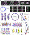
(A) 2D classification of smaller NINJ1 rings reconstituted with liposomes, showing heterogeneous rings. (B) Magnified images of 2 classes from (A). (C) Left: irregular large rings in detergents as represented by a low-resolution 3D reconstruction. These rings were segmented into short segments as single particles. Right: 2D classification of NINJ1 segments, showing clear secondary structure details. A pair of arrows point to TM α-hairpins in two neighboring subunits. (D) A magnified 2D class of NINJ1 segments. (E) Final electron density map of NINJ1 ring segment at 4.3 Å resolution, shown in three views. (F,G) Modeled NINJ1 rings from the natural curvature of the ring segment (F) and from an acuter curvature to construct smaller rings (G). (H) Cartoon representation of a NINJ1 monomeric subunit. Amphipathic helices α1 and α2 are in orange, and TM helices α3-α4 are in deep purple. Its relative orientations in a NINJ1 ring are illustrated. α3 and α4 are kinked at Gly residues. (I) Ribbon representation of 5 NINJ1 subunits in the NINJ1 ring segment. The middle subunit has a transparent surface over the ribbon diagram. The α1 helix extends over to the neighboring subunit to form a chain. (J) Ribbon representations (top) and electrostatic potentials (bottom) of NINJ1 concave side (left) and convex side (right) for the TM helices alone. (K) Electrostatic potential of the hydrophilic, convex NINJ1 surface shown in (I). (L) Model of a longer NINJ1 ring segment viewed from the extracellular side by propagating the ring segment oligomer, showing the clear curvature, the hydrophobic concave face with higher density, and the hydrophilic convex face with lower density. The subunit in (H) has to rotate by ~45° along the vertical axis, followed by a ~90° rotation along the horizontal axis to arrive at the orientation here. (M) Stacking in the NINJ1 double filament structure and the resulting straight TM helices and straight filament (PDB ID: 8CQR). See also Figures S2 and S3.
Figure 3.
Figure 3.. NINJ1 rings in THP-1 cells
All scale bars: 10 μM. (A,B) Fluorescence imaging of NINJ1 KO THP-1 cells reconstituted with NINJ1-GFP for GFP (NINJ1, green), anti-ASC (cyan), and Hoechst (DNA), for vehicle (A) or with Doxycycline induction (B). Cells were either untreated, primed with LPS (4 h) or primed with LPS (4 h) and treated with nigericin (1 h). Dox-induced cells showed NINJ1 in both untreated and LPS primed cells. After treatment with nigericin, NINJ1 intensity decreased. (C) Live cells fluorescence and phase images of the same THP-1 cells primed with LPS (top) or also treated with nigericin before membrane rupture (middle) and after membrane rupture (bottom). (D) Time lapse NINJ1-GFP fluorescence (green) and phase images of the same THP-1 cells imaged for 66 min with 2 min intervals. NINJ1 shedding occurred throughout, and complete cell membrane rupture was captured between 58 and 60 min. All data are representative of three or more independent experiments. See also Figure S3, S4 and Movie S1.
Figure 4.
Figure 4.. NINJ1-encircled membrane disks in activated THP-1 supernatant
(A) Western blots of NINJ1-GFP THP-1 cell pellet and supernatant fractions using anti-NINJ1 on a non-reducing SDS-PAGE. Treatment conditions for each lane are shown. Both NINJ1 oligomers were released to the supernatant after 1 h treatment with nigericin. * indicates non-specific band. Red boxes mark NINJ1 bands. (B) Ultracentrifugation pelleting of NINJ1-GFP supernatant from activated THP-1 cells. (C) Negative staining EM images of the supernatant of NINJ1-GFP THP-1 cells after ultracentrifugation, showing membrane disk like particles (yellow arrowheads). (D) Confocal imaging of NINJ1-GFP (green) in THP-1 cell supernatant co-stained with BODIPY lipid stain (magenta). Three NINJ1-containing structures are labeled for (E-G). The imaging showed that NINJ1-GFP rings were filled with lipids stained with BODIPY. (E-G) Line profiles of three ring structures in the field view shown in (D) for both GFP (green) and BODIPY (magenta) channels. All data are representative of three or more independent experiments.
Figure 5.
Figure 5.. Mutational analysis of NINJ1
(A) Ribbon presentation of NINJ1 monomeric subunit highlighting the different types of mutants that were tested. 1) Mutagenesis of Gly residues at the TM kinks, shown in magenta. 2) Mutagenesis of residues located at the hydrophilic face, shown in green. 3) Mutagenesis of residues located at the hydrophobic face, shown in cyan. 4) Mutagenesis of residues in a charged cluster as potential PI(4)P-binding site, shown in red. (B) Sequence alignment of NINJ1 and NINJ2 with mutated residues highlighted according to colors in (A). Residues in NINJ2 that were mutated to corresponding residues in NINJ1 are highlighted in yellow (Figures S7A–D). (C) Cytotoxicity assay of NINJ1 and mutants in HEK293T cells measured based on % of LDH release, at 24 h post transfection. The data are representative of three or more independent experiments. Statistical significance was assessed by 2-tailed t-test. ***, ** and * denote p < 0.0001, 0.001 and 0.01, respectively. (D) A table summarizing the mutational analysis in vitro and in cells. The mutants were scored based on the following assays: 1) oligomerization by EM imaging, 2) oligomerization by sucrose gradient, 3) liposome rupture, 4) cell death (LDH release), and 5) plasma membrane localization by confocal imaging. See also Figures S5–S7.
Figure 6.
Figure 6.. MINFLUX microscopy demonstrates that NINJ1 forms a heterogeneous population of ring-like structures during pyroptosis
LPS-primed primary mouse bone marrow-derived macrophages were stimulated to undergo pyroptosis with nigericin (20 μM, 30–60 min) or left untreated. Cells were fixed in 4% paraformaldehyde and 0.1% glutaraldehyde, followed by immunolabelling with anti-NINJ1 antibody (rabbit monoclonal clone 25, Genentech, Inc) and Alexa Fluor 647-conjugated whole IgG secondary antibody. Samples were then imaged by MINFLUX. (A) 2D-MINFLUX images (confocal inset) of LPS-primed macrophages without (control; left) or with pyroptosis stimulation (LPS + nigericin; right). Small dashed, white boxes indicate images shown in (B). Scale bars: 500 nm. (B-C) 3D-MINFLUX camera-perspective renderings of heterogenous NINJ1 structures in LPS-primed cells (B) or cells stimulated to undergo pyroptosis (C). The white cube in each has an edge length of 100 nm. The rainbow-colored scale indicates distance in the Z-plane, which ranges from −350 to +350 nm (B) or −400 to +450 nm (C). (D) Representative structures of each classification category, ring-like (i), discrete puncta (ii), or other (iii). Scale bar: 40 nm. (E) Largest measured inner diameter of the identified ring-like structures in the 2D plane ranged from 6 to 178 nm, shown with a median of 32.0 nm (interquartile range 24.0, 48.0 nm). (F) Pie charts for NINJ1 structures counted and classified as ring-like (i), discrete puncta (ii), or other (iii) for LPS primed control macrophages and pyroptotic macrophages at different time points. Data are representative of three independent experiments except that the pyroptosis (15 min) data in (F) are representative of two independent experiments. See also Figure S7, Movies S2 and S3.
Figure 7.
Figure 7.
A schematic model for the proposed mechanism of NINJ1 activation and membrane rupture

References

    1. Araki T, and Milbrandt J (1996). Ninjurin, a novel adhesion molecule, is induced by nerve injury and promotes axonal growth. Neuron 17, 353–361. - PubMed
    1. Araki T, Zimonjic DB, Popescu NC, and Milbrandt J (1997). Mechanism of homophilic binding mediated by ninjurin, a novel widely expressed adhesion molecule. J Biol Chem 272, 21373–21380. - PubMed
    1. Kayagaki N, Kornfeld OS, Lee BL, Stowe IB, O’Rourke K, Li Q, Sandoval W, Yan D, Kang J, Xu M, et al. (2021). NINJ1 mediates plasma membrane rupture during lytic cell death. Nature 591, 131–136. - PubMed
    1. Kim SW, Lee HK, Seol SI, Davaanyam D, Lee H, and Lee JK (2020). Ninjurin 1 dodecamer peptide containing the N-terminal adhesion motif (N-NAM) exerts proangiogenic effects in HUVECs and in the postischemic brain. Scientific reports 10, 16656. - PMC - PubMed
    1. Ifergan I, Kebir H, Terouz S, Alvarez JI, Lecuyer MA, Gendron S, Bourbonniere L, Dunay IR, Bouthillier A, Moumdjian R, et al. (2011). Role of Ninjurin-1 in the migration of myeloid cells to central nervous system inflammatory lesions. Annals of neurology 70, 751–763. - PubMed

Publication types

Substances