Simulator-Based Versus Traditional Training of Fundus Biomicroscopy for Medical Students: A Prospective Randomized Trial
- PMID: 38615132
- PMCID: PMC11109054
- DOI: 10.1007/s40123-024-00944-9
Simulator-Based Versus Traditional Training of Fundus Biomicroscopy for Medical Students: A Prospective Randomized Trial
Abstract
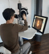
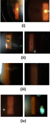
Introduction: Simulation training is an important component of medical education. In former studies, diagnostic simulation training for direct and indirect funduscopy was already proven to be an effective training method. In this prospective controlled trial, we investigated the effect of simulator-based fundus biomicroscopy training.
Methods: After completing a 1-week ophthalmology clerkship, medical students at Saarland University Medical Center (n = 30) were block-randomized into two groups: The traditional group received supervised training examining the fundus of classmates using a slit lamp; the simulator group was trained using the Slit Lamp Simulator. All participants had to pass an Objective Structured Clinical Examination (OSCE); two masked ophthalmological faculty trainers graded the students' skills when examining patient's fundus using a slit lamp. A subjective assessment form and post-assessment surveys were obtained. Data were described using median (interquartile range [IQR]).
Results: Twenty-five students (n = 14 in the simulator group, n = 11 in the traditional group) (n = 11) were eligible for statistical analysis. Interrater reliability was verified as significant for the overall score as well as for all subtasks (≤ 0.002) except subtask 1 (p = 0.12). The overall performance of medical students in the fundus biomicroscopy OSCE was statistically ranked significantly higher in the simulator group (27.0 [5.25]/28.0 [3.0] vs. 20.0 [7.5]/16.0 [10.0]) by both observers with an interrater reliability of IRR < 0.001 and a significance level of p = 0.003 for observer 1 and p < 0.001 for observer 2. For all subtasks, the scores given to students trained using the simulator were consistently higher than those given to students trained traditionally. The students' post-assessment forms confirmed these results. Students could learn the practical backgrounds of fundus biomicroscopy (p = 0.04), the identification (p < 0.001), and localization (p < 0.001) of pathologies significantly better with the simulator.
Conclusions: Traditional supervised methods are well complemented by simulation training. Our data indicate that the simulator helps with first patient contacts and enhances students' capacity to examine the fundus biomicroscopically.
Keywords: Augmented reality; Education; Fundus biomicroscopy; Ophthalmology; Simulation; Training.
© 2024. The Author(s).
Conflict of interest statement
Frank Koch, Berthold Seitz, Elias Flockerzi, Yaser Abu Dail, Tim Berger, Albéric Sneyers, and Svenja Deuchler are gratuitous consultants for Haag-Streit Simulation. Hanns Ackermann and Claudia Buedel have no competing interests to declare.
Figures


References
LinkOut - more resources
Full Text Sources

