Review
. 2024 Jun:72:103123.
doi: 10.1016/j.redox.2024.103123. Epub 2024 Mar 12.

Redox regulation of macrophages

Affiliations
Review

Redox regulation of macrophages

Nhien Tran et al. Redox Biol. 2024 Jun.

Abstract

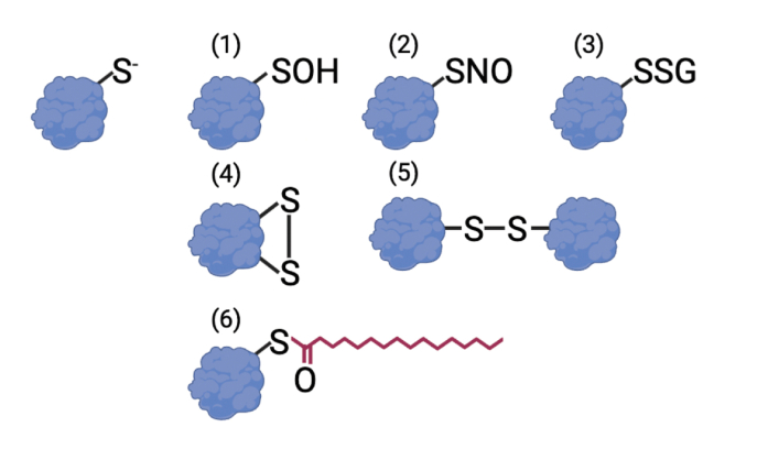
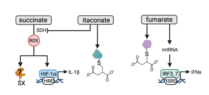
Redox signaling, a mode of signal transduction that involves the transfer of electrons from a nucleophilic to electrophilic molecule, has emerged as an essential regulator of inflammatory macrophages. Redox reactions are driven by reactive oxygen/nitrogen species (ROS and RNS) and redox-sensitive metabolites such as fumarate and itaconate, which can post-translationally modify specific cysteine residues in target proteins. In the past decade our understanding of how ROS, RNS, and redox-sensitive metabolites control macrophage function has expanded dramatically. In this review, we discuss the latest evidence of how ROS, RNS, and metabolites regulate macrophage function and how this is dysregulated with disease. We highlight the key tools to assess redox signaling and important questions that remain.


Conflict of interest statement

Declaration of competing interest None.

Figures

Fig. 1
Fig. 1
Reversible post-translational modification (PTM) of cysteine residues. Free thiolates (top left) can undergo a range of reversible PTMs. These include sulfenylation (1), S-nitrosylation (2), glutathionylation (3), intra- (4), and inter- (5) molecular disulfide bonds, and palmitoylation (6).
Fig. 2
Fig. 2
Cysteine PTM of proteins of the NF-κB pathway. NF-κB is a master transcription factor that plays a central role in regulation of inflammatory gene expression in macrophages, therefore, its activity must be carefully regulated. Several components of the NF-κB pathway are redox-regulated. ROS can oxidize cysteine (Cys)62 of the p50 subunit to inhibit NF-κB DNA binding and transcriptional activity (1). Cys62 of p50 can also undergo glutathionylation and S-nitrosylation, both of which impair DNA binding and recognition. S-nitrosylation of Cys38 of the p65 subunit reduces p50-p65 heterodimer formation and decreases NF-κB DNA binding while sulfhydration of Cys38 increases DNA binding and transcription activity of NF-κB in macrophages (2). Glutathionylation Cys179 of inhibitor of NF-κB (IκB) kinase (IKK)β suppresses its kinase activity thereby dampening NF-κB activity (3). An intermolecular disulfide bond between Cys54 and Cys347 of NF-κB essential modulator (NEMO), which is a subunit of IKK, is critical for the stabilization of NEMO, assembly of the IKK complex, and NF-κB activation (4).
Fig. 3
Fig. 3
Cysteine PTMs in the regulation of pattern recognition receptors (PRRs). PRRs such as TLRs, the cyclic GMP-AMP synthase-stimulator of interferon genes (cGAS-STING) pathway, and nucleotide oligomerization domain (NOD)–like receptors (NLRs) are redox-regulated. Glutathionylation of myeloid differentiation protein 88 (MyD88) adaptor-like protein (MAL; Cys91; 1) and palmitoylation of Cys113 of MyD88 (2) enhance TLR activity. Palmitoylation of Cys88 and Cys91 of STING (3) is essential for STING signaling. Palmitoylation of several cysteine residues on NOD1 (Cys558, 567, & 952; 4) and NOD2 (Cys 395 & 1033; 5) is required for NOD1/2 membrane recruitment during bacterial challenge in macrophages.
Fig. 4
Fig. 4
Redox regulation of NLRP3. ROS modulate the NLRP3 inflammasome, a protein complex that mediates IL-1β processing and secretion. ROS enhance NIMA-related kinase 7 (NEK7) phosphorylation and its interaction with NLRP3 through a poorly defined mechanism. Additionally, deglutathionylation of Cys253 on NEK7 is important for its interaction with NLRP3 (1). Itaconate derivatives targets Cys548 of the NLRP3 inflammasome to prevent its interaction with NEK7 (2). ROS oxidize Cys192 of GSDMD to enhance its pore-forming activity (3) while palmitoylation of Cys192 of GSDMD leads to membrane translocation of GSDMD and the subsequent pore-formation and pyroptosis. Furthermore, ROS promote cleavage of GSDMD, mobilization to the plasma membrane, oligomerization and eventually pore formation. GSDMD is also regulated by succination and alkylation by itaconate.
Fig. 5
Fig. 5
Redox regulation of macrophages by metabolites. Metabolites also contribute to redox signaling in macrophages. Oxidation of succinate by succinate dehydrogenase (SDH), promotes complex I-derived mtROS production, and inflammation in a hypoxia inducible factor (HIF)-1α-dependent manner. It remains to be determined if mtROS driven by succinate oxidation promotes oxidation of specific cysteine residues in target proteins to modulate inflammatory function. Itaconate has potent immunomodulatory effects in macrophages. Two major mechanisms underpinning the immunomodulatory activity of itaconate are SDH inhibition and 2,3-dicarboxypropylation or alkylation of cysteine residues. Several alkylation targets of itaconate have emerged. These include cysteines on KEAP1, the NLRP3 inflammasome, gasdermin D (GSDMD), residues on several metabolic enzymes, including glyceraldehyde 3-phosphate dehydrogenase (GAPDH), aldolase A (ALDOA), and lactate dehydrogenase A (LDHA). Fumarate reacts with thiols in cysteine residues in a PTM termed succination. Many of the protein succination targets of fumarate are shared with the alkylation targets of itaconate including KEAP1, GSDMD, and GAPDH. Fumarate can also promote the release of mtRNA, and retinoic acid-inducible gene I (RIG-I) and melanoma differentiation-associated protein 5 (MDA5) activity to promote interferon signaling.

References

    1. Forman H.J., Ursini F., Maiorino M. An overview of mechanisms of redox signaling. J. Mol. Cell. Cardiol. 2014;73:2–9. doi: 10.1016/j.yjmcc.2014.01.018. - DOI - PMC - PubMed
    1. McCord J.M., Fridovich I. Superoxide dismutase. An enzymic function for erythrocuprein (hemocuprein) J. Biol. Chem. 1969;244:6049–6055. - PubMed
    1. Bae Y.S., et al. Epidermal growth factor (EGF)-induced generation of hydrogen peroxide. Role in EGF receptor-mediated tyrosine phosphorylation. J. Biol. Chem. 1997;272:217–221. doi: 10.1074/jbc.272.1.217. - DOI - PubMed
    1. Beltran B., Orsi A., Clementi E., Moncada S. Oxidative stress and S-nitrosylation of proteins in cells. Br. J. Pharmacol. 2000;129:953–960. doi: 10.1038/sj.bjp.0703147. - DOI - PMC - PubMed
    1. Sundaresan M., Yu Z.X., Ferrans V.J., Irani K., Finkel T. Requirement for generation of H2O2 for platelet-derived growth factor signal transduction. Science. 1995;270:296–299. doi: 10.1126/science.270.5234.296. - DOI - PubMed

Publication types

LinkOut - more resources