. 2025 Mar:69:125-138.
doi: 10.1016/j.jare.2024.04.010. Epub 2024 Apr 12.

Urolithin A inhibits breast cancer progression via activating TFEB-mediated mitophagy in tumor macrophages

Affiliations

Urolithin A inhibits breast cancer progression via activating TFEB-mediated mitophagy in tumor macrophages

Bowen Zheng et al. J Adv Res. 2025 Mar.

Erratum in

Abstract

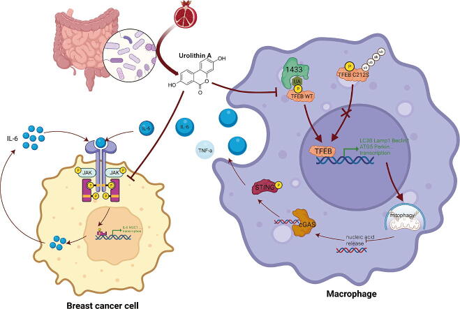
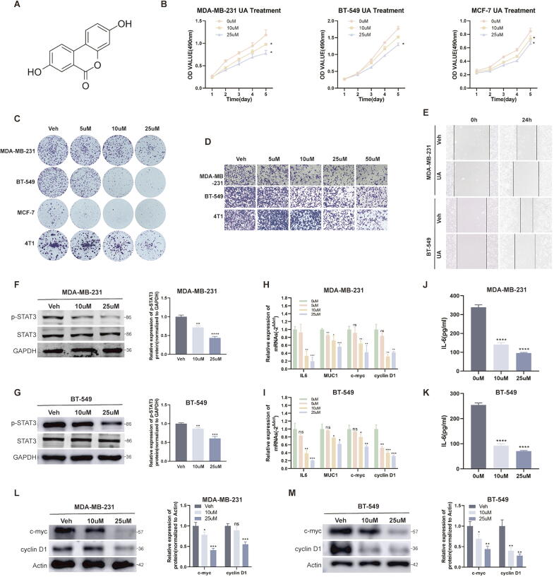
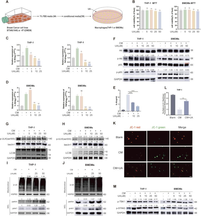
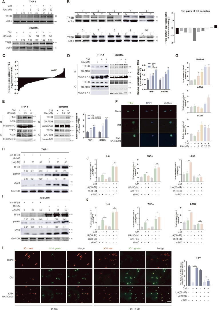
Introduction: Urolithin A (UA) is a naturally occurring compound that is converted from ellagitannin-like precursors in pomegranates and nuts by intestinal flora. Previous studies have found that UA exerts tumor-suppressive effects through antitumor cell proliferation and promotion of memory T-cell expansion, but its role in tumor-associated macrophages remains unknown.

Objectives: Our study aims to reveal how UA affects tumor macrophages and tumor cells to inhibit breast cancer progression.

Methods: Observe the effect of UA treatment on breast cancer progression though in vivo and in vitro experiments. Western blot and PCR assays were performed to discover that UA affects tumor macrophage autophagy and inflammation. Co-ip and Molecular docking were used to explore specific molecular mechanisms.

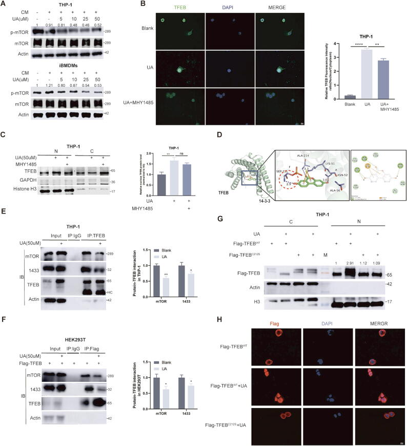
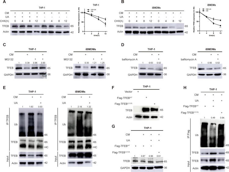
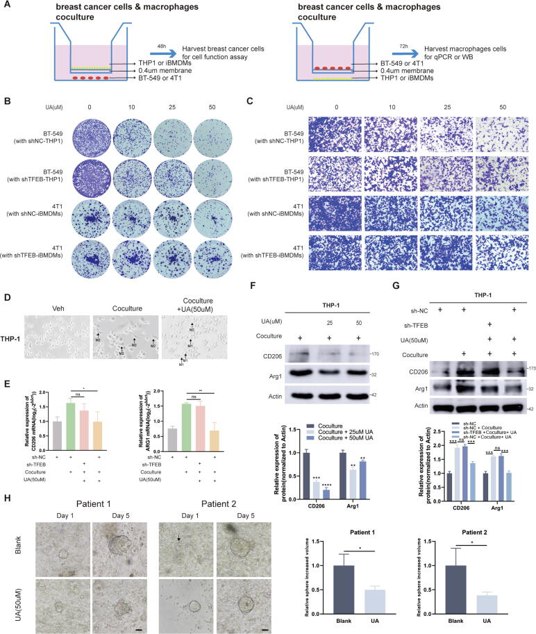
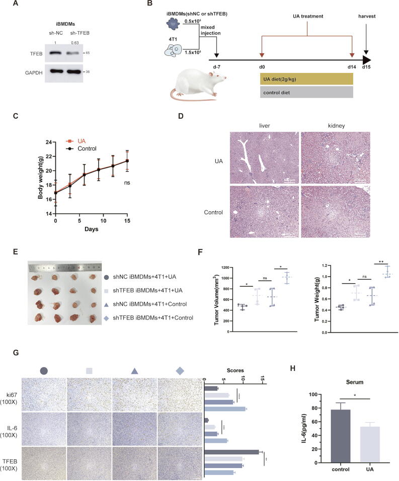
Results: We observed that UA treatment could simultaneously inhibit harmful inflammatory factors, especially for InterleuKin-6 (IL-6) and tumor necrosis factor α (TNF-α), in both breast cancer cells and tumor-associated macrophages, thereby improving the tumor microenvironment and delaying tumor progression. Mechanistically, UA induced the key regulator of autophagy, transcription factor EB (TFEB), into the nucleus in a partially mTOR-dependent manner and inhibited the ubiquitination degradation of TFEB, which facilitated the clearance of damaged mitochondria via the mitophagy-lysosomal pathway in macrophages under tumor supernatant stress, and reduced the deleterious inflammatory factors induced by the release of nucleic acid from damaged mitochondria. Molecular docking and experimental studies suggest that UA block the recognition of TFEB by 1433 and induce TFEB nuclear localization. Notably, UA treatment demonstrated inhibitory effects on tumor progression in multiple breast cancer models.

Conclusion: Our study elucidated the anti-breast cancer effect of UA from the perspective of tumor-associated macrophages. Specifically, TFEB is a crucial downstream target in macrophages.

Keywords: Breast cancer; Mitophagy; TFEB; Tumor-associated macrophages; Urolithin A.


Conflict of interest statement

Declaration of Competing Interest The authors declare that they have no known competing financial interests or personal relationships that could have appeared to influence the work reported in this paper.

Figures

None
Graphical abstract
Fig. 1
Fig. 1
UA inhibits breast cancer cell proliferation and migration. A Chemical structural formula of UA. B MTT assays were performed to compare the proliferative capacity of breast cancer cells after UA treatment at different concentrations. C Colony formation assays were performed to detect the proliferative capacity of breast cancer cells after UA treatment at different concentrations. D, E Transwell migration and wound healing assays were conducted to detect the migration capacity of breast cancer cells after UA treatment. F, G Total STAT3 and phosphorylated STAT3 protein levels in breast cancer cells were detected by western blot after UA treatment. H, I The expression level of relevant mRNA after UA treatment were detected by real-time quantitative PCR. J, K IL-6 abundance in tumor supernatants after UA treatment was detected by ELISA assay. L, M c-myc and cyclin D1 protein levels in breast cancer cells were detected by western blot after UA treatment. Data are presented as mean ± SEM; Student's t-test was performed using Graphpad Prism. *P < 0.05, **P < 0.01, ***P < 0.001, ****P < 0.0001.
Fig. 2
Fig. 2
UA activates mitophagy in tumor macrophages to inhibit IL-6 secretion. A The process of acquisition of tumor-conditioned media and intervention in macrophages. B MTT assays were performed to compare the cell viability after UA treatment. C, D IL-6 and TNF-α mRNA levels in THP-1 and BMDMs after UA and CM treatment were detected by real-time quantitative PCR. E IL-6 abundance in THP-1 cell supernatants after UA and CM treatment was detected by ELISA assay. F NF-κb pathway-associated protein levels in THP-1 and BMDMs were detected by western blot after UA and CM treatment. G, H Macroautophagy-associated protein levels in THP-1 and BMDMs were detected by western blot after UA and CM treatment. I, J Mitophagy-associated protein levels in THP-1 and BMDMs were detected by western blot after UA and CM treatment. (WCL = whole cell lysate) K, L JC-1 assays were performed to detect mitochondrial membrane potential changes after UA and CM treatment. M Phosphorylated STING and TBK protein levels in THP-1 and BMDMs were detected by western blot. Data are presented as mean ± SEM; Student's t-test was performed using Graphpad Prism. *P < 0.05, **P < 0.01, ***P < 0.001, ****P < 0.0001.
Fig. 3
Fig. 3
UA-mediated mitophagy is dependent on TFEB nuclear translocation. A TFEB protein levels in THP-1 and iBMDMs were detected by western blot after 36 h UA and CM treatment. B Comparison of TFEB protein levels in 10 pairs of breast cancer and normal tissues. (N = normal and T = tumor) C Comparison of TFEB mRNA expression levels in 68 pairs of breast cancer and normal tissues. D, E TFEB protein levels in cytoplasm and nucleus were detected by western blot after 8 h UA and CM treatment. (N = nucleus and C = cytoplasm) F An IF assay further confirmed that UA treatment affected the subcellular localization of TFEB. The fluorescence intensity ratio of the nucleus and cytoplasmic fraction was calculated using the ImageJ software. G Macroautophagy-associated mRNA levels in THP-1 and iBMDMs after 24 h UA and CM treatment were detected by real-time quantitative PCR. H, I Knockdown of TFEB followed by UA treatment and detection of LC3B, parkin, and TFEB protein levels in THP-1 and iBMDMs. sh-TFEB is the shRNA targeting TFEB, and sh-NC is the shRNA for the negative control. Not described later. J, K Knockdown of TFEB followed by UA treatment and detection of IL-6, TNF-α, LC3B mRNA levels in THP-1 and iBMDMs. L JC-1 assays were performed to detect mitochondrial membrane potential changes in THP-1. Data are presented as mean ± SEM; Student's t-test (D, E, G) and one-way ANOVA (J, K, L) were performed using Graphpad Prism. *P < 0.05, **P < 0.01, ***P < 0.001, ****P < 0.0001.
Fig. 4
Fig. 4
UA blocks recognition of phosphorylated TFEB by 1433. A Protein levels of mTOR and p-mTOR were detected by WB after CM and UA treatment. B Immunofluorescence observation of TFEB subcellular localization. C Protein level of TFEB in the nucleus and cytoplasmic components was detected by WB. (N = nucleus and C = cytoplasm) D Molecular docking of UA and TFEB-1433 complexes using Autodock software. E, F Observe changes in TFEB and 1433 protein binding after UA treatment. (HC = heavy chain) G THP-1 cells were infiltrated with viral fluids containing wild-type (WT) TFEB and C212S mutant TFEB plasmids, and changes in TFEB protein levels in the nucleus and cytoplasm were observed by WB after UA treatment. (N = nucleus and C = cytoplasm) H THP-1 cells were infiltrated with viral fluids containing wild-type TFEB and C212S mutant TFEB plasmids, then immunofluorescence experiments were performed to observe the subcellular localization of TFEB after UA treatment. Data are presented as mean ± SEM; Student's t-test (E, F) and one-way ANOVA (B, C) were performed using Graphpad Prism. *P < 0.05, **P < 0.01, ***P < 0.001, ****P < 0.0001.
Fig. 5
Fig. 5
UA inhibits TFEB ubiquitination degradation under CM stress. A, B CHX treatments were performed to observe the half-life of TFEB protein in THP-1 and iBMDMs. C, D CM pretreatment was performed for 36 h, MG132 or BAF treatment was performed, and observed the protein level of TFEB. E CM and UA were pretreated for 36 h, the lysis products were immunoprecipitated with TFEB antibody, and western blotting was performed to observe the ubiquitination level of TFEB. (HC = heavy chain) F, G THP-1 cells were infiltrated with viral fluids containing wild-type TFEB and C212S mutant TFEB plasmids. Observed the protein levels of TFEB in UA, CM-treated, and non-treated groups. H THP-1 cells were infiltrated with viral fluids containing wild-type TFEB and C212S mutant TFEB plasmids, then pretreated with CM and UA for 36 h, the lysis products were immunoprecipitated with TFEB antibody, and western blotting was performed to observe the ubiquitination level of TFEB. Data are presented as mean ± SEM; Student's t-test was performed using Graphpad Prism. *P < 0.05, **P < 0.01, ***P < 0.001, ****P < 0.0001.
Fig. 6
Fig. 6
UA treatment inhibits breast cancer in tumor macrophage co-culture and organoid models. A Schematic showing the cancer cells cocultured with macrophages in a transwell chamber of 0.4-um pore size. B Colony formation assays were performed to detect the proliferative capacity of BT-549 and 4 T1 cells after coculturing with macrophages and UA treatment. C Transwell migration assays were conducted to detect the migration capacity of BT-549 and 4 T1 cells after coculturing with macrophages and UA treatment. D Morphologic changes of THP-1 after co-culture and UA treatment was observed by a light microscopy. E CD206 and Arg1 mRNA levels in THP-1 were detected by real-time quantitative PCR after co-culture and UA treatment. F, G CD206, and Arg1 protein levels in THP-1 were detected by western blot after co-culture and UA treatment. H The effect of UA treatment on the growth of organoid cell spheres of origin in breast cancer patients was observed by a light microscopy (Shows images at day two and day seven). Data are presented as mean ± SEM; Student's t-test (F, H) and one-way ANOVA (E, G) were performed using Graphpad Prism. *P < 0.05, **P < 0.01, ***P < 0.001, ****P < 0.0001.
Fig. 7
Fig. 7
UA treatment inhibits tumor growth in BALB/c mice. A Constructing iBMDMs stably expressing sh-NC and sh-TFEB, knockdown efficiency was detected by western blot. B Schematic diagram of mice injected with mixed cells and treated with UA. C The weight of the mice varied with time. D HE staining of liver and kidney in UA-treated and control mice. E Tumor images for each group. F Volume and weight of tumors in each group. (The grouping is consistent with the icon of Fig. 5E) G Immunohistochemical images of various groups of tumors, including ki-67, IL-6, and TFEB, as well as scores of different indicators. (The grouping is consistent with the icon of Fig. 5E) H ELISA assay measures the IL-6 levels in mice plasma. Data are presented as mean ± SEM; Student's t-test (C, H) and one-way ANOVA (F, G) were performed using Graphpad Prism. *P < 0.05, **P < 0.01, ***P < 0.001, ****P < 0.0001.

Similar articles

Cited by

References

    1. Giaquinto A.N., Sung H., Miller K.D., Kramer J.L., Newman L.A., Minihan A., et al. Breast cancer statistics, 2022. CA Cancer J Clin. 2022 doi: 10.3322/caac.21754. - DOI - PubMed
    1. Zheng T., Liu H., Hong Y., Cao Y., Xia Q., Qin C., et al. Promotion of liquid-to-solid phase transition of cGAS by baicalein suppresses lung tumorigenesis. Sig Transduct Target Ther. 2023;8:133. doi: 10.1038/s41392-023-01326-6. - DOI - PMC - PubMed
    1. Ryu D., Mouchiroud L., Andreux P.A., Katsyuba E., Moullan N., Nicolet-Dit-Félix A.A., et al. Urolithin a induces mitophagy and prolongs lifespan in C. elegans and increases muscle function in rodents. Nat Med. 2016;22:879–888. doi: 10.1038/nm.4132. - DOI - PubMed
    1. Singh A., D’Amico D., Andreux P.A., Fouassier A.M., Blanco-Bose W., Evans M., et al. Urolithin a improves muscle strength, exercise performance, and biomarkers of mitochondrial health in a randomized trial in middle-aged adults. Cell Rep Med. 2022;3 doi: 10.1016/j.xcrm.2022.100633. - DOI - PMC - PubMed
    1. Mohammed Saleem Y.I., Albassam H., Selim M. Urolithin a induces prostate cancer cell death in p53-dependent and in p53-independent manner. Eur J Nutr. 2020;59:1607–1618. doi: 10.1007/s00394-019-02016-2. - DOI - PubMed

MeSH terms

Substances

LinkOut - more resources